Neurally adjusted ventilatory assist in pediatrics:
why, when, and how?
Introduction
In pediatrics, good synchrony in controlled assisted ventilation is not always possible and may delay recovery, prolong mechanical ventilation (MV), and
contribute to loss of muscle strength and increased calorie expenditure.(1)
In controlled assisted ventilation, the trigger (drive) is a decisive factor in
the release of the assisted cycle, as it is regulated by the pressure diference or low diference in the system. Very sensitive triggers induce hyperventilation and atrophy of respiratory muscles, whereas less sensitive systems require more efort, inducing hypoventilation, excessive energy expenditure, and discomfort. Even with adequate sensitivity, there is a delay in the release of the assisted cycle resulting from the interval between the central nerve impulse and the respiratory muscle contraction to initiate the trigger. Air leakage around the tracheal tube is a limiting factor that may not be perceived or compensated for by the device, requiring even greater efort by the child.
Neurally adjusted ventilatory assist (NAVA; Maquet®
, Sweden) is a minimally invasive technology that releases proportional pressure cycling in response to electrical activity of the diaphragm (EAdi), adapting ventilatory
support to the patient’s actual demand.(2) hus, the patient, through his neural
drive, regulates the frequency of cycles and the volume to be released in each of them, with the beneits of avoiding hyper- or hypoventilation of support, preserving the EAdi, increasing the interaction with the ventilator, not being inluenced by air leaks around the tracheal tube, and, especially, incorporating the natural variability of breathing. In short, the mechanical ventilator in the NAVA mode divides the load with the diaphragm to support the ventilation in a synchronized and proportional way and can be used in an invasive or non-invasive way (NIV-NAVA).
he NAVA mode was irst used in Latin America in 2009 with an adult population, and since then, only 15 pediatric studies have been published, where
the neonatal population predominates.(2,3) Justiications for underutilization of
the NAVA mode in pediatric intensive care units (ICU) include theoretical and unconirmed concepts in large studies, high cost, lack of reference values for levels of electrical activity, and the impact of this monitoring and the ventilatory strategy on clinical outcomes. here is a lack of understanding of the ventilatory and monitoring possibilities that this tool can ofer the clinician at the bedside. Lívia Barboza Andrade1,2, Rodrigo Guellner
Ghedini2, Alexandre Simões Dias2, Jefferson Pedro Piva2
1. Instituto de Medicina Integral Prof. Fernando Figueira) - Recife (PE), Brazil.
2. Pediatric Intensive Care Unit, Hospital de Clínicas de Porto Alegre), Universidade Federal do Rio Grande do Sul - Porto Alegre (RS), Brazil.
Conflicts of interest: None.
Submitted on June 8, 2017 Accepted on July 20, 2017
Corresponding author: Lívia Barboza Andrade
Instituto de Medicina Integral Prof. Fernando Figueira
Rua dos Coelhos, 300 - Boa Vista Zip code: 50070-550 - Recife (PE), Brazil E-mail: [email protected]
Responsible editor: Thiago Costa Lisboa
Assistência ventilatória ajustada neuralmente em pediatria: por
que, quando e como?
Why monitor the electrical activity of the diaphragm?
In spontaneous ventilation, the tidal volume generated is proportional to the intensity of contractility of the respiratory muscles, especially the diaphragm. he intensity of this contraction results from the interaction of several factors: aferent information on lung inlation and delation, arterial gases, and diaphragm contractile capacity (sedation and atrophy), among others. herefore, the neural respiratory drive identiies and responds to various factors, generating an EAdi proportional to the ventilation requirements. he EAdi signal is measured and used to trigger the assisted inspiration, releasing an inspiratory pressure proportional to the electrical activity. he ventilatory cycle ends when a 30% reduction in the
EAdi peak is observed.(2) his allows for synchronization
between the electrical activity of the patient and the pressure generated in the ventilator in terms of time and proportionality.
Regardless of ventilation under NAVA, the EAdi waveform can be used to monitor neural respiratory rate, which presents a cyclic characteristic with a pattern of variations between maximum (phasic EAdi) and minimum values (tonic EAdi), whose mean in infants and children varies between 8 and 20 microvolts, with a tendency to
higher values in non-invasive modes(2,4) (Figure 1).
Tonic EAdi persists until the end of expiration above the baseline and is usually absent in healthy adults and children older than 1 year old. In newborns and infants, it is higher to actively maintain the lung volume at the end of the expiration above the volume of relaxation, thus preventing alveolar derecruitment. Other mechanisms are involved in this process, such as rapid respiratory rate with short expiratory time and delayed expiratory low by constriction of the larynx. In intubated children, the tracheal cannula prevents laryngeal braking, further reinforcing the importance of the tonic activity of the diaphragm, which can be evaluated continuously through
the minimal EAdi.(6)
Neural inspiratory eforts (sighs), as well as periods of lat EAdi during central apnea, can also be observed. Compared with adults, the signal in children shows high variability, with a higher tonic activity in pre-term and in non-invasively ventilated patients.
A large study in Canada evaluated EAdi in children undergoing conventional ventilation, in the acute phase of the disease, in the pre- and post-extubation period, and in the ICU discharge. Peak EAdi values were markedly
suppressed in the acute phase (3.6μV) and increased to
4.8μV in the pre-extubation period. here were periods
of total diaphragm inactivity in the acute phase, even with low levels of care. Shortly after extubation, the EAdi
Figure 1 - Demonstration of pressure, flow, and electrical activity curves of the diaphragm. (A) Maximum electrical activity of the diaphragm, showing phasic activity of the diaphragm, and minimal electrical activity of the diaphragm, or tonic activity. (B) Proportional increase in airway
pressure in response to the corresponding increase in electrical activity of the diaphragm. EAdi - electrical activity of the diaphragm; flow - flow; paw - airway
increased to 15μV and remained high (13 - 15μV) until discharge from the ICU. Children with lung disease had higher electrical activity, while low EAdi in the acute phase may be caused by the use of sedation and over-assistance
of MV.(7)
In Finland, EAdi was measured in 81 children (with lung disease and post-surgery) in NAVA mode and 1 hour post-extubation. When ventilated, the NAVA level was
adjusted to maintain peak EAdi between 5 and 15μV.
Children with pulmonary disease presented higher EAdi levels than post-surgical patients at all stages of treatment. After extubation, children with pulmonary disease have,
on average, 20μV compared to post-surgery children, who
presented 9μV.(8)
here are several citations in cross-sectional studies and case series on EAdi monitoring, such as in cases of diaphragm paralysis, central hypoventilation, preterm weaning, infants with viral bronchiolitis, children with
diicult weaning, and respiratory control disorders.(2,9)
Monitoring the EAdi allows clinicians to adapt ventilatory parameters in an individualized way, avoiding the over-assistance and consequent diaphragm atrophy (injury due to disuse). he increase in peak EAdi levels suggests insuicient ventilatory support; in contrast, a strong tonic activity may relect the child’s efort to increase his lung volume.
NAVA mode: mean airway pressure and lung protection
In spontaneous breathing, as lung inlation progresses, pulmonary stretch receptors behave as sensors that inform adequate inspiratory volume and “turn of” inspiration.
In NAVA mode, in which neural inspiration also controls delivery of care, the ventilatory cycle may be discontinued when neural exhalation begins. Some studies show that in spontaneous breathing, children have lower airway mean pressures and tidal volumes very similar to
those found in NAVA mode.(10-12) he justiication for
this behavior is the relex control of the ventilator, which promotes better comfort and synchronization due to lower electrical activity and, consequently, lower mean pressure.
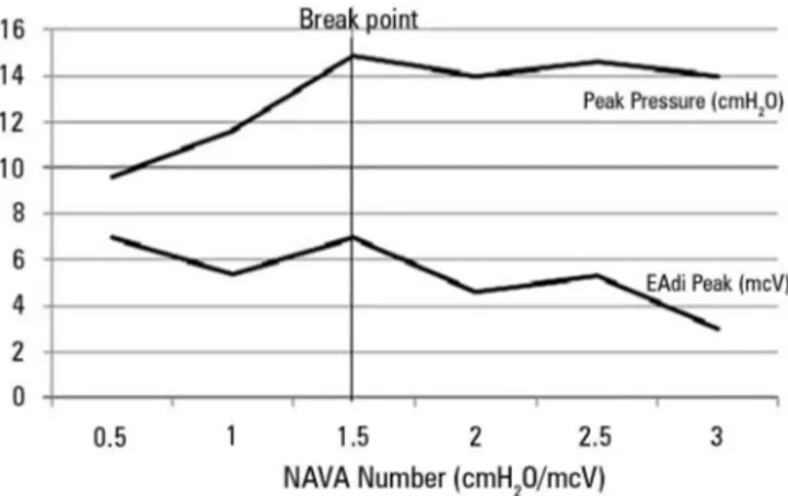
In one study, premature infants presented downregulation of EAdi to avoid overdistension when submitted to a gradual increase in the NAVA level
(0.5cmH2O every 3 minutes) until reaching 4cmH2O. In
the initial portion of the experiment, an increase in the positive inspiratory pressure (PIP) proportional to the increase in assistance was observed, which occurred to a
certain extent where the pressure did not increase. he authors (Figure 2) called this point a breakpoint (a plateau was observed in the PIP). he behavior of tidal volume
also followed a similar pattern.(13)
Figure 2 - Increase in the positive inspiratory pressure and the electrical activity of the diaphragm according to the level of neurally adjusted ventilatory
assist increases until the breaking point (1.5cmH2O/mcV) in premature infants.
NAVA - neurally adjusted ventilatory assist. Source: Stein H, Alosh H, Ethington P, White DB. Prospective crossover comparison between NAVA and pressure control ventilation in premature neonates less than 1500 grams. J Perinatol 2013;33(6):452-6.(13)
Another study showed reduced respiratory muscle load and lower PIP when premature infants were ventilated in NAVA mode compared to synchronized intermittent mandatory ventilation (SIMV) associated with pressure
support ventilation.(14)
he reduction of the pressures observed in the abovementioned studies was associated with the reduction
of the partial pressure of carbon dioxide (PaCO2), the
improvement of oxygen partial pressure/inspired oxygen
fraction (PaO2/FiO2), and the time of weaning, without
hemodynamic impact.
Variability of breathing
In contrast to constant ventilation in conventional modes, the variability of pressures and volumes in neural ventilation is high, as it relects the respiratory center
output.(15) Biological systems are characterized by their
intrinsic variability, called noisiness, which is opposed to monotonic behaviors observed in mechanical systems. Reduction in respiratory variability is associated with
adverse outcomes.(15,16)
PIP = [Level of NAVA x Δ EAdi (max - min)] + PEEP rhythmicity of the respiratory pattern compared to
healthy controls in spontaneous breathing. NAVA was the mode that presented greater variability, resembling the
controls.(15) In children who were sick, greater comfort was
also observed when ventilated in NAVA, instead of PSV; still, there was better synchrony, reduction of ventilatory
drive, and increased respiration variability.(17)
Patient-ventilator interaction
Asynchrony between the patient and ventilator is considered an important cause of cyanotic episodes and can result in large tidal volumes, air trapping, blood pressure luctuations, and worsening of oxygenation. Similar to what occurs in adults, 16 studies involving infants and children observed that the interaction is better in NAVA mode compared to controlled modes. However, asynchrony indices are quite varied in these studies: 12 to 73% in conventional modes compared to 0 to 20%
in NAVA mode.(2) his better assistance is due to more
sensitive and accurate drive mechanisms, correct cycling, and proportionality of efort assistance.
Practical aspects in the use of NAVA in pediatrics
he EAdi signal is picked up by electrodes embedded in the distal part of the catheter, positioned at the level of
the crural diaphragm. he passage of the catheter has been described as safe and easy, allowing its use for infusion
of diet, without interfering in the signal quality.(18,19) In
the insertion, it is suggested to use the measurements of the distances between the nose, the lobe of the ear, and xiphoid appendix in the formula indicated by the manufacturer. he catheter is adequate when the central electrode is at the height of the diaphragm and is visible on the ventilator screen with the presence of blue signals in the central curves (Figure 3).
After conirmation of the positioning, with good capture of the EAdi signal, titration of the NAVA level begins, and
minimum assistance values between 0.5 and 2cmH2O/μV
(up to 4 in children) are recommended. Lower values are not interpreted as ventilatory drives. he magnitude of the mechanical assistance varies with each breath according to the EAdi and the gain factor (NAVA level). In practical terms, the “NAVA level” is the factor to be multiplied in the EAdi to generate a certain inspiratory pressure. Setting a very low NAVA level requires an excessive diaphragm load to generate PIP, while high NAVA values require less efort and induce muscle atrophy. he mathematical equation of the relationship between PIP and EAdi can be expressed as follows:
Figure 3 - (A) Lines marked in blue on the electrocardiographic tracing demonstrate adequate positioning of the catheter for measuring diaphragm electrical activity. (B) Simultaneous recording of electrical activity. (1) Schematic of the positioning of the catheter and its outputs for feeding and coupling with the neurally adjusted ventilation assist cable. (2) Probe in the esophagogastric position. (C) Neurally adjusted
ventilation assist cable that attaches to the mechanical ventilator. Source: Adapted from Stein H, Firestone K, Rimesberg P. Synchronized mechanical ventilation using
Table 1 - Pediatric studies investigating the use of neurally adjusted ventilatory assist in an invasive manner compared with controlled ventilation in pneumatic modes
Author Number of
patients Type of study Outcomes Results
Clement et al.(9) 33 Crossover Ventilator response time, inspiratory efforts, and breathing work
NAVA demonstrated a shorter response time, reduced trigger, reduced workload (lower pressure/ time product)
Alander et al.(11) 18 Crossover Index of asynchrony (analysis of ineffective efforts and self-trigger), analysis of airway pressures, vital signs
IA (NAVA) = 08 IA (CMV) = 28 Lower PIP and MAP
de la Oliva et al.(17) 12 Non-randomized crossover
Index of asynchrony (ineffective effort and self-trigger analysis), respiratory variability, COMFORT score
IA (NAVA) = 2 IA (CMV) = 12
Better variability and comfort scores
Breatnach et al.(20) 16 Crossover Asynchrony (trigger and cycling), analysis of airway pressures
Better synchrony, reduced PIP and MAP levels in NAVA mode
Bordessoule et al.(21) 10 Case series Index of asynchrony (ineffective effort and self-trigger analysis), respiratory variability
IA (NAVA) = 11 IA (CMV) = 25
NAVA has greater variability of EAdi that is brought about in ventilator pressure variability
Vignaux et al.(22) 19 Crossover, randomized, prospective
Index of asynchrony (analysis of ineffective efforts and self-trigger)
IA (NAVA) = 4 IA (CMV) = 29
Kallio et al.(23) 170 Randomized clinical trial
Ventilation time, ICU stay, required amount of sedation, ventilation parameters
Lower MV time and pediatric ICU stay.
Sedation was lower in NAVA in clinical patients (no significance in surgical patients).
Lower FiO2 and PIP
NAVA - neurally adjusted ventilatory assist; IA - index of asynchrony; CMV - conventional mechanical ventilation; PIP - positive inspiratory pressure; MAP - mean airway pressure; EAdi - electrical activity of the diaphragm; ICU - intensive care unit; FiO2 - inspired oxygen fraction.
he target EAdi peak should be between 5 and 15μV,
considering the breathing luctuations. hus, positive
end-expiratory pressure (PEEP), FiO2, and NAVA level
are the only predeined parameters. For safety, upper pressure limits must be deined and backup ventilation must be ready, which automatically activates if EAdi does not occur.
Pediatric studies
Table 1 summarizes the main pediatric studies comparing NAVA with pneumatic ventilatory modes. No studies with NIV-NAVA were included.
Final comments
Current studies indicate that neural ventilation in infants and children is better tolerated compared to conventional ventilatory modes. It appears to be safe, it has better patient-ventilator interaction, provides comfort, requires a lower level of sedation, shortens length of stay, and ofers monitoring of electrical activity. However, its long-term role is still uncertain, especially regarding the duration of mechanical ventilation, length of stay, and mortality in children.
REFERENCES
1. Hudson MB, Smuder AJ, Nelson WB, Bruells CS, Levine S, Powers SK. Both high level pressure support ventilation and controlled mechanical ventilation induce diaphragm dysfunction and atrophy. Crit Care Med. 2012;40(4):1254-60.
2. Beck J, Emeriuad G, Liu Y, Sinderby C. Neurally-adjusted ventilator assist (NAVA) in children: a systematic review. Minerva Anestesiol. 2016;82(8):874-83.
3. Beck J, Tucci M, Emeriaud G, Lacroix J, Sinderby C. Prolonged neural expiratory time induced by mechanical ventilation in infants. Pediatr Res. 2004;55(5):747-54.
4. Larouche A, Massicotte E, Constantin G, Ducharme-Crevier L, Essouri S, Sinderby C, et al. Tonic diaphragmatic activity in critically ill children with and without ventilatory support. Pediatr Pulmonol. 2015;50(12):1304-12. 5. Stein H, Firestone K, Rimensberger PC. Synchronized mechanical
ventilation using electrical activity of the diaphragm in neonates. Clin Perinatol. 2012;39(3):525-42.
6. Emeriaud G, Beck J, Tucci M, Lacroix J, Sindberg C. Diaphragm electrical activity during expiration in mechanically ventilated infants. Ped Res. 2006;59(5):705-10.
8. Kallio M, Peltoniemi O, Anttila E, Jounio U, Pokka T, Kontiokari T. Electrical activity of the diaphragm during neurally adjusted ventilatory assist in pediatric patients. Pediatr Pulmonol. 2015;50(9):925-31.
9. Clement KC, Thurman TL, Holt SJ, Heulitt MJ. Neurally triggered breaths reduce trigger delay and improve ventilator response times in ventilated infants with bronchiolitis. Intensive Care Med. 2011;37(11):1826-32. 10. Beck J, Reilly M, Grasselli G, Mirabella L, Slutsky AS, Dunn MS, et al.
Patient-ventilator interaction during neutrally adjusted ventilatory assist in very low birth weight infants. Pediatr Res. 2009;65(6):663-8.
11. Alander M, Peltoniemi O, Pokka T, Kontiokari T. Comparison of pressure-, flow-, and NAVA- triggering in pediatric and neonatal ventilatory care. Pediatr Pulmonol. 2012;47(1):76-83.
12. Bengtsson JA, Edberg KE. Neurally adjusted ventilatory assist in children: an observational study. Pediatr Crit Care Med. 2010;11(2):253-7. 13. Stein H, Alosh H, Ethington P, White DB. Prospective crossover comparison
between NAVA and pressure control ventilation in premature neonates less than 1500 grams. J Perinatol. 2013;33(6):452-6.
14. Lee J, Kim HS, Sohn JA, Lee JA, Choi CW, Kim EK, et al. Randomized crossover study of neutrally adjusted ventilatory assist in preterm infants. J Pediatr. 2012;161(5):808-13.
15. Baudin F, Wu HT, Bordessoule A, Beck J, Jouvet P, Frasch MG, et al. Impact of ventilatory modes on the breathing variability in mechanically ventilated infants. Front Pediatr. 2014;2:132.
16. Mhanna MJ. Impact of ventilatory modes on the brething variability in mechanically ventilated infants: a commentary. Front Pediatr. 2015;2:147.
17. de la Oliva P, Schüffelmann C, Gómez-Zamora A, Villar J, Kacmarek RM. Asynchrony, neural drive, ventilatory variability and CONFORT: NAVA versus pressure support in pediatric patients. A non-randomized cross-over trial. Intensive Care Med. 2012;38(5):838-46.
18. Duyndam A, Bol BS, Kronn A, Tibboel D, Ista E. Neurally adjusted ventilatory assist: assessing the comfort and feasibility of use in neonates and children. Nurs Crit Care. 2013;18(2):86-92.
19. Green ML, Walsh BK, Wolf GK, Arnold JH. Electrocardiographic guidance for the placement of gastric feeding tubes: a pediatric case series. Respir Care. 2011;56(4):467-71.
20. Breatnach C, Conlon NP, Stack M, Healy M, O’Hare BP. A prospective crossover comparison of neurally adjusted ventilatory assist and pressure-support ventilation in a pediatric and neonatal intensive care unit population. Pediatr Crit Care Med. 2010;11(1):7-11.
21. Bordessoule A, Emeriaud G, Morneau S, Jouvet P, Beck J. Neurally adjusted ventilatory assist improves patient-ventilator interaction in infants as compared with conventional ventilation. Pediatr Res. 2012;72(2):194-202. 22. Vignaux L, Grazioli S, Piquilloud L, Bochaton N, Karam O, Jaecklin T, et
al. Optimizing patient ventilator synchrony during invasive ventilator assist in children and infants remains a difficult task. Pediatr Crit Care Med. 2013;14(7):316-25.