Correlation between Bhalla score and spirometry in children and
adolescents with Cystic Fibrosis
FLAVIA FAJARDO LINHARES PEREIRA1*, CASSIODA CUNHA IBIAPINA2, CRISTINA GONÇALVES ALVIM3, PAULO AUGUSTO MOREIRA CAMARGOS4,
REGINALDO FIGUEIREDO5, JESIANA FERREIRA PEDROSA6
1 Masters in child and adolescent Health at the Federal University of Minas gerais (UFMg). Specialization in radiology and diagnostic imaging at the Santa casa de Belo Horizonte/Faculty of Medical Sciences of Minas
gerais. Residency in pediatrics at the Hospital general israel Pinheiro (iPSeMg). Degree in medicine at the Federal University of Juiz de Fora
2 Doctor of Health Sciences, UFMg, Professor of the Faculty of Medicine, UFMg 3 PhD by UFMg and Professor college of Medicine, UFMg
4 Post-doctorate at the Université Pierre et Marie curie and professor at the Faculty of Medicine at UFMg. 5 Doctor of medicine at the Paulista School of Medicine (UniFeSP) and professor at the Faculty of Medicine at UFMg. 6 Master at UFMg and professor at the Faculty of Medicine, UFMg.
SUMMARY
Work conducted at the Federal University of Minas gerais, Belo Horizonte, Mg,
Brazil
Article received: 10/23/2012
Accepted for publication: 1/9/2014
*Correspondence:
Hospital das clínicas da Universidade Federal de Minas gerais address: av. Prof. alfredo Balena, 190 Sala 4061 - Santa eigênia Belo Horizonte, Mg, Brazil ZiP code: 30130-100 Phone: +55 31 3409-9773 Mobile: +55 31 9128-5463
Fax: +55 31 3248 9664 [email protected]
http://dx.doi.org/10.1590/1806-9282.60.03.009
Conflict of interest: none
Objective: to correlate the findings of high resolution computed tomography of the chest based on the Bhalla score with the clinical data and spirometry in children and adolescents with cystic fibrosis, and to study the concordance be-tween two radiologists for the Bhalla score and its categories.
Methods: we evaluated the medical records of 23 patients from the outpatient clinic. The items evaluated included age, weight, height, height/age Z-score, weight/ age Z-score, body mass index (BMI), O2 saturation, spirometry and Bhalla score.
Results: the patients had a mean age of 17.4 years ± 5.7 years, with fifteen fe-males and eight fe-males. There was good correlation between Bhalla score and spi-rometry (FVC-r =0.718, p<0.001; FEV1-r=0.830, p<0.001; FEF25-75%-r =0.786, p<0.001; FEV1/FVC-r=0.714, p<0.001). It was also noted that some patients with FEF25-75%> 70% already had changes in their final Bhalla score. In the analy-sis of the concordance between the examiners a Kappa coefficient of 0.81 (p <0.001) was found, and an intraclass correlation coefficient of 0.98.
Conclusion: a good correlation between Bhalla scores with spirometry con-firmed its usefulness in evaluating and monitoring patients with cystic fibrosis, given it can be used both in patients who are unable to perform spirometry as well as for a pooled analysis of the two examinations since the HRCT scans show early changes in patients with normal function tests.
Uniterms: cystic fibrosis, tomography, spirometry, child, adolescent.
I
NTRODUCTIONIndividuals with cystic fibrosis (CF) undergo constant multi-professional monitoring and require standardization of treatment in order for the most subtle improvements or de-teriorations in their clinical condition can be noted, with the minimum possible variation between examiners.
In this context, various authors have been interested in developing scores to evaluate the development of the disease, known as scores of the severity of cystic fibrosis and scores for evaluating radiological and tomographic changes.1-6 The use
of scores enables the longitudinal evaluation of patients and standardized comparisons between them, and is therefore useful both at the clinical and research areas.1-3
Since publication of the classic work by Shwachman and Kulczycki in 1958, various other scores have been de-veloped and published. 1-6
As pulmonary insufficiency is related to the majori-ty of deaths for patients with CF, a specific evaluation of pulmonary impairment would be recommendable and, given these circumstances, some authors have been inter-ested in developing scores based on radiological findings using chest radiography or tomography.7
in cystic fibrosis, given that it is recommended for evalu-ation of the lung parenchyma.
The major concern at present in relation to the use of HRCT relates to the issue of ionizing radiation and especial-ly the amount of radiation to which the patient is exposed throughout their life, resulting from multiple examinations, particularly when considering a chronic pulmonary disease such as Cystic Fibrosis. Within this perspective, studies have been conducted in an attempt to reduce doses while main-taining the quality of the images.8,9
A classic work by Lucaya et al. evaluated the effect of the reduction of this dose on the quality of the images and standards the so-called “low dose HRCT”. Low dose HRCT presents a significant reduction in the dose of ra-diation received by the patient (72% when using 50 mAs and 80% when using 34 mAs) compared to the exam with the usual dose (180 mAs).23 It is important to note that
the said study was conducted with children and young adults submitted to low dose HRCT, and not necessari-ly suffering from cystic fibrosis.
Monitoring pulmonary changes, however, is not only based on imaging examinations. Tests of pulmonary func-tion have also been used with this objective over the years.2
In the literature it is possible to find works comparing the different clinical/radiological and tomographic scores and pulmonary function tests. In the majority of cases, a positive correlation can be noted between them, with the exception of patients with diseases considered as mild, who may present alterations in the tomography examinations and normal pulmonary function tests.8-10 Comparing the
different evaluation parameters of pneumonopathy in CF is important for understanding the evolution of the chang-es and chang-establishing criteria for realizing preliminary and therapeutic interventions at the appropriate time.
In recent years, owing to advancements in nutritional support, appropriate treatment of chronic colonization and exacerbations as well as recent neonatal triage, the survival curve of cystic fibrosis patients has improved. On the other hand, there is no standardized protocol about the age for conducting the first tomography or the frequency at which this should be conducted. This issue is crucial considering the amount of ionizing radiation to which the patient is ex-posed throughout life, as many of them undergo regular ra-diological examinations. Therefore, studies that evaluate the correlation between pulmonary function and chest to-mography present high clinical applicability.
The objective of this study was to correlate structural pulmonary changes from chest tomography using the Bhal-la score with the pulmonary function evaluated using spi-rometry in children and adolescents with cystic fibrosis.
M
ETHODSThe study was planned with a review of all of the medi-cal record for patients being monitored at the Cystic Fi-brosis Outpatient Clinic at the Federal University of Minas Gerais (AFC/UFMG). In the AFC/UFMG service proto-col, the realization of the first tomography is recommend-ed at six years, an age at which the majority of children are also able to undertaken spirometry.
The inclusion criteria were defined as the realization of at least one chest tomography examination and one spirometry examination in the same month. The exclu-sion criteria were the interval between the tomography and spirometry with an interval of over 1 month, spiral CT, without high resolution or non-recovery of examina-tions, given that the study was retrospective.
High Resolution Computed Tomography
The HRCT examinations were conducted at the Federal University of Minas Gerais Hospital das Clínicas using Somaton AR-T (Siemens) and Auklet (Toshiba) devices. The images were obtained during inhalation for all patients and upon exhalation for some, using 1.5 to 2.0 mm col-limation, 10 mm intervals and 120 kV and 160 mAs. The images were reconstructed using a high resolution algo-rithm and documented with appropriate windows for evaluation of pulmonary parenchyma.
As not all patients had been submitted to cross sec-tions during exhalation, only images acquired during in-halation were considered.
HRCT evaluation score
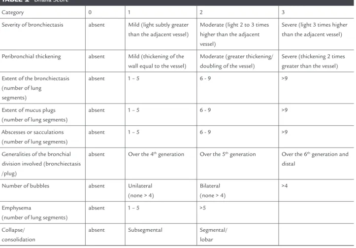
The HRCTs were evaluated independently by two radiol-ogists, using the Bhalla score.6 The items evaluated were:
a) severity of bronchiectasis; b) peribronchial thickening; c) extent of bronchiectasis (number of lung segments); d) extent of mucus plugs (number of lung segments); e) abscesses or sacculations (number of lung segments); f) generalities of the bronchial division involved (bronchi-ectasis/plug); g) number of bubbles; h) emphysema (num-ber of lung segments); i) collapse/consolidation.
TABLE 1 Bhalla Score
Category 0 1 2 3
Severity of bronchiectasis absent Mild (light subtly greater
than the adjacent vessel)
Moderate (light 2 to 3 times higher than the adjacent vessel)
Severe (light 3 times higher than the adjacent vessel)
Peribronchial thickening absent Mild (thickening of the
wall equal to the vessel)
Moderate (greater thickening/ doubling of the vessel)
Severe (thickening 2 times greater than the vessel)
Extent of the bronchiectasis (number of lung
segments)
absent 1 – 5 6 - 9 >9
Extent of mucus plugs (number of lung segments)
absent 1 – 5 6 - 9 >9
Abscesses or sacculations (number of lung segments)
absent 1 – 5 6 - 9 >9
Generalities of the bronchial division involved (bronchiectasis /plug)
absent Over the 4th generation Over the 5th generation Over the 6th generation and
distal
Number of bubbles absent Unilateral
(none > 4)
Bilateral (none > 4)
>4
Emphysema
(number of lung segments)
absent 1 – 5 >5
Collapse/ consolidation
absent Subsegmental Segmental/
lobar
Clinical evaluation
Weight, height, BMI, height/age and weight/age Z-scores and O2 were used for clinical evaluation.
Pulmonary function tests
The pulmonary function tests were conducted at the Pulmonary Function Laboratory at the Federal Univer-sity of Minas Gerais Hospital das Clínicas and the fol-lowing data was used in the analysis: a) forced vital ca-pacity (FVC); b) forced expiratory volume in one second (FEV1); c) forced expiratory flow between 25 and 75% (FEF 25-75%); d) FEV1/FVC ratio x 100. The recommen-dations of the American Thoracic Society and Polgar, Knudson and Pereira equations were used as reference values.11-14
Statistical analysis
The concordance between the two radiologists for the fi-nal Bhalla score, as well as for each category it composes was analyzed by calculating the kappa coefficient.
The correlation between the Bhalla score and its com-ponents, the pulmonary function tests and other charac-teristics of interest were tested using the Spearman cor-relation coefficient, given that the assumption of normality was violated.
The average evaluation by the two radiologists was used for the Bhalla score.
R
ESULTSThe records of 144 patients were analyzed. After analyz-ing the data collected, 53 patients had realized tomogra-phy and spirometry . Patients that had conducted spiral chest tomography, without high resolution (N=3), tients whose films were not available for analysis and pa-tients whose interval between tomography and spirome-try was greater than 30 days (N=27) were excluded from the 53 eligible patients (N=27). The final sample was there-fore constituted by 23 patients.
D
ISCUSSIONThe results found in this study prove the hypothesis that the Bhalla score represents a good correlation with pul-monary function tests and can be an ally in the evalua-tion of pulmonary structural changes.
In a study using the same score, Demirkazik et al. eval-uated 14 patients from a group of 40 that had HRCTs compared to the clinical and radiographic categories of the Shwachman-Kulczycki score and pulmonary func-tion tests. The age range varied from five months to 18 years, with all patients undergoing pulmonary function tests aged over six years. A good correlation was found between the Bhalla score and FVC (r = 0.71 and p = 0.004) and the FEV1 (r = 0.66 and p = 0.01). These findings cor-respond to the present study, which presented a strong and positive correlation for FVC (r = 0.718, p < 0.001) and FEV1 (r = 0.830, p < 0.001).10
On the other hand, Helbich et al. evaluated the to-mography of 107 patients with an average age of 14.5 ± 7.3 years at four different times. There was a weak corre-lation (r = 0.25 and p < 0.0001) between the tomography and the pulmonary function tests, and while the HRCT showed significant structural changes, the spirometry manifested minimal progression or slight improvement on the standard.15
More recently, with the objective of investigating the relationship between non-invasive inflammatory markers, function tests and pulmonary structural changes, Robroeks
et al. studied 34 patients with average age of 12.6 years ± 4.4 using the score described by Brody et al.9 and found a
strong correlation between the final score and the FVC (r = -0.73, p< 0.001), FEV1 (r = -077, p < 0.001) and FEV1/ FVC (r = -0.43, p < 0.001).11 Likewise, in 22 patients with
an average age of 22 years ± 5.9 and using a modified Bhal-la score, Dodd et al. found a significant correlation between the final score and the FEV1 (r = -0.40, p < 0,05).18
Further-more, Marchant et al., in an evaluation of 16 patients aged under 12 years with the objective of comparing the Bhal-la and Nathanson scores found a significant correBhal-lation between the final Bhalla score and the FEV1 (r = -0.65, p = 0.012) and FVC (r = -0.57, p = 0.032).19
However, analyzing the scores and function tests of each patient in isolation and taking into consideration that the FEF 25-75% is the parameter that causes most ear-ly changes in CF, it was verified that 11 of the 23 patients studied presented an FEF 25-75% above 70%. Despite the final Bhalla score being over 16 in these cases or being clas-sified as good or excellent, the letter T was added to the fi-nal score of 7 patients, indicating the presence of peribron-chial thickening, and bronchiectasis was recorded in 4
TABLE 2 Characteristics of the population studied
Characteristics n Mean SD
Age (years) 23 17.45 5.75
Weight (kg) 23 43.42 13.66
Height (cm) 23 152.1 14.8
Height/age Z-score 23 -1.3 1.5
Weight/age Z-score 23 -1.6 2.6
BMI 23 18.5 3.1
O2 Saturation 23 95.3 2.1
Shwachman - Kulczycki Score 23 80 16.1
Bhalla Score (R1) 23 16.8 7
Bhalla Score (R2) 23 17 6.6
Bhalla Score (R1 R2 average) 23 16.9 6.7
FVC% 23 87.1 20.5
FEV1% 23 74.2 23.7
FEF 25-75% 23 54.9 32.8
FEV1/FVC x 100 23 74.2 12.6
SD: standard deviation.
The median age of the population studied was 16.5 years.
Cultures
Nine of the patients studied (39.1%) were colonized by
Pseudomonas aeruginosa, being three (13%) by the mucoid strain, fifteen (65.2%) by Staphylococcus aureus, and three (13%) were not colonized.
Concordance between radiologists
A very good concordance between radiologists was found for the Bhalla score, with a kappa coefficient of 0.81 and an intraclass correlation coefficient (ICC) of 0.98, which classified it as adequate (ICC equal to or greater than 0.80).
Relationship between Bhalla scores and pulmonary function tests
A statistically significant positive correlation was identi-fied between the Bhalla score and pulmonary function tests.
TABLE 3 Correlation between the Bhalla score and pulmonary function tests
Characteristics Bhalla Score
r P-value
FVC% 0.718 <0.001
FEV% 0.830 <0.001
FEF 25-75% 0.786 <0.001
patients by the two radiologists.16,17 The findings in this
study are reflected in the literature and a study by March-ant et al., given that all of the 16 patients head bronchiec-tasis, including 5 who presented normal pulmonary func-tion tests, showing that the examinafunc-tions should be complementary and should not substitute one another. 19
Within the same perspective, De Jong et al. evaluated the HRCTs and pulmonary function tests of 48 patients that were conducted when they had an average age of 11.05 years ± 3.30 and repeated after a two year interval. The HRCTs were evaluated using five different scores (Castile, Brody, Helbich, Santamaria and Bhalla).19 The
authors verified that while there was deterioration in the HRCT findings in all scores used, the function tests re-mained unchanged or showed improvements. Further-more, some patients with normal pulmonary function tests presented structural changes in the HRCT.
This last finding reinforces the importance of HRCT in monitoring patients with CF, suggesting its use in or-der to detect early structural changes regardless of the re-sults of pulmonary function tests.
The concordance between examiners for the total Bhalla score in the present study was considered very good by the kappa coefficient (0.81, p < 0.001) and the ICC was considered adequate (0.98). These are similar results to those of Cademartiri et al., in which the Bhalla score was used to independently evaluate 145 tomographies by three radiologists.21 The average age of patients was 15.6 years
± 8,4. The ICC was 0.99 between observers 1 and 2, 1 and 3, 2 and 3 and between all three observers, with p = 0.001 for all analyses.
C
ONCLUSIONThe evaluation of pulmonary disease in patients with CF should include different clinical and preliminary methods, enabling an improvement in the effectiveness of treatment aiming at containing the progression of the disease.
In recent years, greater ease of access to computed to-mography has enabled this test to be incorporated into the routine assessment of patients, presenting essential infor-mation about the structural changes that occur in the air-ways and pulmonary parenchyma, with greater accuracy than assessment by plain radiography. However, the high dose of ionizing radiation, mainly due to the repetition of examinations, raises questions regarding the best time to start implementation, as well as its frequency.
We should reiterate the finding of peribronchial thick-ening and mucous plugs in patients with preserved lung function (FEF 25-75 > 70%), confirming the need for a
joint analysis between high-resolution CT and spirome-try for better monitoring and early intervention.
In regards to frequency, it is reasonable to infer that patients with stable and unchanged pulmonary function could repeat the tests at longer intervals than those show-ing a decrease in spirometry values. And this is undoubt-edly a result with great clinical applicability. It is impor-tant to stress that spirometry should not replace the realization of HRCT.
Furthermore, good concordance between examiners for the Bhalla score demonstrates its usefulness as an eval-uation criterion for tomographic changes during longi-tudinal follow-up of patients with CF.
In relation to the limitations of the study it is impor-tant to note that this is a retrospective study in a single referral service with a relatively small number of patients, which may limit the generalization of results. Neverthe-less, based on the results, we cannot definitively exclude the extrapolation of the findings in this study, though it certainly should be a matter to be explored further in fu-ture studies with a prospective design.
Finally, the notion of complementarity of clinical, ra-diological and functional information will contribute to the development of more discerning monitoring proto-cols aimed at the overall wellbeing of individuals with CF. These protocols, should consider patients with CF as well as other chronic diseases, who will be submitted to nu-merous radiological tests and must therefore be protect-ed from the effects of ionizing radiation, either by rprotect-educ- reduc-ing the dose durreduc-ing the exams or more careful requests for such. It should be noted that the standardization of the frequency should not be instituted by service proto-cols and that each patient should be evaluated on an in-dividual basis, although spirometry could be an impor-tant ally in the decision making regarding the time to perform examination.
R
ESUMOCorrelação entre escore de Bhalla e espirometria em crian-ças e adolescentes com fibrose cística
Métodos: foram avaliados os prontuários e os exames de 23 pacientes do ambulatório. Os itens avaliados foram idade, peso, altura, escore Z altura/idade, escore Z peso/ idade, índice de massa corpórea (IMC), saturação de O2,
espirometria e escore de Bhalla.
Resultados: os pacientes avaliados tinham média de idade de 17,4±5,7 anos, sendo 15 do sexo feminino e 8 do sexo masculino. Houve boa correlação entre o escore de Bhal-la e a espirometria (CVF–r = 0,718, p < 0,001; VEF1–r =
0,830, p < 0,001; FEF 25-75%–r = 0,786, p < 0,001; VEF1/
CVF–r = 0,714, p < 0,001). Nota-se, ainda, que alguns pa-cientes com FEF 25-75% > 70% já apresentavam altera-ções na nota final do escore de Bhalla. Na análise da con-cordância entre os examinadores, foi encontrado coeficiente kappa de 0,81 (p < 0,001) e coeficiente de cor-relação intraclasse de 0,98.
Conclusão: a boa correlação do escore de Bhalla com as provas de função pulmonar confirma a sua utilidade na avaliação e no acompanhamento dos pacientes com FC, podendo ser utilizado tanto para pacientes que são inca-pazes de realizar a espirometria quanto para uma análise em conjunto dos dois exames, uma vez que a TCAR mos-tra alterações precoces em pacientes com espirometrias normais.
Unitermos: fibrose cística; tomografia; espirometria; criança; adolescente.
R
EFERENCES1. Shwachman H, Kulczycki LL. Long-term study of one hundred five patients with cystic fibrosis; studies made over a five- to fourteen-year period. AMA J Dis Child. 1958;96(1):6-15.
2. Brasfield D. Hicks G. Soong S. Tiller RE. The chest roentgenogram in cystic fibrosis: a new screening system. Pediatrics. 1979;63(1):24-9.
3. Bhalla M, Turcios N, Aponte V, Jenkins M, Leitman BS, McCauley DI et al. Cystic fibrosis: scoring system with thin-section CT. Radiology. 1991;179(3):783-8.
4. Assis I, Camargos PAM, Reis FJC, Sulmonett N, Carneiro APS. Assessing correlations between spirometry and Shwachman-Kulczycki score in children and adolescents. Pediatr Pulmonol. 2003;36(4):305-9.
5. Stollar F, Adde FV, Cunha MT, Leone C, Rodrigues JC. Shwachman-Kulczycki score still useful to monitor cystic fibrosis severity. Clinics (São Paulo). 2011;66(6):979-83.
6. Van Beek EJ, Hill C, Woodhouse N, Fichele S, Fleming S, Howe B et al. Assessment of lung disease in children with cystic fibrosis using hyperpolarized 3-Helium MRI: comparison with Shwachman score, Chrispin-Norman score and spirometry. Eur Radiol. 2007;17(4):1018-24. 7. O’Sullivan BP, Freedman SD. Cystic fibrosis. Lancet.
2009;373(9678):1891-904.
8. Lucaya J, Piqueras J, García-Peña P, Enríquez G, García-Macías M, Sotil J. Low-dose high-resolution CT of the chest in children and young adults: dose, cooperation, artifact incidence, and image quality. AJR Am J Roentgenol. 2000;175(4):985-92.
9. Long FR. High-resolution computed tomography of the lung in children with cystic fibrosis: technical factors. Proc Am Thorac Soc. 2007;4(4):306-9. 10. Freire ID, Abreu e Silva FA, Araújo MA. Comparison among pulmonary
function test results, the Shwachman-Kulczycki score and the Brasfield score in patients with cystic fibrosis. J Bras Pneumol. 2008;34(5):280-7. 11. Goris ML, Zhu HJ, Blankenberg F, Chan F, Robinson TE. An automated
approach to quantitative air trapping measurements in mild cystic fibrosis. Chest. 2003;123(5):1655-63.
12. Demirkazik FB, Ariyürek OM, Ozçelik U, Göçmen A, Hassanabad HK, Kiper N. High resolution CT in children with cystic fibrosis: correlation with pulmonary functions and radiographic scores. Eur J Radiol. 2001;37(1):54-9.
13. American Thoracic Society. Standardization of spirometry, 1994 update. Am J Respir Crit Care Med. 1995;152(3):1107-36.
14. Polgar G, Promadhat V. Pulmonary function testing in children: techniques and standards. Philadelphia: WB Saunders; 1971.
15. Miller MR, Hankinson J, Brusasco V, Burgos F, Casaburi R, Coates A et al. Standardisation of spirometry. Eur Respir J. 2005;26(2):319-38.
16. Sociedade Brasileira de Pneumologia e Tisiologia. Diretrizes para teste de função pulmonar. J Bras Pneumol. 2002;28(Supl 3):S1-S238.
17. Helbich TH, Heinz-Peer G, Fleishmann D, Wojnarowski C, Wunderbaldinger P, Huber S et al. Evolution of CT findings in patients with cystic fibrosis. AJR Am J Roentgenol. 1999;173(1):81-8.
18. Robroeks CM, Roozeboom MH, de Jong PA, Tiddens HA, Jöbsis Q, Hendriks HJ et al. Structural lung changes, lung function, and non-invasive inflammatory markers in cystic fibrosis. Pediatr Allergy Immunol. 2010;21(3):493-500.
19. Brody AS, Molina PL, Klein JS, Rothman BS, Ramagopal M, Swartz DR. High-resolution computed tomography of the chest in children with cystic fibrosis: support for use as an outcome surrogate. Pediatr Radiol. 1999;29(10):731-5.
20. Dodd JD, Barry SC, Barry RB, Gallagher CG, Skehan SJ, Masterson JB. Thin-section CT in patients with cystic fibrosis: correlation with peak exercise capacity and body mass index. Radiology. 2006;240(1):236-45.
21. Marchant JM, Masel JP, Dickinson FL, Masters IB, Chang AB. Application of chest high-resolution computer tomography in young children with cystic fibrosis. Pediatr Pulmonol. 2001;31(1):24-9.
22. De Jong PA, Nakano Y, Lequin MH, Mayo JR, Woods R, Paré PD et al. Progressive damage on high resolution computed tomography despite stable lung function in cystic fibrosis. Eur Respir J 2004;23(1):93-7.