MARCHI SEN ETAL.
1 9 8 REV ASSOC MED BRAS 2014; 60(3):198-199
at bedside
What are the benefits of endoscopic ultrasound in the staging of
pancreatic cancer?
Q
UAIS OS BENEFÍCIOS DA ECOENDOSCOPIA NO ESTADIAMENTO DO CÂNCER DE PÂNCREAS?
SÍLVIA MANSUR REIMÃO1, JARBAS FARACO MALDONADO LOUREIRO1, ROGÉRIO COLAIACOVO1, RICARDO GANC1, ELIAS JIRJOSS ILIAS2, LUCIO GIOVANNI BATISTA ROSSINI1
1endoscopists, Centro Francobrasileiro de ecoendoscopia, school of Medical sciences of santa Casa Misericórdia de são Paulo, são Paulo, sP, brazil 2Phd-Professor at the surgery department of brotherhood of santa Casa de Misericórdia de são Paulo, são Paulo, brazil
http://dx.doi.org/10.1590/1806-9282.60.03.006
C
ASE REPORTFemale patient, 73 years old, with a history of jaundice, epi-gastric pain radiating to the back, and weight loss of 15 kg in three months. She was referred to our hospital with a possible diagnosis of cancer in the head of the pancreas, with evidence of increased pancreatic head and dilatation of intra- and extra-hepatic biliary ducts on abdominal com-puted tomography and magnetic resonance cholangiogra-phy. Due to obstructive jaundice, a retrograde cholangio-pancreatography was performed, showing partial stenosis of the distal common bile duct with dilatation of bile ducts, which was drained by inserting two biliary plastic stents. An endoscopic ultrasound was requested to further study the pancreas and adjacent structures.
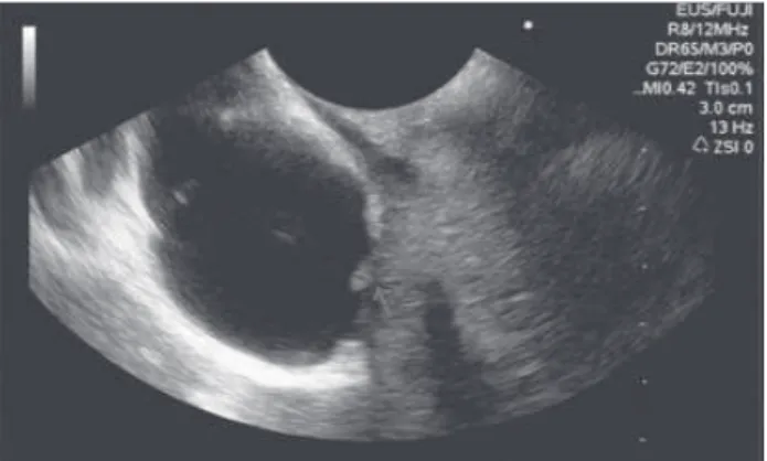
During the investigation, a hypoechoic heterogeneous lesion measuring 35 x 30 mm and presenting ill-deined margins was seen in the pancreatic head. There were also signs of vascular involvement, with loss of acoustic inter-face with the wall of the portal vein and absence of vas-cular low (thrombosis). Furthermore, dilatation of the main pancreatic duct (6 mm) in the regions of body and tail of the pancreas upstream to the lesion described abo-ve was obserabo-ved. Echo-guided punctures were performed in the lesion for histological clariication.
FIGURE 1 Details of the vascular invasion of the portal-splenome-senteric confluence.
FIGURE 2 Negative Doppler suggesting portal thrombosis.
FIGURE 3 Echo-guided puncture to obtain sample material from the lesion.
D
ISCUSSIONPancreatic cancer is associated with poor prognosis. Af-ter diagnosis, the survival rate is about 3% at 5 years and only 15% of tumors are resectable. Surgical resection of the tumor is still the only curative treatment and, there-fore, an accurate preoperative staging is mandatory to
WHATARETHEBENEFITSOFENDOSCOPICULTRASOUNDINTHESTAGINGOFPANCREATICCANCER?
REV ASSOC MED BRAS 2014; 60(3):198-199 199
In the absence of distant metastases, which contrain-dicate surgery, assessment of vascular invasion is the most important parameter to determine the resectability of the
lesion. Invasion is found in 21 to 64% of cases.2
Endoscopic ultrasound is a test that complements the evaluation made by other imaging studies (CT or MRI), providing an accurate assessment of peripancrea-tic vasculature and the relationship between the tumor and adjacent structures. Studies have shown that the sen-sitivity to detect vascular invasion through endoscopic ultrasound ranges from 50 to 100%, with specificity
between 58 and 100%.2 In addition to the image data, it
enables the realization of intra-pancreatic and lymph
node biopsies1,3 that can change the therapeutic approach.
Fine needle aspiration (FNA) has a diagnostic accuracy approaching 90%. Obtaining histological material from biopsy plays an important role to initiate palliative or
neoadjuvant treatment.5
As to arterial blood investigation, the iniltration of large vessels such as the celiac trunk, superior
mesente-ric or hepatic arteries, is also a contraindication to
surgi-cal treatment. The superior mesenteric vessels are the ves-sels most often involved in this type of cancer, due to its close relationship with the head, uncinate process and
body of the pancreas.2
In the venous study, visualization of tumor throm-bus or the involvement of more than 25% of the circum-ference of the portal vein or superior mesenteric vein are
criteria for irresectability.4
In conclusion, endoscopic ultrasound is a comple-mentary method for staging patients with pancreatic can-cer, providing data on the involvement of blood vessels and other peripancreatic structures, and allows histolo-gical deinition of the lesion, which is essential for achie-ving adjuvanticity.
R
EFERENCES1. Ahmad NA, Kochman ML, Lewis JD, Kadish S, Morris JB, Rosato EF, et al. Endosonography is superior to angiography in the preoperative assessment of vascular involvement among patients with pancreatic carcinoma. J Clin Gastroenterol 2001;32(1):54-8.
2. Buchs NC, Chilcott M, Poletti PA, Buhler LH, Morel P. Vascular invasion in pancreatic cancer: imaging modalities, preoperative diagnosis and surgical management. World J Gastroenterol 2010;16(7):818-31.
3. De Angelis C, Brizzi RF, Pellicano R. Endoscopic ultrasonography for pancreatic cancer: current and future perspectivers. J Gastroenterol Oncol 2013;4(2):220-30.
4. Callery MP, Chang KJ, Fishman EK, Talamonti MS, Traverso LW, Linehan DC. Pretreatment assessment of resectable and borderline resectable pancreatic cancer: expertise consensus statement. Ann Surg Oncol 2009;16(7):1727-33. 5. Garcia JI, Noia JL, Muñoz JED. Endoscopic ultrasound in the diagnosis and