MARCHI SEN ETAL.
196 REV ASSOC MED BRAS 2014; 60(3):196-197
Image In medIcIne
Gouverneur’s syndrome in a patient with abdominal pain: mind
Crohn’s disease!
S
ÍNDROMEDEG
OUVERNEUREMUMPACIENTECOMDORABDOMINAL:
CUIDADOCOMADOENÇADEC
ROHN!
JULIO MARIA FONSECA CHEBLI1*, ANDRE AVARESE FIGUEIREDO2, PEDRO DUARTE GABURRI3
1 Inlammatory Bowel disease center, Juiz de Fora, mg, Brazil
2 Teaching Hospital of the Federal University of Juiz de Fora, Juiz de Fora, mg, Brazil 3 School of medicine, University of Juiz de Fora, minas gerais, mg, Brazil
Study conducted at the Federal University of Juiz de Fora, Juiz de Fora, mg, Brazil
*Correspondence:
Rua maria José Leal , 296 Juiz de Fora, mg, Brazil – ZIP code 36036-247 Phone / Fax: +55 32 3216-7122 [email protected]
http://dx.doi.org/10.1590/1806-9282.60.03.005 Conflict of interest: none
Gouverneur’s syndrome, which describes suprapubic pain, frequency, dysuria and tenesmus, is the hallmark of en-terovesical fistula.1 However, Gouverneur’s syndrome as
the initial presentation of Crohn’s disease (CD) has not been previously reported in literature.
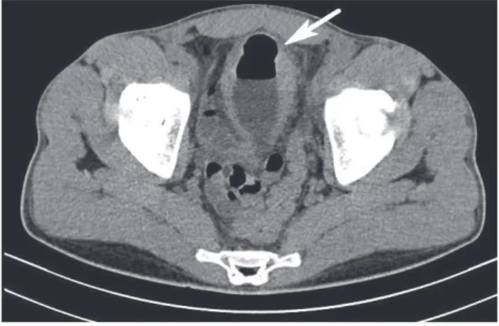
A 26 year old man was admitted in January 2013 complaining of suprapubic pain, urinary frequency, dys-uria and tenesmus over the past 3 weeks. He also report-ed colic abdominal pain during the past 4 months. The physical examination yielded discrete anemia. A diag-nostic hypothesis of Gouverneur’s syndrome related to enterovesical fistula was considered. Urine analysis showed pyuria and significant bacteriuria, and urine cul-ture showed Escherichia coli. Contrast-enhanced comput-ed tomography of the abdomen revealcomput-ed the presence of gas in the bladder (Figure 1, white arrow). Cystosco-py revealed a localized area of erythema, edema and con-gestion on the bladder dome.
Skip lesions, cobblestone mucosa and ulcerations with clear margins surrounded by normal mucosa, sug-gestive of Crohn’s colitis, were visualized at left colon on ileocolonoscopy. Endoscopic biopsies displayed lympho-plasmacytic infiltration with submucosal involvement, in addition to fissuring ulcerations in the sigmoid colon. Surgical management involved left colectomy with colo-colonic anastomosis and closing primary of the bladder defect using absorbable suture. The patient had an uneventful postoperative course. One month later he was
started on induction therapy with infliximab followed by maintenance with 5 mg/kg every 8 weeks. The patient re-mained asymptomatic 6 months later.
Crohn’s disease accounts for 10% of enterovesical fistu-la and is the commonest cause of ileovesical fistufistu-lae.1
How-ever, colovesical fistula is a rare condition in CD.2 Although
the underlying illness is usually of intestinal origin, the ma-jority of patients with enterovesical fistula are referred be-cause of urinary symptoms, since the flow through the fis-tula predominantly occurs from the bowel to the bladder due to high compliance of the bladder and low intravesical pressure.2 The cardinal feature of enterovesical fistulae may
be described as Gouverneur’s syndrome.3 Other findings
in-clude abnormal urinalysis findings, pneumaturia, fecal-uria, and recurrent urinary tract infections. Preoperative imaging should characterize the anatomical abnormality and exclude underlying malignancy, as well as point out the fistulous track. Computerized tomography is an excel-lent image modality for diagnosing enterovesical fistula. Cystoscopy is a crucial method of the investigation as it can suggest the presence of a fistula and also be useful in excluding the rare case of urological neoplasm.1
GOUVERNEUR’SSYNDROMEINAPATIENTWITHABDOMINALPAIN: MIND CROHN’SDISEASE!
REV ASSOC MED BRAS 2014; 60(3):196-197 197
FIGURE 1 Contrast-enhanced computed tomography of the
abdomen showing the presence of gas in the bladder (white arrow).
R
EFERENCES1. Scozzari G, Arezzo A, Morino M. Enterovesical fistulas: diagnosis and management. Tech Coloproctol 2010;14:293-300.
2. Gruner JS, Sehon JK, Johnson LW. Diagnosis and management of enterovesical fistulas in patients with Crohn’s disease. Am Surg 2002; 68:714-9. 3. Garcea G, Majid I, Sutton CD, Pattenden CJ, Thomas WM. Diagnosis and