279
IMAGE IN MEDICINE
Rev Assoc Med Bras 2012; 58(3):279-280
Nephrocalcinosis in a patient with Sjögren’s syndrome/systemic
lupus erythematosus
AMANDA CAROLINA DAMASCENO ZANUTO1, TATIARA BUENO1, VINICIUS DAHER ALVARES DELFINO2, ALTAIR JACOB MOCELIN2
1 Nephrology Residents, Hospital Evangélico de Londrina, Londrina, PR, Brazil
2 Nephrology Department, Hospital Evangélico de Londrina, Londrina, PR, Brazil
Study conducted at Universidade Estadual de Londrina, Londrina, PR, Brazil
Correspondence to: Vinicius D. A. Delfino, Departamento de Nefrologia, Centro de Ciências da Saúde, Universidade Estadual de Londrina, Av. Robert Koch, 60 Vila Operária, 86038-440, Londrina – PR, Brazil – [email protected]
©2012 Elsevier Editora Ltda. All rights reserved.
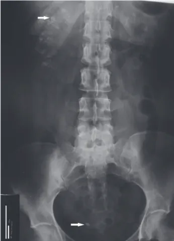
Figure 1 – Abdominal X-ray of the patient showing clustered calcifications in projection of the kidneys, more evident at the right side (upper arrow identifies one of these clusters). An 8-mm right distal ureteral stone is present (lower arrow). A 46-year-old white woman has been followed in this
nephrology clinic since 1995 due to recurrent nephroli-thiasis and medullary nephrocalcinosis resulting from renal tubular acidosis (urinary pH of 8 in the context of systemic normal anion gap acidosis; serum potassium at lower limit, and 24-hour urine with normocalciuria but no detectable citrate) due to Sjögren’s syndrome (SS). During follow-up, her adherence to alkali therapy had been suboptimal due to gastric intolerance to Shohl’s solution, potassium citrate, and sodium bicarbonate. As consequence, she had progression in her nephrocal-cinosis and passed several urinary calculi (biochemical analysis showed them to be composed of calcium phos-phate and calcium oxalate). Currently, she presents
nor-mal estimated GFR (MDRD, 82 mL/min/1.73 m2) and
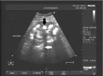
no signiicant proteinuria, despite having presented, ten years ater the diagnosis of SS, onset of systemic lupus er-ythematosus (SLE) with severe difuse proliferative class IV lupus nephritis, treated successfully with corticoste-roids and intravenous ciclophosphamide. Figure 1 shows the radiological appearance and Figure 2 depicts the ul-trasonographic aspect of the patient’s nephrocalcinosis.
Nephrocalcinosis is characterized by the presence of calcium deposits in the renal parenchyma; nephrolithia-sis represents calciication within the lumen of the col-lecting system, ureter, and bladder1. Recent observation
suggests that nephrocalcinosis and calcium nephrolithi-asis are to be considered independent pathologies, and that nephrocalcinosis may cause calcium nephrolithiasis only in certain conditions2. Both types of renal
calciica-tion may be present in patients with SS. Nephrocalcinosis may be classiied as cortical or medullary, according to the anatomic area involved3. When associated with SS, it
is typically medullary and secondary to distal renal tubu-lar acidosis (RTA). It is believed that the renal acidiica-tion problem seen in patients with SS is due to immune-mediated loss of proton regulation4. In addition to RTA,
other disorders such as hyperparathyroidism, medullary sponge kidney, renal papillary necrosis, renal tubercu-losis, hyperoxaluria, milk-alkali syndrome, sarcoidosis,
immobilization, and other conditions associated with hypercalcemia and hypercalciuria, may cause medullary nephrocalcinosis.
Nephrocalcinosis is usually identiiable by X-ray, but both ultrasonography (US) and computed tomogra-phy (CT) can detect it earlier than ordinary abdominal
X-ray1. US is considered to be an excellent diagnostic
method for detection and monitoring of
280
IMAGEINMEDICINE
Rev Assoc Med Bras 2012; 58(3):279-280
normally echogenic cortex is believed to be speciic to medullary nephrocalcinosis3,5. Despite being an excellent
diagnostic method as well, CT (usually performed with-out radiocontrast) involves a considerable dose of radia-tion, which may limit its use for the follow-up of patients with nephrocalcinosis. Indeed, CT was found to be un-necessary for the clinical management of the patient.
Finally, the importance of investigating SS in patients with medullary nephrocalcinosis or nephrolithiasis as-sociated with renal tubular acidosis, especially women, regardless of the presence of keratitis sicca or systemic symptoms, is emphasized, because it may be the irst manifestation of the syndrome. herapy for correction of acidosis is intended to control the nephrocalcinosis and preserve renal function6. his case corroborates that
al-though RTA is a treatable urologic condition, incomplete treatment can lead to progression of nephrocalcinosis.
REFERENCES
1. Vanrenterghem Y, Kuypres D, Verswijvel G, Schepens D. Renal cortical nephrocalcinosis. Nephrol Dial Transplant. 2000;15:1080-2.
2. Vervaet BA, Verhulst A, D’Haese PC, De Broe ME. Nephrocalcinosis: new insights into mechanisms and consequences. Nephrol Dial Transplant. 2009;24: 2030-5.
3. Hoppe B, Kemper MJ. Diagnostic examination of the child with urolithiais or nephrocalcinosis. Pediatr Nephrol. 2010;25:403-13.
4. Evan AP, Lingeman J, Coe F, Shao Y, Miller N, Matlaga B, et al. Renal histopa-thology of stone-forming patients with distal renal tubular acidosis. Kidney Int. 2007;71:795-801.
5. Simões A, Domingos F, Prata MM. Nephrocalcinosis induced by furosem-ide in an adult patient with incomplete renal tubular acidosis. Nephrol Dial Transplant. 2001;16:1073-4.
6. Soriano JR. Renal tubular acidosis: the clinical entity. J Am SocNephrol. 2002;13:2160-70.