Cardiac-Specific Activation of IKK2 Leads
to Defects in Heart Development and
Embryonic Lethality
Bärbel Kraut1☯, Harald J. Maier1☯, Enikö Kókai1☯, Katja Fiedler1, Thomas Boettger2, Annett Illing3, Sawa Kostin2, Paul Walther4, Thomas Braun2, Thomas Wirth1
*
1Institute of Physiological Chemistry, University of Ulm, Ulm, Germany,2Max Planck Institute for Heart and Lung Research, Bad Nauheim, Germany,3Institute of Molecular Medicine, University of Ulm, Ulm,
Germany,4Core Facility Electron Microscopy, University of Ulm, Ulm, Germany
☯These authors contributed equally to this work.
Abstract
The transcription factor NF-κB has been associated with a range of pathological conditions
of the heart, mainly based on its function as a master regulator of inflammation and pro-survival factor. Here, we addressed the question what effects activation of NF-κB can have
during murine heart development. We expressed a constitutively active (CA) mutant of IKK2, the kinase activating canonical NF-κB signaling, specifically in cardiomyocytes under
the control of theα-myosin heavy chain promoter. Expression of IKK2-CA resulted in
embryonic lethality around E13. Embryos showed defects in compact zone formation and the contractile apparatus, and overall were characterized by widespread inflammation with infiltration of myeloid cells. Gene expression analysis suggested an interferon type I signa-ture, with increased expression of interferon regulatory factors. While apoptosis of cardio-myocytes was only increased at later stages, their proliferation was decreased early on, providing an explanation for the disturbed compact zone formation. Mechanistically, this could be explained by activation of the JAK/STAT axis and increased expression of the cell cycle inhibitor p21. A rescue experiment with an IκBαsuperrepressor demonstrated that the
phenotype was dependent on NF-κB. We conclude that activation of NF-κB is detrimental
during normal heart development due to excessive activation of pro-inflammatory pathways.
Introduction
NF-κB is a pleiotropic transcription factor that has been associated with diverse biological functions such as cell proliferation, cell survival, immunity, and inflammation. Several patho-logical heart conditions, among them myocarditis, cardiac hypertrophy, adverse cardiac remodeling, and heart failure, have been associated with activation NF-κB signaling as well [1].
a11111
OPEN ACCESS
Citation:Kraut B, Maier HJ, Kókai E, Fiedler K, Boettger T, Illing A, et al. (2015) Cardiac-Specific Activation of IKK2 Leads to Defects in Heart Development and Embryonic Lethality. PLoS ONE 10(11): e0141591. doi:10.1371/journal.pone.0141591
Editor:Diego Fraidenraich, Rutgers University -New Jersey Medical School, UNITED STATES
Received:August 21, 2015
Accepted:October 9, 2015
Published:November 5, 2015
Copyright:© 2015 Kraut et al. This is an open access article distributed under the terms of the
Creative Commons Attribution License, which permits unrestricted use, distribution, and reproduction in any medium, provided the original author and source are credited.
Data Availability Statement:Microarray data are available in the ArrayExpress database (www.ebi.ac. uk/arrayexpress) under accession number E-MTAB-3971 (http://www.ebi.ac.uk/arrayexpress/experiments/ E-MTAB-3971).
Funding:The authors have no support or funding to report.
NF-κB is a dimeric transcription factor composed of different combinations of the subunits p50, p52, RelA, RelB, and c-Rel. Under baseline conditions, these NF-κB dimers remain inac-tive and sequestered in the cytoplasm by inhibitoryκB (IκB) proteins. Different signals, such as engagement of cytokine or Toll-like receptors, trigger a signaling cascade that eventually converges on and activates the IκB kinase complex (IKK). This complex is composed of the catalytic subunits IKK1 (also known as IKKα) and IKK2 (IKKβ), and the regulatory subunit NEMO (IKKγ). IKK in turn can phosphorylate IκB proteins and thereby mark them for ubi-quitin-mediated degradation. This releases NF-κB dimers, which are now free to translocate to the nucleus and activate specific target genes [2].
Murine heart development begins with the specification of cardiac progenitor cells at E6.0 and the formation of the cardiac crescent at E7.5 [3]. Beginning at E8.0, a linear heart tube is formed, sarcomeres are established, and a heart beat can be detected. After cardiac looping with the establishment of left-right asymmetry around E9.0, the heart tube differentiates into the four chambers and undergoes trabeculation and expansion (E10.5—E12.5), followed by further maturation of essential components such as the heart valves and the great vessels (E13.5 onwards).
Genetic and genomic approaches have identified a multitude of genes essential for mamma-lian heart development. In particular, they have revealed a complex network of cardiogenic transcription factors that orchestrate heart development [3]. Components of the IKK/ NF-κB signaling pathway appear not to be essential for heart development: For example, mice defi-cient in IKK2 (Ikbkb-/-) undergo normal heart development and only die due to hepatocyte apoptosis between E12.5 and E14.5 [4–6]. Similarly, a conditional ablation of IKK2 in the heart—using the myosin light chain 2V promoter which is specifically active in cardiomyo-cytes—does not affect normal heart development [7], neither does direct interference with the NF-κB pathway by expressing a transgenic IκBαsuperrepressor [8].
Still, IKK/ NF-κB signaling may be of great importance under pathologic conditions, since IKK/ NF-κB is activated during many adverse events that are common during gestation, such as infection. In this study, we wanted to address the consequences of the activation of IKK/ NF-κB in the developing heart as a model for adverse events during gestation.
Materials and Methods
Mice
Mice were kept under specific pathogen-free conditions at the animal facility of the University of Ulm. The tetO.IKK2-CA [9],α-MyHC.tTA [10], and IκBα-3M mice [11] have been described previously. Animals were killed by cervical dislocation under isoflurane anesthesia. All experiments were in accordance with institutional guidelines and German animal protec-tion laws and were approved by the regional government authority (Regierungspräsidium Tübingen).
Histology and immunofluorescence microscopy
For paraffin sections, whole embryos were fixed in buffered formalin, dehydrated, embedded in paraffin, and cut in 3μm sections on a rotary microtome (Thermo Scientific). Sections were
deparaffinized and rehydrated, and then used for H&E staining. Where appropriate, sections were boiled in citrate buffer for 10 minutes prior to immune staining. For cryosections, embryos snap-frozen in liquid nitrogen were cut in 4μm sections on a cryotome (Leica).
1:200 (Dako antibody diluent) for 1 h at room temperature, with antibodies againstα-SMA (Sigma F3777), IKK1/2 (Santa Cruz sc-7607, lot H2208), cardiac Troponin T (Abcam 8295), RelA (NeoMarkers RB-1638-P1), sarcomeric actinin (Sigma A7811), desmin (Cell Signaling 4024), F4/80 (eBioscience 14-4801-85, lot E04273-301), Icam-1 (Santa Cruz sc-18853, lot A2808), Sca-1 (eBioscience 14-5981-82, lot E020936). Secondary antibodies coupled with Alexa Fluor 488 or 596 were purchased from Invitrogen/ Molecular Probes, and were incu-bated for one hour at room temperature. Co-staining was performed with 4',6-diamidino-2-phenylindole (DAPI) for nuclear staining. For detection of apoptosis, an in situ TUNEL detection kit (Roche) was used. Fluorescent samples were analyzed on a Zeiss Axiovert 200M microscope equipped with a digital camera (Zeiss AxioCam MR3) and Axiovision software. Other staining were evaluated on a Leica DM5500B microscope equipped with a DFC420C camera (Leica).
Murine cardiomyocyte isolation
Excised hearts were digested in a calcium-free buffer supplemented with 6.5 mg/mL Liberase DH (Roche) and 3.5 mg/mL trypsin (Sigma). Digestion was stopped with FCS, and the cell sus-pension was filtered through a 100μm mesh and centrifuged.
BrdU labeling and flow cytometry
Pregnant females were given BrdU (100 mg/ kg body weight i.p.) 4 hours before harvesting the embryos. FACS of digested whole hearts was performed on a FACSCanto™II equipped with FACSDiva 6.2 software (BD).
Protein extracts and Western blotting
Tissue was snap-frozen in liquid nitrogen, pulverized, and resuspended in a buffer containing 4% SDS, 100 mM Tris-HCl, and protease/ phosphatase inhibitors (Roche). 10 to 20μg of
pro-tein were separated on 4–12% gradient gels (Invitrogen). Proteins were transferred to a PVDF or nitrocellulose membrane with a semi-dry blotter (Bio-Rad). Membranes were blocked for 1 h (RT) in TBS, 0.1% Tween-20 with 5% w/v nonfat dry milk, incubated with primary antibody over night (6°C) in TBS, 0.1% Tween-20 with 5% nonfat dry milk or 5% BSA, and then incu-bated with HRP-coupled secondary antibody for 1 h (RT). After application chemilumines-cence reagent, membranes were exposed to x-ray films. Primary antibodies against the following antigens were used: human IKK2 (Abcam Y466), IKK1/2 (Santa Cruz sc-7607, lot H2208), ERK2 (Santa Cruz sc-154, lot F0210), p21 (Santa Cruz sc-6246, lot A1209), Stat1 (Cell Signaling 9172, lot 14), phospho-Stat1 (Cell Signaling 9167, lot 6), and luciferase (Promega G7451).
RNA extraction and quantitative PCR
Heart tissue was snap-frozen in liquid nitrogen, pulverized and processed with the Qiagen RNeasy Fibrous Tissue kit. The Transcriptor High Fidelity cDNA Synthesis Kit (Roche) was used for cDNA synthesis. Quantitative PCR was done on a LightCycler 480 system, using the Universal Probe Library (Roche).Rpl13was used as reference gene for relative quantification. Primer sequences are available on request.
Gene expression profiling
GeneChip protocol. Labeled samples were hybridized to Affymetrix GeneChip1
Mouse Gene 1.0 ST Array and further processed. Arrays were scanned with an Affymetrix GeneChip Scan-ner 3000 7G and data were analyzed by the RMA algorithm using the Affymetrix Expression Console and the GeneSifter1microarray data analysis system. Microarray data are available in the ArrayExpress database (www.ebi.ac.uk/arrayexpress) under accession number E-MTAB-3971 (http://www.ebi.ac.uk/arrayexpress/experiments/E-MTAB-3971).
Statistical analysis
Values are given as arithmetic mean + (or +/-) standard error of the mean (SEM). Means of two groups were compared by the student’s t test or, when indicated, by the Mann-Whitney test. Welch correction was applied for unequal variances. Means of multiple groups were com-pared by ANOVA. All tests were two-tailed. p values<0.05 were deemed significant (p<0.05, p<0.01,p<0.001).
Results
Expression of Constitutively Active IKK2 in the Heart Causes Embryonic
Lethality
In order to activate NF-κB in the developing myocardium, constitutively active IKK2
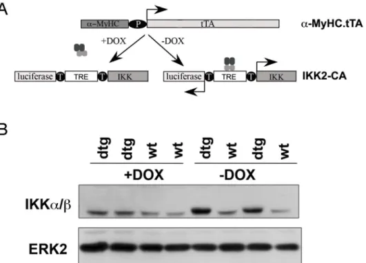
(IKK2-CA) was expressed specifically in cardiomyocytes. To this end, mice expressing the tet-racycline transactivator (tTA) under the control of the cardiomyocyte-specificα-myosin heavy chain (α-MyHC) promoter [10] were crossed with mice carrying a constitutively active IKK2 transgene (IKK2-CA) and a luciferase reporter gene regulated by a bidirectional, tTA-respon-sive promoter (Fig 1A) [9]. As expected, expression of the IKK2 transgene was only seen in hearts of double transgenic (IKKMyHC) embryos and could be silenced by the administration of doxycycline to pregnant mothers (Fig 1B).
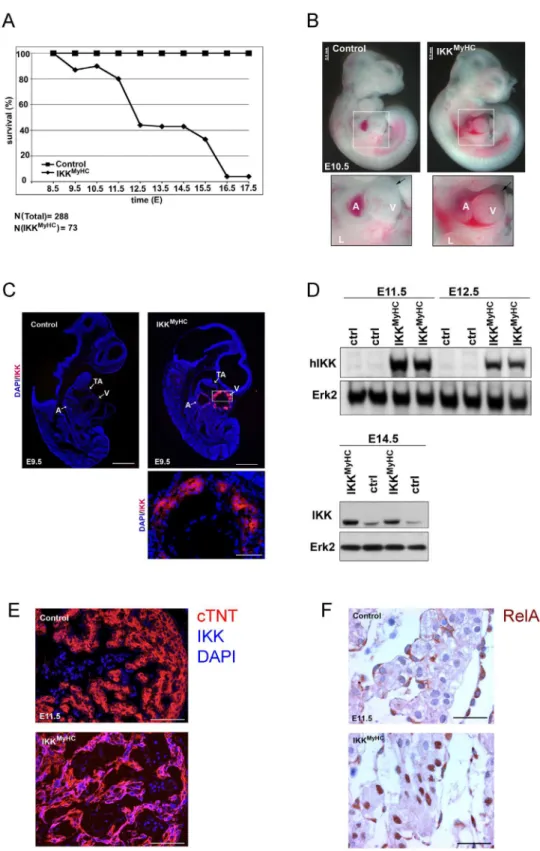
When mice were bred in the absence of doxycycline, i.e. under conditions that allow the transgene to be expressed, the number of double transgenic animals was strongly reduced in comparison to single transgenic and non-transgenic animals: Only 3.3% (instead of the expected 25%) of the born animals (4 out of 122) carried both transgenes (Table 1). When the transgene expression was shut off during development by administering doxycycline to the pregnant females, the ratio was normalized to the expected 25% [12]. These observations sug-gested that the expression of constitutively active IKK2 resulted in embryonic lethality.
To determine the time point of lethality, the viability of embryos was analyzed at different time points of gestation. IKKMyHCembryos started to die as early as E9.5 (Fig 2A). The exact time point of death was somewhat variable between individual embryos, but 50% of the ani-mals were dead between E12.5 and E13.5. Less than 4% of IKKMyHCembryos survived longer than E17.5. Closer examination of the heart showed pericardial edema and hemorrhages at the ventricles by E10.5; in several cases the pericardial sac was filled with blood (Fig 2B).
Since the lethality was observed as early as day E9.5, we confirmed by immunofluorescence that the transgene IKK2-CA was actually expressed at this early stage (Fig 2C). Transgene expression was also detectable by Western blot at later time points and was stronger than endogenous IKK2 expression (Fig 2D). Expression was restricted to cardiomyocytes, as shown by immunofluorescence costaining of sections at E11.5 with an antibody against the human IKK2 transgene and an antibody against the cardiomyocyte-specific protein cardiac troponin T
(Fig 2E). Next, we wanted to analyze whether IKK2-CA expression actually activated NF-κB
found in cardiomyocytes of IKKMyHCembryos, whereas this was not the case for controls
(Fig 2F).
IKK
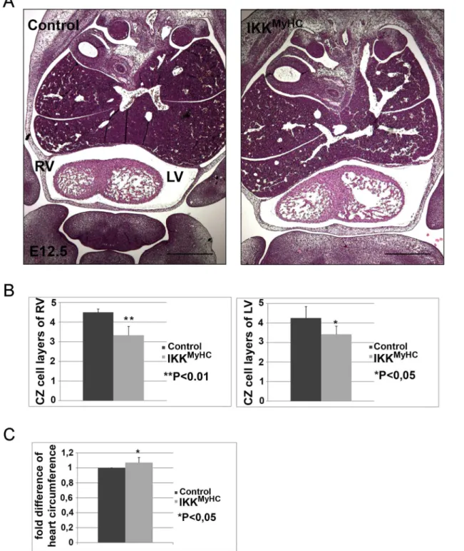
MyHCembryonic hearts show defects in compact zone formation
Embryonic hearts are characterized by the presence of an inner trabecular and an outer com-pact zone. Systematic histological examination of the IKKMyHCembryonic hearts at E12.5
Table 1. Expression of constitutively active IKK2 in cardiomyocytes results in embryonic lethality.
MyHC-tTA IKK2-CA Numbers observed Numbers expected % observed % expected
- - 40 30.5 32.8 25
+ - 40 30.5 32.8 25
- + 38 30.5 31.1 25
+ + 4 30.5 3.4 25
Total 122 122 100 100
The table shows the absolute number and the percentage of animals born with the indicated genotypes for the transgenes MyHC.tTA and IKK2-CA (observed), and the corresponding expected number and percentage (expected). The observed number of animals positive for both MyHC-tTA and IKK2-CA (4) is much lower than expected (30.5).
doi:10.1371/journal.pone.0141591.t001
Fig 1. Cardiomyocyte-specific expression of IKK2-CA in embryos.(A) In order to direct the expression of constitutively active IKK2 (IKK2-CA) to the heart, mice expressing the tetracycline-transactivator (tTA) under theα-myosin heavy chain promoter (α-MyHC) were crossed with mice bearing an IKK2-CA (IKK) and a
luciferase allele under the control of a bidirectional tTA-responsive promoter (TRE, tetracycline response element). The transgene IKK2-CA is expressed only in the absence of doxycycline (-DOX), whereas its expression is blocked in the presence of doxycycline (+DOX). (B) Western blot of embryonic heart extracts at day E12.5. The transgene IKK2-CA is expressed exclusively in double transgenic (dtg) IKKMyHCanimals in
the absence of doxycycline (-DOX). The antibody used also detects endogenous IKK, which migrates slightly below the transgene and is readily detectable in wild type animals (wt) as well as IKKMyHCanimals (dtg)
treated with doxycycline. An antibody against ERK2 was used as a loading control.
Fig 2. Cardiomyocyte-specific expression of IKK2-CA is embryonically lethal.(A) Survival rate of IKKMyHCembryos with expression of IKK2-CA transgene (i.e. in the absence of doxycycline). Pregnant mothers were killed at the indicated day of embryonic development, embryos were checked for viability by assessing the presence of a heart beat, and the genotype of the embryos was assessed both by PCR and by measuring luciferase activity. A total of 73 IKKMyHCembryos were scored out of 288 embryos in total at the
showed a significant reduction in the thickness of the compact zone: The compact zones of both ventricles comprised fewer cell layers in comparison to wild type animals (Fig 3A and 3B). Furthermore, measurement of ventricle circumference revealed that the hearts of
IKK-MyHCanimals were slightly enlarged (Fig 3C).
IKK
MyHCembryonic hearts show defects in the contractile apparatus
The enlargement of embryonic IKKMyHChearts prompted us to examine the contractile appa-ratus more closely. As evident from sarcomericα-actinin staining, hearts of IKKMyHCembryos at E12.5 exhibited a defective organization of the contractile filaments (Fig 4A). Whereas con-trol hearts showed the characteristic striped pattern of sarcomericα-actinin, the staining of IKKMyHChearts was much more diffuse, with only few cardiomyocytes retaining the regular striped pattern. Similarly, the staining for the Z disc protein desmin was more diffuse and scat-tered in IKKMyHChearts as compared to control hearts (Fig 4B). Ultrastructural analysis by transmission electron microscopy confirmed the morphological abnormalities of IKKMyHC car-diomyocytes (Fig 4C). Control embryos showed the typical pattern of myofibers with normally developed sarcomeres. In contrast, IKKMyHCanimals exhibited generally thinner and more dis-organized myofibers.
Embryonic IKK
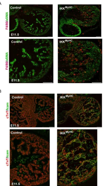
MyHChearts exhibit inflammation
Since the IKK/ NF-κB axis is one of the master regulators of inflammation in adults, embryonic IKKMyHChearts were analyzed for features of inflammation. Indeed, IKKMyHChearts at E11.5 manifested a strong infiltration of macrophages, as visualized by staining for the macrophage marker F4/80 (Fig 5A). Also, adhesion molecules like ICAM-1 were strongly expressed in car-diomyocytes (Fig 5B). This indicated that IKKMyHChearts were characterized by continuous recruitment of inflammatory cells.
Cardiomyocyte apoptosis is enhanced in embryonic IKK
MyHChearts at
later stages
The reduced thickness of the compact zone of embryonic IKKMyHChearts could be the result of enhanced cell death and/ or decreased proliferation. Embryonic hearts were examined for apoptosis at days E10.5, E12.5 and E14.5 using TUNEL (terminal deoxynucleotidyltransferase-mediated UTP end labeling) assays (Fig 6A). Apoptosis was prominent in IKKMyHCembryos only at later time points, thus putting into question its causal role for the reduced thickness of
(arrow, lower right); the pericardium is filled with blood (lower right). A: atrium, V: ventricle, L: limb. Scale bar: 0.5 mm. (C) Immunofluorescence analysis of IKK2-CA transgene expression in control (upper left) and IKKMyHC(upper right) embryos at E9.5. IKK2-CA expression (red) was exclusively detected in the heart of
IKKMyHCembryos. Nuclear staining with DAPI (blue). Scale bar 500
μm. Lower panel: Magnification of the
ventricle of an IKKMyHCembryo. Scale bar: 100μm. A: atrium, V: ventricle, TA: truncus arteriosus. (D) Upper
panel: Western blot analysis of heart extracts from E11.5 and E12.5 embryos. An antibody against human IKK (hIKK) was used to detect the IKK transgene, which is of human origin. IKK2-CA was exclusively detected in double transgenic embryos (IKKMyHC), but not in control embryos (ctrl). Expression of Erk2 was
used as a loading control. Lower panel: Western blot analysis of heart extracts of E14.5 animals with an antibody detecting both human (transgenic) and murine (endogenous) IKK. Erk2 was used as a loading control. (E) Immunofluorescence staining of E11.5 cryosections with antibodies against cardiac troponin T (cTNT, red), IKK and DAPI (blue), showing a colocalization of the cardiomyocyte-specific cTNT and the IKK transgene in IKKMyHCembryos. Scale bar 200μm. (F) Immunohistochemical analysis at E11.5 shows that
RelA/p65 in IKKMyHCcardiomyocytes, but not in control cardiomyocytes, is localized in the nucleus,
suggesting NF-κB activation. Scale bar: 310μm.
Fig 3. Cardiomyocyte-specific expression of IKK2-CA leads to defects in compact zone formation.(A) Hematoxylin/ eosin staining of frontal paraffin sections of control (left) and IKKMyHCembryos (right) at E12.5. RV, right ventricle; LV, left ventricle. Scale bar: 500μm. Both ventricles of the IKKMyHCembryo
show fewer cell layers in the compact zone, and the IKKMyHCheart is generally dilated. (B) Quantification of the number of compact zone cell layers of the
right and left ventricles of control and IKKMyHChearts. N = 6 embryos per group; shown are the arithmetic means +SEM.
*P<0.05,**P<0.01. (C) Quantification of total heart circumference of control and IKKMyHChearts. N = 6 embryos per group; shown are the arithmetic means +SEM.
*P<0.05.
Fig 4. Cardiomyocyte-specific IKK/NF-κB activation induces myofiber degeneration and loss of typical cardiomyocyte proteins.(A) Control and IKKMyHCsections were stained for sarcomeric
α-actinin
(green) and DAPI (blue). Scale bar: 100μm. (B) Control and IKKMyHCsections were stained for desmin
(yellow) and DAPI (blue). Scale bar: 100μm. (C) Electron microscopic analyses of control and IKKMyHCheart
sections. Cardiomyocytes of control animals show normally developed myofibers with typical sarcomeres (left). IKKMyHCcardiomyocytes show general disorganization and a reduced width of myofibers (right). Scale
bar: 1μm.
the compact zone. In contrast, proliferation appeared to be reduced already at day E11.5, as revealed by BrdU labeling of cardiomyocytes (Fig 6B).
Gene Expression Analysis of embryonic IKK
MyHChearts
For a more comprehensive understanding of gene expression changes in embryonic IKKMyHC hearts we performed microarray analysis of embryonic ventricles at day E12.5 (S1 Table). Con-stitutive activation of IKK/ NF-κB resulted in a interferon type 1 signature, with a strong
Fig 5. Cardiomyocyte-specific IKK/NF-κB activation up-regulates cell-adhesion molecules and induces the recruitment of macrophages.(A) Immunofluorescence staining of heart cryosections from control and IKKMyHCembryonic hearts shows infiltration with F4/80 positive macrophages. Macrophages
were stained with an antibody against F4/80 (red) and cardiomyocytes were stained with an antibody against
α-smooth muscle actin (αSMA, green). Scale bar: 100μm. (B) Immunofluorescence staining of heart
cryosections from control and IKKMyHCembryonic hearts show Icam-1 expression in IKKMyHCanimals, but
not in control animals at E11.5. Colocalization of Icam-1 (green) and the cardiomyocyte-specific cTNT (red) is visible as yellow staining. Scale bar: 100μm.
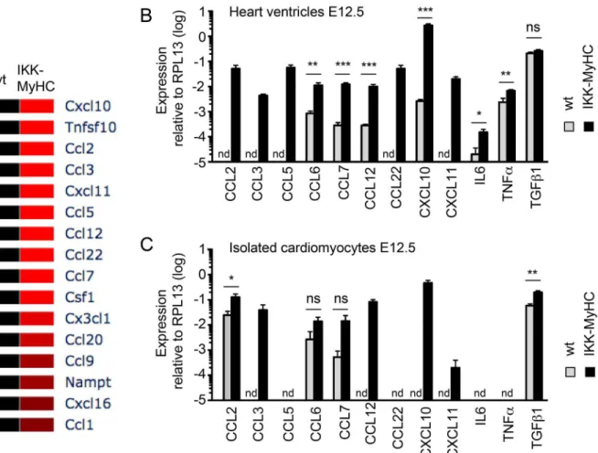
upregulation of interferon-regulatory factors (in particular Irf7) and the excessive expression of interferon-inducible genes like Sca1 (Ly6a/e) and Bst2. The gene expression signature of embryonic IKKMyHChearts also suggested a strong activation of Toll-like receptor (e.g. Tlr3) and Jak/ Stat signaling (e.g. Stat1, Stat2). Genes encoding chemokines (e.g. Ccl2), as well as cell adhesion molecules (e.g. Madcam1) were strongly expressed, reflecting the inflammation observed in IKKMyHChearts.Fig 7gives an overview of the deregulated cytokines and their val-idation via quantitative PCR.
Several genes related to cardiac muscle architecture and function (e.g. triadin, titin, and dys-trophin) were downregulated. Both apoptotic (such as Trail) and anti-apoptotic genes were upregulated in IKKMyHChearts. Transcripts of several cell cycle regulators showed a stronger
Fig 6. Cardiomyocyte undergo apoptosis only in later stages of IKK2-CA expression, but show early defects in proliferation.(A) TUNEL (terminal deoxynucleotidyltransferase-mediated UTP end labeling) assays were performed at E10.5, E12.5 and E14.5 for the indicated genotypes. TUNEL-positive cells show green fluorescence. An antibody againstα-myosin heavy chain was used to specifically stain cardiomyocytes
(red); nuclei were stained with DAPI (blue). RV, right ventricle; LV, left ventricle. Scale bar: 500μm. The right
panels show higher magnifications of the areas indicated in the middle panels. (B) Pregnant mice were injected with BrdU and their embryos (E10.5) were harvested 4 hours later. Cardiomyocytes were isolated and analyzed in FACS by staining with antibodies against the early murine cardiomyocyte marker Alcam (CD166)[13] and against BrdU. IKKMyHCcardiomyocytes (grey, filled) generally show a shift to the left in
comparison to control cardiomyocytes (black line), indicating a lower BrdU staining and thus less proliferation.
expression in IKKMyHChearts, in particular the gene encoding p21, consistent with a reduction in proliferation.
Regulators of proliferation and differentiation are disturbed in embryonic
IKK
MyHChearts
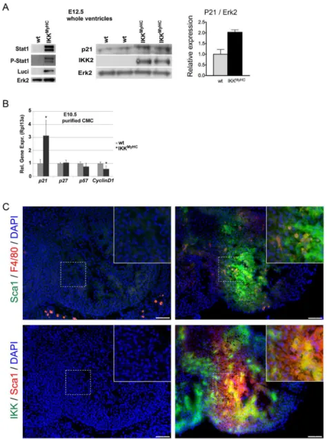
The activation of the Jak/ Stat axis suggested by the gene expression analysis was indeed con-firmed on the protein level: Stat1 expression and phosphorylation was strongly enhanced in the IKKMyHCembryos at E12.5 (Fig 8A). Also, the cell-cycle inhibitor p21, a target of Stat1, was upregulated at the protein level in embryonic IKKMyHChearts at E12.5 (Fig 8A).
In order to minimize the effect of infiltrating cells on these results, embryonic cardiomyo-cytes were isolated at E10.5, purified, and analyzed by quantitative PCR. Again, the transcript encoding p21 was upregulated (Fig 8B). In addition, the cyclin D1 transcript was
downregulated.
Furthermore, we tried to elucidate the significance of the strong upregulation of the stem cell marker Sca1 (Ly6a) in the gene expression analysis. Indeed, Sca1-positive cells were a prominent feature of embryonic IKKMyHC, but not control hearts at day E11.5 (Fig 8C). There
Fig 7. Cytokine profile of IKKMyHChearts and IKKMyHCcardiomyocytes.(A) Heat map of cytokines expressed in IKKMyHChearts at E12.5 (>2-fold
regulation, p<0.05) as detected by microarray (N = 3 for wild type (wt), N = 4 for transgenics (IKK-MyHC). (B) Expression of the indicated cytokines as assessed by real-time quantitative PCR from IKKMyHC(black bars) and control (grey bars) heart ventricles at E12.5. Shown are the means +SEM; N = 6,
*P<0.05,**P<0.01,***P<0.001 (t test), nd, not detected. (C) Expression of the indicated cytokines as assessed by real-time quantitative PCR from
IKKMyHC(black bars) and control (grey bars) cardiomyocytes isolated at E12.5. Shown are the means +SEM; N = 6,
*P<0.05,**P<0.01,***P<0.001 (t test), nd, not detected.
was only a very limited overlap between Sca1-positive cells and F4/80-positive cells, suggesting that macrophages were not primarily responsible for the observed Sca1 expression (Fig 8C, upper panel). In part, the Sca1-positive population also expressed the transgene IKK2-CA, sug-gesting that they constitute or contain a precursor population of cardiomyocytes with a possi-ble function in regeneration (Fig 8C, lower panel).
Fig 8. Dysregulation of regulators of proliferation and differentiation in embryonic IKKMyHChearts.(A) The expression of Stat1, phospho-Stat1, p21 and the transgenes IKK2 and luciferase was determined by Western blot, with Erk2 shown as loading control. The diagram on the right shows a quantification of the p21 immunoblot, normalized to the expression of ERK2. (B) Expression of the indicated cell cycle modulators was determined at the mRNA level using real-time quantitative PCR in purified cardiomyocytes from IKKMyHC
(black bars) and control (grey bars) embryos at E10.5. Shown are the means +SEM; N = 5,*P<0.05. (C) Upper panel: Staining of embryonic heart sections at E11.5 for Sca1 (green) and F4/80 (red). Lower panel: Staining of embryonic heart sections at E11.5 for the IKK2 transgene (green) and the progenitor marker Sca1 (red). DAPI was used to stain nuclei. Insets show a higher magnification of the indicated areas.
The Embryonic Lethality of IKK
MyHCEmbryos Is Dependent on NF-
κ
B
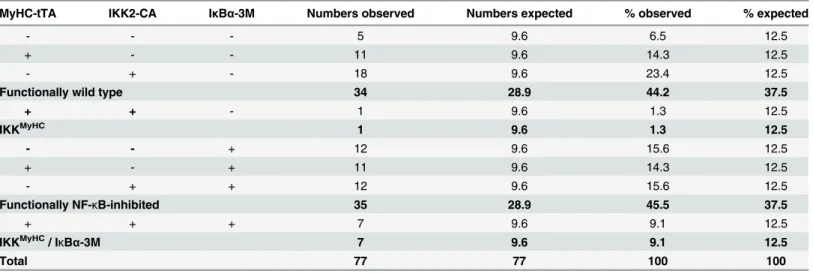
Finally, we wanted to address the question whether the embryonic lethality of IKKMyHC embryos was in fact due to NF-κB activation or potential other pathways activated by the IKK complex. For this end, we crossed IKKMyHCanimals with animals expressing a mutated form of IκBα-(IκBα(S32A, S36A, Y42F)), which is impervious to phosphorylation-induced degra-dation and thus acts as a superrepressor of NF-κB signaling[11]. Indeed, whereas only very few IKKMyHCanimals survived embryonic and postnatal development until weaning (constituting 1.3% of the surviving offspring instead of the expected 12.5%), IKKMyHCanimals expressing the IκBα-superrepressor did survive at almost normal Mendelian ratios (constituting 9.1% of the surviving offspring instead of the expected 12.5%), thus supporting the notion that IKK2 exerts its lethal effects via NF-κB signaling (Table 2).
Discussion
Previously, we showed that activation of IKK/ NF-κB signaling in the adult animal led to inflammatory cardiomyopathy and heart failure [12]. Our present study now expands on these results by illuminating the consequences of IKK2 activation under the specific conditions of embryonic heart development.
Cardiac-specific expression of a constitutively active IKK2 mutant during mouse develop-ment resulted in embryonic lethality. The rather long time window during which the embryos died, ranging from E9.5 (shortly after the beginning of the activity of theα-myosin heavy chain promoter) through E16.5 with half of the embryos dead by E12.5, suggests that IKK2 activation does not interfere with a specific, chronologically strictly defined event, but rather with the gen-eral conditions required for normal heart development during a longer period of time. Alterna-tively, the long time window might be due to variability in the strength of transgene expression and the number of cells that express it. In any case, the rescue experiments with an IκBα super-repressor suggest that the lethality IKK2 exerts is mediated by NF-κB.
Table 2. Embryonic lethality induced by constitutively active IKK2 can be prevented by an IκBαsuperrepressor.
MyHC-tTA IKK2-CA IκBα-3M Numbers observed Numbers expected % observed % expected
- - - 5 9.6 6.5 12.5
+ - - 11 9.6 14.3 12.5
- + - 18 9.6 23.4 12.5
Functionally wild type 34 28.9 44.2 37.5
+ + - 1 9.6 1.3 12.5
IKKMyHC 1 9.6 1.3 12.5
- - + 12 9.6 15.6 12.5
+ - + 11 9.6 14.3 12.5
- + + 12 9.6 15.6 12.5
Functionally NF-κB-inhibited 35 28.9 45.5 37.5
+ + + 7 9.6 9.1 12.5
IKKMyHC/ I
κBα-3M 7 9.6 9.1 12.5
Total 77 77 100 100
The table shows the absolute number and percentage of animals born with the indicated actual and functional genotype (observed), and the
corresponding expected number and percentage (expected). Only one IKKMyHCanimal was identified, whereas seven IKKMyHC/ IκBα-3M mice could be
detected, indicating a rescue of the lethality by the IκBα-3M transgene.
The most prominent finding in IKKMyHChearts was certainly the strong inflammatory response observed upon IKK/ NF-κB activation. The inflammatory infiltrate was characterized by F4/80-positive cells and thus monocytes/ macrophages. Gene expression analysis revealed that the signature observed in IKKMyHChearts very much resembled an interferon type I response as observed in an anti-viral response. This is consistent with the notion that IKK/
NF-κB signaling is central to the development of myocarditis, both virally induced and autoim-mune [14–17]. The activation of the transcription factor STAT1, which is prominent in our model, is also known to be an early event in viral myocarditis [18].
In addition to the inflammation observed in IKKMyHChearts, the defect in compact zone for-mation was a prominent finding. Several biological processes could explain this defect—in partic-ular an increase in cell death or a decrease in proliferation. Whereas cell death was indeed increased in IKKMyHCembryos, this was a rather late phenomenon and therefore most likely not responsible for the disease process. In contrast, proliferation was reduced in IKKMyHCembryonic hearts already at an earlier time point, suggesting that it might be responsible for the thinning of the compact zone. Indeed, proliferation is a crucial event during this time of heart development, and any disturbance might have catastrophic consequences for the developing heart [3]. This is of course much in contrast to the observations in the adult animals, where proliferation is of minor or no relevance due to the largely post-mitotic status of the adult heart [12].
We also identified a potential link between the observed inflammation and the reduction in proliferation: Among the pathways activated in this response is the JAK/ STAT pathway, which via its target gene p21 might directly inhibit proliferation [19]. The cause of the decrease in proliferation might thus indeed lie in the inflammatory environment that is so characteristic for IKKMyHChearts.
An interesting finding in IKKMyHCembryonic hearts is the emergence of a Sca1-positive cell population. Recently, Sca1-positive cells have been identified as a source for myocardial renewal, albeit in the adult heart [20]. It remains to be seen whether the cells observed in
IKK-MyHCembryonic hearts fulfill a similar and thus beneficial role, or whether they in contrast
rep-resent a lack of differentiation of cardiomyocyte progenitors, or even a dedifferentiation of cardiomyocytes. Dedifferentiation might indeed be one of the effects of IKK2, since the con-tractile apparatus, a hallmark of cardiomyocyte differentiation, was severely affected upon IKK/ NF-κB activation as shown by electron microscopy and immunofluorescence analyses of typical markers such as sarcomeric actinin.
Since infections are a major cause for adverse events during gestation in humans, our find-ings might be of relevance for a better understanding of the molecular processes in affected embryos and fetuses. According to our study, IKK/ NF-κB is a key regulator of the anti-viral response in cardiomyocytes, a finding that is very interesting in the light that viral infections (e.g. by CMV, parvovirus B19, and rubella virus) are a major problem during gestation.
Supporting Information
S1 Table. Microarray analysis reveals widespread changes in gene expression in IKKMyHC embryos.The table shows the full name, the gene ID and the fold induction of genes, arranged according to their function.
(XLS)
Acknowledgments
Author Contributions
Conceived and designed the experiments: TW. Performed the experiments: BK HJM EK KF AI. Analyzed the data: BK HJM EK SK T. Braun TW. Contributed reagents/materials/analysis tools: T. Boettger PW. Wrote the paper: HJM TW.
References
1. Gordon JW, Shaw JA, Kirshenbaum LA. Multiple facets of kappaB in the heart: to be or not to NF-kappaB. Circulation research. 2011; 108(9):1122–32. doi:10.1161/CIRCRESAHA.110.226928PMID:
21527742.
2. Hinz M, Scheidereit C. The IkappaB kinase complex in NF-kappaB regulation and beyond. EMBO reports. 2014; 15(1):46–61. doi:10.1002/embr.201337983PMID:24375677; PubMed Central PMCID:
PMC4303448.
3. Clowes C, Boylan MG, Ridge LA, Barnes E, Wright JA, Hentges KE. The functional diversity of essen-tial genes required for mammalian cardiac development. Genesis. 2014; 52(8):713–37. doi:10.1002/
dvg.22794PMID:24866031; PubMed Central PMCID: PMC4141749.
4. Li Q, Van Antwerp D, Mercurio F, Lee KF, Verma IM. Severe liver degeneration in mice lacking the Ikap-paB kinase 2 gene. Science. 1999; 284(5412):321–5. PMID:10195897.
5. Li ZW, Chu W, Hu Y, Delhase M, Deerinck T, Ellisman M, et al. The IKKbeta subunit of IkappaB kinase (IKK) is essential for nuclear factor kappaB activation and prevention of apoptosis. The Journal of experimental medicine. 1999; 189(11):1839–45. PMID:10359587; PubMed Central PMCID:
PMC2193082.
6. Tanaka M, Fuentes ME, Yamaguchi K, Durnin MH, Dalrymple SA, Hardy KL, et al. Embryonic lethality, liver degeneration, and impaired NF-kappa B activation in IKK-beta-deficient mice. Immunity. 1999; 10(4):421–9. PMID:10229185.
7. Hikoso S, Yamaguchi O, Nakano Y, Takeda T, Omiya S, Mizote I, et al. The I{kappa}B kinase {beta}/ nuclear factor {kappa}B signaling pathway protects the heart from hemodynamic stress mediated by the regulation of manganese superoxide dismutase expression. Circulation research. 2009; 105(1): 70–9. doi:10.1161/CIRCRESAHA.108.193318PMID:19478205.
8. Dawn B, Xuan YT, Marian M, Flaherty MP, Murphree SS, Smith TL, et al. Cardiac-specific abrogation of NF- kappa B activation in mice by transdominant expression of a mutant I kappa B alpha. Journal of molecular and cellular cardiology. 2001; 33(1):161–73. PMID:11133232.
9. Herrmann O, Baumann B, de Lorenzi R, Muhammad S, Zhang W, Kleesiek J, et al. IKK mediates ische-mia-induced neuronal death. Nature medicine. 2005; 11(12):1322–9. PMID:16286924.
10. Yu Z, Redfern CS, Fishman GI. Conditional transgene expression in the heart. Circulation research. 1996; 79(4):691–7. PMID:8831492.
11. Brown M, McGuinness M, Wright T, Ren X, Wang Y, Boivin GP, et al. Cardiac-specific blockade of NF-kappaB in cardiac pathophysiology: differences between acute and chronic stimuli in vivo. American journal of physiology Heart and circulatory physiology. 2005; 289(1):H466–76. PMID:15695559. 12. Maier HJ, Schips TG, Wietelmann A, Kruger M, Brunner C, Sauter M, et al. Cardiomyocyte-specific
IkappaB kinase (IKK)/NF-kappaB activation induces reversible inflammatory cardiomyopathy and heart failure. Proceedings of the National Academy of Sciences of the United States of America. 2012; 109(29):11794–9. doi:10.1073/pnas.1116584109PMID:22753500; PubMed Central PMCID:
PMC3406816.
13. Hirata H, Murakami Y, Miyamoto Y, Tosaka M, Inoue K, Nagahashi A, et al. ALCAM (CD166) is a sur-face marker for early murine cardiomyocytes. Cells, tissues, organs. 2006; 184(3–4):172–80. PMID:
17409743.
14. Alter P, Rupp H, Maisch B. Activated nuclear transcription factor kappaB in patients with myocarditis and dilated cardiomyopathy—relation to inflammation and cardiac function. Biochemical and
biophysi-cal research communications. 2006; 339(1):180–7. PMID:16297880.
15. O'Donnell SM, Hansberger MW, Connolly JL, Chappell JD, Watson MJ, Pierce JM, et al. Organ-specific roles for transcription factor NF-kappaB in reovirus-induced apoptosis and disease. The Journal of clini-cal investigation. 2005; 115(9):2341–50. PMID:16100570; PubMed Central PMCID: PMC1184036. 16. Yamamoto K, Shioi T, Uchiyama K, Miyamoto T, Sasayama S, Matsumori A. Attenuation of
17. Yokoseki O, Suzuki J, Kitabayashi H, Watanabe N, Wada Y, Aoki M, et al. cis Element decoy against nuclear factor-kappaB attenuates development of experimental autoimmune myocarditis in rats. Circu-lation research. 2001; 89(10):899–906. PMID:11701617.
18. Ruppert V, Meyer T, Pankuweit S, Jonsdottir T, Maisch B. Activation of STAT1 transcription factor pre-cedes up-regulation of coxsackievirus-adenovirus receptor during viral myocarditis. Cardiovascular pathology: the official journal of the Society for Cardiovascular Pathology. 2008; 17(2):81–92. doi:10.
1016/j.carpath.2007.07.004PMID:18329552.
19. Chin YE, Kitagawa M, Su WC, You ZH, Iwamoto Y, Fu XY. Cell growth arrest and induction of cyclin-dependent kinase inhibitor p21 WAF1/CIP1 mediated by STAT1. Science. 1996; 272(5262):719–22.
PMID:8614832.
20. Uchida S, De Gaspari P, Kostin S, Jenniches K, Kilic A, Izumiya Y, et al. Sca1-derived cells are a source of myocardial renewal in the murine adult heart. Stem cell reports. 2013; 1(5):397–410. doi:10.