p110
a
Ameliorates Cardiac Dysfunction and Improves
Survival in Polymicrobial Sepsis
Chuanfu Li1, Fang Hua2, Tuanzhu Ha1, Krishna Singh3, Chen Lu1, John Kalbfleisch4, Kevin F. Breuel5, Tiffany Ford5, Race L. Kao1,2, Ming Gao1, Tammy R. Ozment1, David L. Williams1*
1Department of Surgery, Quillen College of Medicine, East Tennessee State University, Johnson City, Tennessee, United States of America,2Department of Emergency Medicine, Emory University School of Medicine Atlanta, Georgia, United States of America,3Department of Biomedical Sciences, Quillen College of Medicine, East Tennessee State University, Johnson City, Tennessee, United States of America,4Department of Biometry and Medical Computing, Quillen College of Medicine, East Tennessee State University, Johnson City, Tennessee, United States of America,5Department of Obstetrics and Gynecology, Quillen College of Medicine, East Tennessee State University, Johnson City, Tennessee, United States of America
Abstract
Phosphoinositide-3-kinase (PI3K)/Akt dependent signaling has been shown to improve outcome in sepsis/septic shock. There is also ample evidence that PI3K/Akt dependent signaling plays a crucial role in maintaining normal cardiac function. We hypothesized that PI3K/Akt signaling may ameliorate septic shock by attenuating sepsis-induced cardiac dysfunction. Cardiac function and survival were evaluated in transgenic mice with cardiac myocyte specific expression of constitutively active PI3K isoform, p110a(caPI3K Tg). caPI3K Tg and wild type (WT) mice were subjected to cecal ligation/puncture (CLP) induced sepsis. Wild type CLP mice showed dramatic cardiac dysfunction at 6 hrs. Septic cardiomyopathy was significantly attenuated in caPI3K CLP mice. The time to 100% mortality was 46 hrs in WT CLP mice. In contrast, 80% of the caPI3K mice survived at 46 hrs after CLP (p,0.01) and 50% survived.30 days (p,0.01). Cardiac caPI3K expression prevented expression of an inflammatory phenotype in CLP sepsis. Organ neutrophil infiltration and lung apoptosis were also effectively inhibited by cardiac PI3k p110aexpression. Cardiac high mobility group box–1 (HMGB-1) translocation was also inhibited by caPI3K p110aexpression. We conclude that cardiac specific activation of PI3k/Akt dependent signaling can significantly modify the morbidity and mortality associated with sepsis. Our data also indicate that myocardial function/dysfunction plays a prominent role in the pathogenesis of sepsis and that maintenance of cardiac function during sepsis is essential. Finally, these data suggest that modulation of the PI3K/p110a signaling pathway may be beneficial in the prevention and/or management of septic cardiomyopathy and septic shock.
Citation:Li C, Hua F, Ha T, Singh K, Lu C, et al. (2012) Activation of Myocardial Phosphoinositide-3-Kinase p110aAmeliorates Cardiac Dysfunction and Improves Survival in Polymicrobial Sepsis. PLoS ONE 7(9): e44712. doi:10.1371/journal.pone.0044712
Editor:Charles C. Caldwell, University of Cincinnati, United States of America
ReceivedJune 28, 2012;AcceptedAugust 7, 2012;PublishedSeptember 19, 2012
Copyright:ß2012 Li et al. This is an open-access article distributed under the terms of the Creative Commons Attribution License, which permits unrestricted use, distribution, and reproduction in any medium, provided the original author and source are credited.
Funding:This work was supported by an American Heart Association (AHA) Career Scientist Development grant 0830481N (to F. Hua); East Tennessee State University Research Development Committee awards (to T. Ha and R.L. Kao); Veterans Affairs Merit Review Grant (to K. Singh); National Institutes of Health (NIH) grants HL091405 and HL092459 (to K. Singh); AHA Career Scientist Development grant 11SDF5330002 (to T. Ozment); NIH HL071837 (to C. Li), NIH GM53552 (to D.L. Williams), and NIH GM083016 (to C. Li and D.L. Williams). The funders had no role in study design, data collection and analysis, decision to publish, or preparation of the manuscript.
Competing Interests:The authors have declared that no competing interests exist. * E-mail: [email protected]
Introduction
The critically ill patient frequently develops a complex disease spectrum that may include acute respiratory distress syndrome (ARDS), systemic inflammatory response syndrome (SIRS), sepsis syndrome and/or septic shock and multi-organ dysfunction syndrome (MODS) [1]. In the United States,750,000 patients/
year develop sepsis syndrome [2]. The overall mortality rate is 28.6% (,215,000 deaths/year) [2]. Those patients that survive the
initial event may ultimately succumb to widespread organ dysfunction that can be either acute, due to hyper-inflammatory responses, or more prolonged due to immune dysfunction and infection [3,4]. Indeed, sepsis is a frequent cause of MODS [3,4]. In many cases, the lung is the primary organ affected, however, it is well known that cardiovascular dysfunction is also associated with MODS morbidity and mortality [5–7]. Cardiovascular
dysfunction as a consequence of sepsis has been termed ‘‘septic cardiomyopathy’’ [5–7]. It is now well accepted that septic cardiomyopathy is an emerging problem in the management of the critically ill patient [5].
The phosphoinositide-3-kinases (PI3Ks) are a conserved family of signal transduction enzymes which are involved in regulating cellular proliferation and survival [8,9]. The PI3Ks and the downstream serine/threonine kinase Akt (also known as protein kinase B = PKB) regulate cellular activation, inflammatory re-sponses, chemotaxis and apoptosis [9]. Guha and Mackman have reported that the ‘‘PI3K-Akt pathway imposes a braking mechanism to limit the expression’’ of pro-inflammatory media-tors in LPS treated monocytes [10]. Using anin vivosepsis model,
increases survival outcome in sepsis [11]. In support of this observation, Bommhardt et al reported that mice which consti-tutively over express active Akt in their lymphocytes showed decreased lymphocyte apoptosis, a TH1 cytokine propensity, and a marked improvement in survival outcome in response to sepsis [12]. We and others have reviewed the role of PI3K in the regulation of inflammatory and/or septic responses [13,14]. The data suggest that PI3K/Akt may be an endogenous negative feedback regulator and/or compensatory mechanism which is crucial to the maintenance and integrity of homeostasis during septic and/or inflammatory insults [13,14]. However, the mech-anisms by which PI3K/Akt ameliorates septic sequelae and improves survival outcome have not been fully delineated.
There is ample evidence that PI3k/Akt dependent signaling plays a crucial role in cardiac function [15–17]. We have reported that sepsis decreases myocardial Akt activation [18], which positively correlates with cardiac dysfunction in sepsis. We also reported that preventing sepsis induced changes in Akt activation ameliorates cardiovascular dysfunction in sepsis [18]. Based on these data, we hypothesized that activation of PI3K/Akt dependent signaling will prevent or blunt cardiac dysfunction during sepsis/septic shock. To test this hypothesis, we examined the effect of sepsis/septic shock on cardiac function and survival in transgenic mice with cardiac myocyte specific expression of a constitutively active PI3K isoform p110a(caPI3K Tg).
Materials and Methods
Experimental Animals
The caPI3K p110a transgenic mice, on the C47Bl/6J background, were kindly provided by Dr. Seigo Izumo from Harvard Medical School [19,20]. The mice were maintained and bred in the Division of Laboratory Animal Resources at East Tennessee State University (ETSU). The experiments outlined in this manuscript conform with the Guide for the Care and Use of Laboratory Animals published by the US National Institutes of Health (NIH Publication No. 85–23, revised 1996). The experi-mental protocols as well as all aspects of animal care described in this manuscript were reviewed and approved by the ETSU Committee on Animal Care.
Cecal Ligation and Puncture (CLP)
Cecal ligation and puncture was performed to induce sepsis in mice as previously described [21–23]. Briefly, the mice were anesthetized by isoflurane inhalation and ventilated with room air using a rodent ventilator. A midline incision was made on the anterior abdomen and the cecum was exposed and ligated with a 0 suture. Two punctures were made through the cecum with an 18-gauge needle and feces were extruded from the holes. The abdomen was then closed. Fluid resuscitation (1 ml lactated Ringers) was administered subcutaneously after surgery. Sham surgical operated mice (anesthesia and laparotomy) served as the surgical control group. Mice that were not subjected to surgery or anesthesia served as the normal controls.
Experimental Protocol
Mice were subjected to either CLP or sham surgery at time 0. Cardiovascular function was assessed at 6 hrs post-CLP. In a separate experiment, the same groups were followed for survival and at 16 hrs representative post-CLP mice were taken from each group, euthanized and serum and tissues were harvested for analysis of serum cytokines/chemokines, Akt and glycogen synthase kinase-3b (GSK3b) phosphorylation, tissue apoptosis
and tissue nuclear factor-kB (NFkB) analysis. For HMGB-1 analysis, tissue samples were harvested at 6 and 12 hrs after CLP.
Hemodynamic Measurements
Cardiovascular function was assessed as previously described by our group [18]. Briefly, mice were anesthetized with isoflurane inhalation and ventilated with room air using a rodent ventilator. A microconductance pressure catheter (Millar Instruments Inc., Houston, TX) was positioned in the left ventricle (LV) via the right carotid artery for continuous registration of LV pressure-volume loops [24,25] using the PowerLab system (AD Instruments, Inc., Colorado Springs, CO). A cuvette calibration method was used to convert the conductance voltage into volume units by filling nonconductive cuvettes of known diameter with heparin treated mouse blood. Parallel conductance from surrounding structures was determined by intravenous (external jugular vein) injection of a small bolus (15 ml) of hypertonic saline (15% NaCl). All measurements were performed while ventilation was turned off momentarily. Indices of systolic and diastolic cardiac performance were derived from LV pressure-volume data obtained at steady state.
Echocardiographic Analysis of Cardiac Function
Transthoracic two-dimensional M-mode echocardiograms were obtained using a Toshiba Aplio 80 Imaging System (Tochigi, Japan) equipped with a 12 MHz linear transducer. The mice were anesthetized using a mixture of isoflurane (1.5%) and oxygen (0.5 l/min) and the body temperature was maintained at,37uC
using a heating pad. M-mode tracings were used to measure end-diastolic diameter (LVEDD). Percent fractional shortening (%FS) and ejection fraction (%EF) were calculated as described [26]. All echocardiographic assessments were performed by the same investigator.
Western Blot
Cytoplasmic proteins were isolated from heart, liver, lung and spleen. Immunoblots were performed as described previously [27– 31]. Briefly, the cellular proteins were separated by sodium dodecyl sulfate (SDS)-polyacrylamide gel electrophoresis and transferred onto Hybond ECL membranes (Amersham Pharma-cia, Piscataway, NJ). The ECL membranes were incubated with appropriate primary antibody against phospho-Akt, or anti-Akt; anti-phospho-GSK3b or anti-GSK3b respectively, followed by incubation with peroxidase-conjugated second antibodies (Cell Signaling Technology, Inc. Beverly, MA). The membranes were analyzed by the ECL system (Amersham Pharmacia). The same membranes were stripped and re-probed with anti-GAPDH (glyceraldehyde-3-phosphate dehydrogenase, Biodesign, Saco, Maine) as loading controls. In the case of nuclear HMGB-1, histone 3 was employed as the loading control. The signals were quantified by scanning densitometry and computer-assisted image analysis.
Assessment of Serum Cytokine Levels
interleukin-6 (IL-6), interleukin-10 (IL-10), interleukin-12 p40/p70 (IL-12p40/p70), interleukin-13 (IL-13), interleukin-17 (IL-17), interferon inducible protein-10 (IP-10), keratinocyte chemoattrac-tant (KC), monocyte chemotactic protein-1 (MCP-1), macrophage inflammatory protein-1a (MIP-1a), monokine induced by inter-feron-c (MIG), tumor necrosis factor a (TNFa) and vascular endothelial growth factor (VEGF). Cytokine levels were estab-lished by comparison to a standard curve as per the manufactur-er’s instructions.
Splenocyte Apoptosis
We employed two approaches to assess splenocyte apoptosis in response to sepsis. First, in situ cardiac splenocyte apoptosis was
examined by the TdT mediated dUTP nick end labeling (TUNEL) assay (Boehringer Mannheim, Indianapolis, IN) as described previously [24]. Three slides from each block were evaluated for apoptotic cells using the TUNEL assay. Tunel stains were imaged using a fluorescent microscope. In addition, four slide fields were randomly examined using a defined rectangular field area with 200X magnification. Apoptotic cells were counted in each field. Second, spleens were harvested into ice cold phosphate buffered saline (PBS) 16 hrs after induction of CLP. Individual cells were isolated by teasing the stroma apart with needles, allowing the stroma to settle, and pouring the suspended cells into a fresh tube. The red blood cells were lysed with hypotonic saline. The leukocytes were fixed with ice cold 70% ethanol for at least 18 h at220uC. The cells were washed three times with PBS, and then stained with 50mg/ml propidium iodide with 10mg/ml RNase A for 30 min at 37uC. The samples were then analyzed for the sub G0 population using a FACScalibur flow cytometer with CellQuest software (BD Biosciences, Mountain View, CA).
Tissue Myeloperoxidase (MPO) Activity
Lung tissue was processed as directed by the MPO Fluorometric Detection Kit (Assay Designs, Ann Arbor, MI). Specifically, 50 mg of tissue was weighed out into 1X assay buffer containing 10 mM N-ethylmaleimide (Sigma). The tissue samples were homogenized with a Polytron. After pelleting, the cells were lysed using 0.5% hexadecyltrimethylammonium in 1X assay buffer. Following homogenization and sonication at 50% power for 3–10 s pulses, the homogenates were subjected to two freeze thaw cycles. After clearing of cell debris by centrifugation, the lysates were stored at 280uC until assayed. The samples were assayed according to kit directions and were read using the Modulus Microplate fluores-cent plate reader after 30 min incubation (Turner Biosystems, Sunnyvale, CA).
Tissue NFkB Assay
Nuclear protein was isolated from lung tissues according to previously published methods [21]. Nuclear protein was assayed for NFkB activity using a commercially available kit (Pierce Thermo Scientific, Rockford, IL) according to the manufacturer’s instructions.
Statistics
Survival trends were compared with the log-rank Wilcoxon non-parametric procedures. Phosphorylation, caspase and cyto-kine levels were summarized by the group mean. Using replicate averages for each animal, group means were statistically compared with the one-way analysis of variance (ANOVA) followed by the least significant difference test. Unless otherwise stated data are expressed as mean+s.e.m. A probability level of 0.05 or smaller was used to report statistical significance.
Results
Cardiac Specific Expression of Constitutively Active PI3K p110aAttenuates Sepsis Induced Cardiac Dysfunction
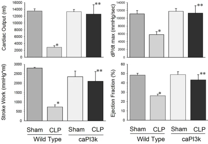
We examined the effect of sepsis on cardiac function in caPI3K mice using two approaches. First, we examined cardiac function using a conductance catheter as previously described [24,25,18]. Second, we employed echocardiography as a non-invasive approach to assess cardiac function in septic mice. Cardiac function was assessed at 6 hrs after CLP induced sepsis. As expected, WT mice subjected to CLP showed significant depression of cardiac function (Table 1andFigure 1). Cardiac output was reduced by 78.5%, stroke work by 73.7%, dp/dt max (mmHg/sec) by 47.6%, and ejection fraction by 45.7%, respec-tively, compared with sham control (Figure 1). However, cardiac dysfunction was attenuated in response to sepsis in mice with cardiac specific expression of constitutively active PI3K p110a, (Table 1and Figure 1). In some cases, expression of the PI3k p110a transgene resulted in a maintenance of normal cardiac function in response to sepsis (Figure 1). Specific examples include cardiac output, dP/dt max, stroke work, ejection fraction, maximum elastance (Emax) and regression of log (pressure) versus time (Tau-Weiss) (Figure 1 and Table 1). End systolic pressure, end systolic volume and stroke volume in caPI3K CLP mice were maintained at levels significantly higher than the WT CLP mice, but they were significantly different from the sham controls.
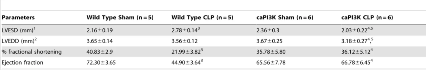
Echocardiographic analysis confirmed that caPI3K Tg mice showed attenuation of cardiac function in response to CLP sepsis (Table 2). Left ventricular end systolic diameter (LVESD) was significantly increased, while %FS and ejection fraction (%EF) were significantly decreased in WT CLP mice, when compared to WT sham and caPI3K CLP groups (Table 2). Interestingly, LVEDD did not change in response to CLP sepsis, but in the caPI3K-CLP group LVEDD was significantly lower than either the caPI3K-sham or WT-CLP groups (Table 2). There was no significant difference in LVESD, %FS and EF between caPI3K-sham and caPI3K-CLP groups.
Cardiac Specific Expression of Constitutively Active PI3K p110aResults in Increased Survival Outcome in CLP Sepsis
To investigate whether preservation of cardiac function during sepsis/septic shock will alter survival outcome, we performed CLP to induce sepsis/septic shock in caPI3K (p110a) mice (n = 10). Age-matched wild type (WT) mice (n = 10) served as control. As shown inFigure 2, caPI3K mice were more resistant to CLP-induced mortality than WT mice. The median survival time in WT mice was,28 hrs and time to 100% mortality was 46 hrs. In
striking contrast, 90% of the caPI3K mice were alive at,28 hrs
and 80% of the caPI3K mice survived at 46 hrs after CLP (Figure 2). Of greater significance, 50% of the caPI3K mice went on to survive indefinitely (.30 days) after CLP.
Constitutive Activation of Myocardial PI3K p110aResults in Increased Phosphorylation of Akt in the Myocardium, but not Other Tissues
difference in myocardial Akt phosphorylation in p110atransgenic mice in the presence or absence of sepsis, however, sepsis did result in a significant decrease in myocardial Akt phosphorylation in wild type mice. We also examined Akt phosphorylation in the liver,
lung and spleen of caPI3K Tg mice in the presence and absence of sepsis. We did not find any differences in Akt phosphorylation in organs other than the heart in the presence or absence of sepsis (data not shown). These data indicate that constitutive activation
Figure 1. Cardiac myocyte specific expression of PI3K p110aattenuated cardiac dysfunction in CLP-induced septic mice.We
performed hemodynamic and echocardiographic analysis at 6 hrs after CLP. Echocardiography confirmed that cardiac function was compromised in WT CLP. In contrast, caPI3K mice with CLP induced sepsis showed an overall maintenance of cardiac function. The bar graphs show the cumulative cardiac function data for the respective control and experimental groups. Six hrs after CLP, left ventricular hemodynamic parameters were examined. N = 5 mice/group. * p,0.05 compared to age-matched WT control. **p,0.05 compared to WT CLP group.
doi:10.1371/journal.pone.0044712.g001
Table 1.Cardiac myocyte specific expression of PI3K p110asignificantly attenuated cardiac dysfunction in caPI3K mice with CLP induced polymicrobial sepsis.
Hemodynamic parameter Wild Type Sham Wild Type CLP caPI3K Sham caPI3K CLP
Heart rate (beats/min) 419.7647.4 280.2650.61 441.8
682.8 552.367.82,3
End systolic pressure (mmHg) 93.6611.16 77.6617.21 96.967.8 104.1615.12
End diastolic pressure (mmHg) 3.360.8 2.460.3 4.261.8 3.461.4
End systolic volume (ml) 37.067.6 28.963.8 38.963.5 28.561.93
End diastolic volume (ml) 66.463.4 37.962.91 69.0
63.4 48.863.82,3
Stroke volume (ml) 32.363.3 10.163.01 34.4
63.2 22.869.72,3
Emax (mmHg/ml) 19.166.4 7.861.71 12.7
64.6 15.362.82
Tau-Weiss (ms) 6.461.3 12.062.51 7.2
60.9 7.262.72
Data are presented as mean6SD. 1p
,0.05 vs wild type sham group. 2p
,0.05 vs wild type CLP group. 3p
,0.05 vs caPI3K sham group. N = 5–6/group.
doi:10.1371/journal.pone.0044712.t001
of myocardial PI3K p110aactivates Akt in the myocardium, but it does not appear to activate Akt in extra-cardiac tissues. This is consistent with previous observations indicating that the effect of the transgene is limited to the heart [20].
Constitutive Activation of Myocardial PI3K p110aResults in Increased Phosphorylation of GSK3bin the
Myocardium, but not Other Tissues
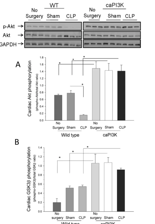
Activated Akt phosphorylates several downstream targets of the PI3K pathway including glycogen synthase kinase-3b (GSK3b) [32,33]. GSK3bis a crucial regulator of many cellular functions, including cell survival and apoptosis [32]. GSK3b is a constitu-tively active enzyme that is inactivated by Akt via phosphorylation of serine 9 [32]. The levels of phosphorylated GSK3b were increased in the myocardium of normal control, sham control and CLP mice expressing the p110atransgene when compared to WT mice (Figure 3B). These data indicate that constitutive activation
of myocardial PI3K p110ainactivates GSK3bin the myocardium. Interestingly, we did not see any significant difference in myocardial GSK3b phosphorylation in p110a transgenic mice in the presence or absence of sepsis. We also examined the phosphorylation of GSK3bin tissues other than the heart. There were no differences in GSK3b phosphorylation in tissues other than the heart,i.e.liver, lung and spleen (data not shown), in the presence or absence of sepsis. These data indicate that constitutive activation of myocardial PI3K p110a inactivates GSK3b in the myocardium in the presence or absence of sepsis, but it does not appear to inactivate GSK3bin extra-cardiac tissues.
Serum Cytokine/chemokine Levels are Maintained at Normal Levels in Septic Mice with Cardiac Specific Expression of PI3K p110a
We examined serum cytokine/chemokine levels at 16 hrs after induction of CLP sepsis (Figs. 4 and 5). As previously reported,
Table 2.Echocardiographic analysis indicates that caPI3K mice maintain cardiac function in response to CLP sepsis.
Parameters Wild Type Sham (n = 5) Wild Type CLP (n = 5) caPI3K Sham (n = 6) caPI3K CLP (n = 6)
LVESD (mm)1 2.16
60.19 2.7860.143 2.36
60.3 2.0360.224,5
LVEDD (mm)2 3.65
60.14 3.5660.12 3.6760.25 3.1860.274,5
% fractional shortening 40.8362.9 21.9963.823 35.78
65.80 36.1265.124
Ejection fraction 72.3063.65 44.9063.643 65.56
67.78 66.7866.454
Data are presented as mean6SD. 1left ventricular end systolic diameter. 2left ventricular end diastolic diameter. 3p
,0.05 vs wild type sham group. 4p
,0.05 vs wild type CLP group. 5p
,0.05 vs caPI3K sham group. N = 5–6/group.
doi:10.1371/journal.pone.0044712.t002
Figure 2. Cardiac myocyte specific expression of PI3K p110asignificantly improves survival outcome in CLP-induced sepsis/septic
shock.caPI3K transgenic mice (N = 10) and age-matched wild type mice (N = 11) were subjected to CLP at time 0. The mice were followed for survival for up to 30 days.
CLP sepsis resulted in a significant increase in serum cytokine levels at 16 hrs post-surgery (Figs. 4 and 5). In striking contrast, expression of cardiac specific PI3K p110ahad a dramatic effect on CLP induced serum cytokine levels. Serum cytokine/chemokine levels were maintained at normal levels in caPI3K CLP mice (Figs. 4 and 5). Specifically, serum IL-1a, 5, 6, 12, IL-17, TNFa, IFN-c, IP-10, MCP-1, MIG and MIP-1a levels in
caPI3K Tg mice were not significantly different from control and sham mice (Figs. 4 and 5). These data indicate that septic caPI3K Tg mice show a predominantly normal inflammatory profile, when compared to WT CLP mice. No significant changes were observed in IL-1b, IL-2, IL-4, IL-13, GM-CSF, FGFbasicor GM-CSF in caPI3K Tg mice in the presence or absence of sepsis (data not shown).
Figure 3. Constitutive activation of myocardial PI3K p110a results in increased phosphorylation of Akt and GSK3b in the
myocardium. A. Akt phosphorylation is increased in the myocardium of caPI3K Tg mice, when compared to WT mice. Six hrs after CLP, the hearts
were harvested and cellular proteins were examined for levels of phospho-Akt/Akt. *p,0.05 compared with indicated group. (n = 6/group).B. Expression of constitutively active PI3K p110aincreases myocardial GSK3bphosphorylation in caPI3K mice. caPI3K mice and age-matched WT mice were subjected to CLP. Six hrs after CLP, the hearts were harvested and cellular proteins were examined for levels of phospho-GSK3b. *p,0.05 compared with indicated group. (n = 6/group).
doi:10.1371/journal.pone.0044712.g003
Attenuation of Serum KC Levels in Septic Mice with Cardiac Specific Expression of PI3K p110a
KC is the murine equivalent of human IL-8, a neutrophil chemoattractant and activator [34]. We observed that CLP in WT mice significantly increased serum KC levels (Fig. 5). However, septic mice with cardiac specific expression of PI3K p110ashowed attenuation (Q49.4%) of serum KC levels. Despite this attenua-tion, serum KC levels in the caPI3K mice were significantly
elevated when compared to control and sham control mice (Fig. 5).
Attenuation of CLP Induced NFkB Activation in caPI3K Mice
Sepsis induced activation of tissue NFkB activity is associated with increased morbidity and mortality [21]. NFkB is also known to play a pivotal role in the regulation of cytokine/chemokine
Figure 4. Cardiac specific expression of PI3K p110aattenuates serum cytokine expression during CLP sepsis.caPI3K Tg mice and
age-matched WT mice were subjected to CLP. Sixteen hours after CLP the mice were euthanized, serum was harvested and analyzed for cytokine/ chemokine levels. *p,0.05 vs WT control; **p,0.05 vs WT sham surgery;#p,0.05 vs WT CLP. (n = 4–6/group).
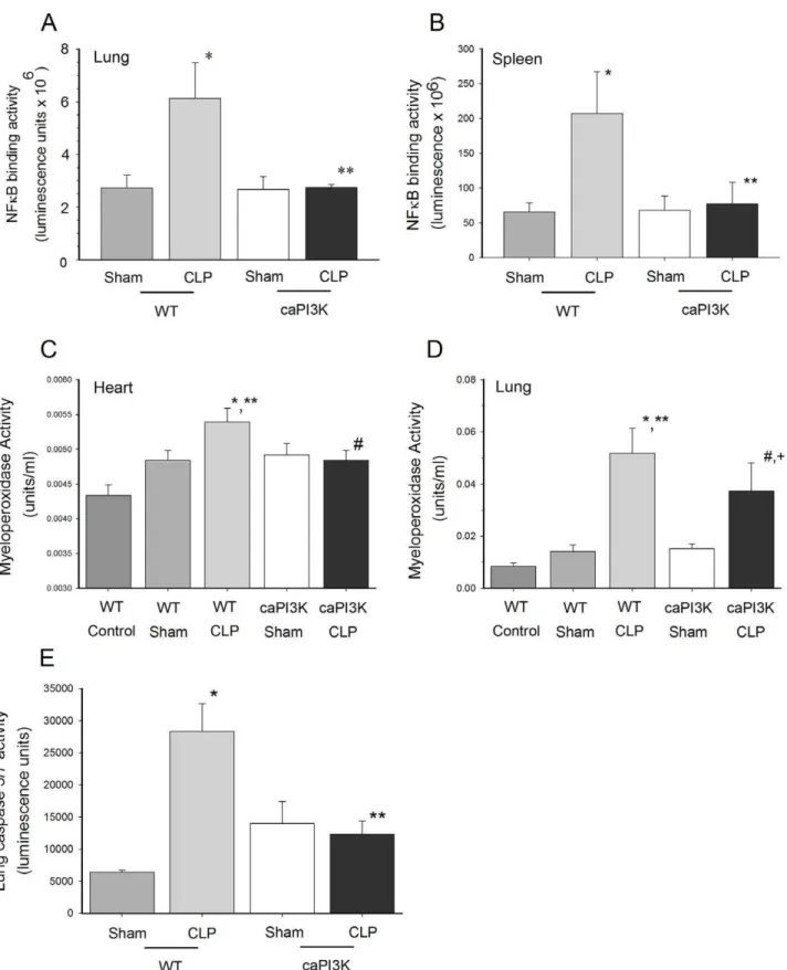
expression [35]. The data in Figures 4 and 5 indicate that caPI3K mice do not show a pro-inflammatory phenotype. This observation prompted us to examine organ NFkB activity in caPI3K mice in response to CLP sepsis. As expected, WT mice showed a significant increase in lung and spleen NFkB activity in response to sepsis (Figure 6A and 6B). However, lung and spleen tissue from mice expressing the p110a transgene did not show nuclear translocation of NFkB components in response to sepsis (Figure 6A and B). Surprisingly, we did not see any change in cardiac NFkB activity in the presence or absence of sepsis (data not shown). Thus, the NFkB confirms and extends the cytokine/
chemokine data which indicates that caPI3K mice show a predominantly normal phenotype with respect to inflammatory mediators.
Attenuation of Tissue MPO Activity in Septic caPI3K Mice Neutrophil accumulation and degranulation in organs during CLP sepsis is thought to mediate some of the pathophysiology of this devastating disease [36]. Even though caPI3K mice showed attenuation of serum KC levels, a neutrophil chemo-attractant, in response to sepsis, it was noted that KC levels were higher in septic caPI3K mice than in control or sham caPI3k mice (Fig. 5). To
Figure 5. Cardiac specific expression of PI3K p110aattenuates serum cytokine expression during CLP sepsis.caPI3K Tg mice and
age-matched WT mice were subjected to CLP. Sixteen hours after CLP the mice were euthanized, serum was harvested and analyzed for cytokine/ chemokine levels. *p,0.05 vs WT control; **p,0.05 vs WT sham surgery;#p,0.05 vs WT CLP;+p
,0.05 vs caPI3K control;@p
,0.05 vs caPI3K sham surgery control. (n = 4–6/group).
doi:10.1371/journal.pone.0044712.g005
Figure 6. Cardiomyocyte expression of the p110atransgene attenuated tissue NFkB activity in response to CLP sepsis.Lung (A) and spleen (B) NFkB activity are shown at the top. caPI3K mice and age-matched WT mice were subjected to CLP. Lung tissue was harvested 16 hrs after CLP. *p,0.05 vs WT control; **p,0.05 vs WT CLP group. (n = 4/group). Cardiac specific expression of PI3K p110aattenuated myeloperoxidase activity in the heart (C) and lung (D) in response to sepsis. caPI3K mice and age-matched WT mice were subjected to CLP. Heart and lung tissue was harvested 16 hrs after CLP. (n = 4–6/group).*p
,0.05 vs WT;**p
,0.05 vs caPI3k control;#
p,0.05 vs WT CLP;+p
,0.05 vs caPI3k sham.E.Lung caspase 3/7 activity was increased in wild type, but not caPI3K Tg, mice in response to CLP sepsis. caPI3K mice and age-matched WT mice were subjected to CLP. Lung tissue was harvested 16 hrs after CLP. (n = 4–6/group). *p,0.05 vs WT control; **p,0.05 vs WT CLP group.
evaluate the effect of transgenic p110aon neutrophils in sepsis, we examined tissue MPO activity as an indicator of tissue neutrophil infiltration and accumulation [37]. Tissue MPO activity was increased in the heart and lung of WT CLP mice at 16 hrs after surgery (Fig. 6C and D). MPO activity in the heart of caPI3K mice was not significantly elevated in response to sepsis and it was significantly lower than the MPO levels in the WT CLP myocardium, indicating that expression of the p110a transgene prevented neutrophil accumulation in the heart. MPO activity was also significantly elevated in the lungs of WT mice at 16 hrs after CLP, indicating neutrophil accumulation (Figure 6D). Lung MPO activity was 27.4% lower (p,0.05) in septic caPI3K mice, when compared to the WT CLP mice. However, lung MPO activity in septic caPI3K Tg mice was increased (p,0.05) when compared to caPI3K Tg sham control mice (Fig. 6D), indicating that myocardial expression of the p110atransgene can attenuate, but did not completely prevent, neutrophil accumulation in the lung.
Cardiac Specific Expression of PI3k p110aPrevented Activation of Tissue Caspase 3/7 and 8 in CLP Mice
Apoptosis is thought to play a significant role in pathophysiology of sepsis [38]. PI3K signaling has been reported to be a pro-survival, anti-apoptotic pathway [11,12]. We examined caspase 3/ 7 and 8 as indicators of the intrinsic and extrinsic apoptotic signaling pathways. CLP significantly increased lung caspase 3/7 activity at 16 hrs (Figure 6E). However, caPI3K Tg mice showed no change in caspase 3/7 activity in response to CLP (Figure 6E). We did not detect any significant difference in lung caspase 8 activity (data not shown). We did not detect changes in heart or liver apoptosis in response to CLP.
Cardiac Specific Expression of the p110a Transgene did not Alter Splenocyte Apoptosis in Response to CLP Sepsis
We [11] and others [12] have reported that CLP sepsis induces splenocyte apoptosis. Activation of PI3K/Akt dependent signaling has been reported to attenuate splenocyte apoptosis in sepsis [11,12]. In this study we examined splenocyte apoptosis at 16 hrs post-CLP. We examined apoptosis by TUNEL staining of splenic tissue (Figure 7A) and flow cytometry of isolated splenocytes (Figure 7B). As expected, CLP sepsis significantly increased splenocyte apoptosis in WT mice. Mice expressing the cardiac specific p110atransgene showed comparable levels of splenocyte apoptosis with both methods, indicating that cardiac specific expression of PI3K p110adid not alter sepsis induced splenocyte apoptosis (Figure 7A and B).
Cardiac Specific Expression of PI3K p110aInhibits Sepsis Induced Release of HMGB-1 from the Nucleus to the Cytoplasm in Cardiac Myocytes
HMGB-1 is a late mediator of septic sequelae that is released from the nucleus into the cytosol and systemic circulation in response to various stimuli including sepsis [39–42]. We examined the effect of myocardial caPI3K on release of nuclear HMGB-1 in cardiac myocytes in response to CLP sepsis (Figure 8). We examined two time intervals, six (top) and twelve (bottom) hours after CLP. As expected, WT CLP mice showed a loss of nuclear HMGB-1 and an increase in cytoplasmic levels at both time intervals (Figure 8). In striking contrast, caPI3K mice, subjected to CLP sepsis, showed no significant translocation of HMGB-1 from the nucleus to the cytoplasm.
Discussion
In this study we found that constitutive activation of myocardial PI3K p110a dramatically attenuates the cardiac dysfunction associated with polymicrobial sepsis. We critically examined a broad range of cardiac functional parameters in order to better understand the role of myocardial PI3K p110aactivation in the attenuation of cardiac dysfunction during sepsis. As shown above, cardiac output was dramatically reduced in wild type mice in response to sepsis, suggesting inadequate perfusion of vital organs as well as inadequate blood return to the left ventricle. However, septic caPI3K mice showed higher heart rates, although a smaller stroke volume was observed in these mice. The decrement in stroke volume was offset by the higher heart rate in septic caPI3K mice resulting in cardiac output that was maintained at normal levels. The data also indicate that blood return to the heart was maintained at or near normal levels in septic caPI3K mice. In response to sepsis, wild type mice showed significant decreases in left ventricular contractility. In contrast, constitutive activation of PI3K p110a in cardiac myocytes resulted in left ventricular contractility that was maintained at normal levels during sepsis. We also observed that myocardial activation of PI3K p110a maintained normal compliance of the left ventricle during sepsis, while ventricular wall stiffness was markedly increased in septic wild type mice. It was also noted that in contrast to wild type mice,
Figure 7. Expression of cardiac specific PI3K p110adid not
significantly alter splenocyte apoptosis in response to CLP
sepsis.Splenocytes were harvested 16 h after induction of CLP sepsis.
Splenocyte apoptosis was assessed by TUNEL staining (A) and flow cytometry (B). *p,0.05 vs control or sham treatment. N = 4–6/group. doi:10.1371/journal.pone.0044712.g007
constitutive activation of cardiomyocyte PI3K p110a resulted in left ventricular elastance and compliance that was maintained at normal levels, thus preserving normal left ventricular function during sepsis. When taken together, these data indicate that sepsis negatively impacts a broad range of left ventricular functional parameters. However, myocardial activation of PI3K p110a prevents or significantly attenuates many of the deleterious effects of polymicrobial sepsis on left ventricular function. The overall maintenance of cardiac function in caPI3K mice strongly correlates with decreased morbidity, attenuation of the pro-inflammatory phenotype and improved survival outcome in sepsis. Akt (aka = protein kinase B) is an important downstream target of PI3K [9,33]. In this study we observed that sepsis attenuated myocardial Akt phosphorylation in wild type mice, but not caPI3k Tg mice. Furthermore, Akt phosphorylation (activation) was only increased in the myocardium of caPI3k Tg mice. Therefore, the effect of the p110a transgene did not significantly influence Akt phosphorylation at extra-cardiac sites. This is consistent with previous reports using the PI3K transgenic mice [20]. Based on these data it is reasonable to conclude that the effect of the
transgene was primarily observed in the heart and that the systemic effect is due to an overall maintenance of left ventricular cardiac function during sepsis. Activated Akt phosphorylates several downstream targets of the PI3K pathway including GSK3b [32,33]. GSK3b is a crucial regulator of many cellular functions, including cell survival and apoptosis [32]. GSK3bis a constitutively active enzyme that is inactivated by Akt via phosphorylation of serine 9 [32]. We found that constitutive activation of myocardial PI3K p110a increased the levels of phosphorylated GSK3b, indicating inhibition of this enzyme in the myocardium. Inhibition of myocardial GSK3b positively correlated with maintenance of cardiovascular function and improved survival outcome in CLP sepsis.
The precise role of circulating cytokines in the pathophysiology of sepsis/septic shock is controversial and there is no definitive cause-and-effect relationship between systemic cytokine levels and survival outcome in sepsis [43]. Nevertheless, an examination of circulating cytokines can provide valuable information on the inflammatory and immunologic phenotype in response to CLP sepsis [11]. Our data clearly show that cardiac specific expression
Figure 8. Cardiac specific expression of PI3K p110ainhibits HMGB-1 release from the nucleus to the cytoplasm in cardiac myocytes.
Nuclear and cytoplasmic HMGB-1 levels are shown at 6 (top) and 12 hrs (bottom) after CLP. caPI3K Tg mice and age-matched WT mice were subjected to CLP. Six and twelve hours after CLP the mice were euthanized and nuclear and cytoplasmic protein was harvested. HMGB-1 was assessed by Western blot. *p,0.05 WT CLP vs WT sham control or caPI3k CLP. N = 4–6/group.
of PI3K p110a prevents cytokine/chemokine over expression in response to sepsis. Indeed, serum cytokine/chemokines in caPI3K mice remained at control levels in response to sepsis. Therefore, caPI3K did not show a pro-inflammatory phenotype in response to sepsis. These data also indicate that we did not see a shift from the Th1 to the Th2 phenotype that is frequently associated with fulminating sepsis [44]. These results also indicate that constitutive activation of p110a inhibited myocardial translocation of HMGB1. HGMB1 is thought to play a role in mediating organ damage in severe sepsis [41,40]. It is reasonable to speculate that the effects of myocardial caPI3K p110a on HMGB-1 may contribute to the protective mechanism. An intriguing aspect of this investigation is that the PI3k p110a transgene was only expressed in cardiomyocytes and is not expressed in immune competent cells, such as macrophages, neutrophils and lympho-cytes. Based on the current model of sepsis, it would have been reasonable to expect that macrophages and other immunocytes from caPI3K mice would respond normally to CLP sepsis by releasing pro-inflammatory mediators with a subsequent shift to a pro-inflammatory phenotype. This should have occurred irrespec-tive of the PI3K p110atransgene effect in the heart. However, our data clearly indicate that this did not occur. Therefore, our data suggest that the current dogma regarding how immune competent cells respond during sepsis may not be entirely accurate. Specifically, our data suggest that maintaining left ventricular function during sepsis may improve perfusion of the peripheral vasculature and thereby attenuate the systemic inflammatory response.
Activation of tissue NFkB activity is associated with increased morbidity and mortality in CLP sepsis [21,45]. Furthermore, we have reported that blunting tissue NFkB nuclear translocation will reduce morbidity and enhance survival outcome in CLP sepsis [45]. NFkB is also known to play a pivotal role in the regulation of cytokine/chemokine expression during septic sequelae [45]. As expected, WT mice showed a significant increase in lung NFkB activity in response to sepsis, which correlated with a strong pro-inflammatory response and rapid mortality. However, lung and splenic tissue from mice expressing the p110atransgene did not show nuclear translocation of NFkB components in response to sepsis. Not surprisingly, the NFkB data confirms and extends the cytokine/chemokine data, indicating that caPI3K mice show a predominantly normal inflammatory phenotype in response to sepsis.
Tissue neutrophil infiltration, adhesion and degranulation are thought to play a prominent role in tissue damage during sepsis [46]. KC is the murine equivalent of human IL-8 [34]. Like IL-8, KC plays a significant role in neutrophil chemotaxis and activation [34]. We found that serum KC levels in septic caPI3K Tg mice were increased relative to the controls, but KC levels were significantly lower in septic caPI3K Tg mice when compared to WT CLP mice. We examined tissue myeloperoxidase (MPO) levels as an index of tissue neutrophil infiltration and sequestra-tion. Cardiomyocyte p110aexpression resulted in a normalization of MPO levels in the heart of CLP mice, indicating that transgene expression prevented neutrophil sequestration in the heart during
sepsis. In the lung of caPI3K CLP mice we observed a pattern of MPO activity that closely mirrored the serum KC response to sepsis. Specifically, the expression of the p110a transgene significantly attenuated sepsis induced neutrophil infiltration into the lung. However, the increase in lung MPO, i.e. neutrophil levels, did not result in increased apoptosis in the lung. Indeed, caspase 3/7 and 8 activity were at control levels in the heart and lung of caPI3k mice in response to sepsis. Interestingly, splenocyte apoptosis, which is a hallmark of CLP sepsis, was increased in both WT and caPI3K mice. This suggests that the beneficial effect of cardiac specific expression of p110a does not prevent sepsis induced splenocyte apoptosis. We have previously reported that systemic activation of PI3K with a pharmacologic agent decreased morbidity, prevented cardiac dysfunction and increased long term survival in CLP sepsis in a manner very similar to that observed in mice expressing the p110atransgene [11,18]. However, there was one significant difference. In those studies we found that systemic activation of PI3K prevented sepsis induced splenocyte apoptosis, while at the same time maintaining cardiac function and enhancing survival outcome. When taken together, these data suggest that CLP sepsis does not mediate splenocyte apoptosis via cardiac dysfunction, rather it is mediated through an alternative mechanism. These data also suggest that transient and systemic activation of PI3K/Akt [11] may be more beneficial in sepsis than cardiac specific PI3K p110aactivation.
Our data indicate that activation of a single isoform of PI3K (p110a), in a single cell type (the cardiac myocyte), in a single organ (heart), can have a profound effect on the morbidity and mortality associated with fulminating sepsis. The precise mecha-nism(s) by which myocardial PI3K p110aactivation induces such dramatic effects at the systemic level are not entirely clear. It is reasonable to speculate that maintaining left ventricular function in sepsis/septic shock would have systemic benefits due to the maintenance of peripheral vascular perfusion, blood pressure and related issues. To the best of our knowledge, this is the first demonstration of a causal relationship between PI3K/p110a, the cardiac myocyte, cardiac function and survival outcome in sepsis. These data have practical implications for the management of sepsis, sepsis syndrome and septic shock. We have reported that transient and systemic activation of PI3K by a pharmacologic agent results in a maintenance of cardiovascular function and decreased morbidity and mortality in sepsis [11,18]. The decreased morbidity and mortality reported in that study is consistent with the data observed in the current study, which employed tissue specific genetic up regulation of PI3K p110a. Therefore, transient pharmacologic up regulation of PI3K/Akt signaling pathways may be beneficial in the prevention and/or management of sepsis and septic sequelae.
Author Contributions
Conceived and designed the experiments: C. Li FH KS DLW. Performed the experiments: FH TH C. Lu KB TF RK MG TRO. Analyzed the data: C. Li TH JK KS DLW. Contributed reagents/materials/analysis tools: C. Li KS KB RK DLW. Wrote the paper: C. Li FH JK DLW.
References
1. Oberholzer A, Oberholzer C, Moldawer LL (2001) Sepsis Syndromes: Understanding the Role of Innate and Acquired Immunity. Shock 16: 83–96. 2. Angus DC, Linde-Zwirble WT, Lidicker J, Clermont G, Carcillo J, et al. (2001)
Epidemiology of severe sepsis in the United States: Analysis of incidence, outcome, and associated costs of care. Crit Care Med 29: 1303–1310. 3. Sauaia A, Moore FA, Moore EE, Lezotte DC (1996) Multiple organ failure
syndrome. World J Surg 20: 392–400.
4. Baue AE (2006) MOF, MODS and SIRS: what is in a name or an acronym? Shock 26: 438–449.
5. Muller-Werdan U, Buerke M, Ebelt H, Heinroth KM, Herklotz A, et al. (2006) Septic cardiomyopathy - A not yet discovered cardiomyopathy? Exp Clin cardiol 11: 226–236.
6. Flynn A, Chokkalingam MB, Mather PJ (2010) Sepsis-induced cardiomyopathy: a review of pathophysiologic mechanisms. Heart Fail Rev 15: 605–611.
7. Vallejo JG (2011) Role of Toll-like receptors in cardiovascular diseases. Clin Sci 121: 1–10.
8. Fruman DA, Cantley LC (2002) Phosphoinositide 3-kinase in immunological systems. Seminars in Immunology 14: 7–18.
9. Cantley LC (2002) The phosphoinositide 3-kinase pathway. Science 296: 1655– 1657.
10. Guha M, Mackman N (2002) The PI3K-Akt pathway limits LPS activation of signaling pathways and expression of inflammatory mediators in human monocytic cells. Journal of Biological Chemistry 277: 32124–32132. 11. Williams DL, Li C, Ha T, Ozment-Skelton T, Kalbfleisch JH, et al. (2004)
Modulation of the phosphoinositide 3-Kinase pathway alters innate resistance to polymicrobial sepsis. J Immunol 172: 449–456.
12. Bommhardt U, Chang KC, Swanson PE, Wagner TH, Tinsley KW, et al. (2004) Akt decreases lymphocyte apoptosis and improves survival in sepsis. J Immunol 172: 7583–7591.
13. Fukao T, Koyasu S (2003) PI3K and negative regulation of TLR signaling. Trends in Immunology 24: 358–363.
14. Williams DL, Ozment-Skelton T, Li C (2006) Modulation of the phosphoino-sitide-3-kinase signaling pathway alters host resistance to sepsis, inflammation, and ischemia/reperfusion injury. Shock 25: 432–439.
15. Condorelli G, Drusco A, Stassi G, Bellacosa A, Roncarati R, et al. (2002) Akt induces enhanced myocardial contractility and cell sizein vivoin transgenic mice.
PNAS 99: 12333–12338.
16. Latronico MVG, Costinean S, Lavitrano ML, Peschle C, Condorelli G (2004) Regulation of Cell Size and Contractile Function by AKT in Cardiomyocytes. Ann NY Acad Sci 1015: 250–260.
17. Rota M, Boni A, Urbanek K, Padin-Iruegas ME, Kajstura TJ, et al. (2005) Nuclear Targeting of Akt Enhances Ventricular Function and Myocyte Contractility. Circ Res 97: 1332–1341.
18. Ha T, Hua F, Grant D, Xia Y, Ma J, et al. (2006) Glucan phosphate attenuates cardiac dysfunction and inhibits cardiac MIF expression and apoptosis in septic mice. Am J Physiol Heart Circ Physiol 291: H1910–H1918.
19. Shioi T, Kang PM, Douglas PS, Hampe J, Yballe CM, et al. (2000) The conserved phosphoinositide 3-kinase pathway determines heart size in mice. EMBO J 19: 2537–2548.
20. Luo J, McMullen JR, Sobkiw CL, Zhang L, Dorfman AL, et al. (2005) Class 1A phosphoinositide 3-kinase regulates heart size and physiological cardiac hypertrophy. Mol Cell Biol 25: 9491–9502.
21. Williams DL, Ha T, Li C, Kalbfleisch JH, Ferguson DA Jr (1999) Early activation of hepatic NFkB and NF-IL6 in polymicrobial sepsis correlates with bacteremia, cytokine expression and mortality. Ann Surg 230: 95–104. 22. Yang S, Chung CS, Ayala A, Chaudry IH, Wang P (2002) Differential
alterations in cardiovascular responses during the progression of polymicrobial sepsis in the mouse. Shock 17: 55–60.
23. Baker CC, Chaudry IH, Gaines HO, Baue AE (1983) Evaluation of factors affecting mortality rate after sepsis in a murine cecal ligation and puncture model. Surgery 94: 331–335.
24. Kao RL, Zhang F, Yiang Z-J, Gao X, Li C (2003) Cellular cardiomyoplasty using autologous satellite cells: From experimental to clinical study. Basic Appl Myol 13: 23–28.
25. Ha T, Hua F, Li Y, Ma J, Gao X, et al. (2006) Blockade of MyD88 attenuates cardiac hypertrophy and decreases cardiac myocyte apoptosis in pressure overload induced cardiac hypertrophy in vivo. Am J Physiol Heart Circ Physiol 290: H985–H994.
26. Subramanian V, Krishnamurthy P, Singh K, Singh M (2007) Lack of osteopontin improves cardiac function in streptozotocin-induced diabetic mice. Am J Physiol Heart Circ Physiol 292: H673–H683.
27. Li C, Browder W, Kao RL (1999) Early activation of transcription factor NF-kB during ischemia in perfused rat heart. Am J Physiol 276: H543–H552. 28. Li C, Kao RL, Ha T, Kelley J, Browder IW, et al. (2001) Early activation of
IKKbduring in vivo myocardial ischemia. Am J Physiol Heart Circ Physiol 280: H1264–H1271.
29. Li C, Ha T, Liu L, Browder W, Kao RL (2000) Adenosine prevents activation of transcription factor NF-kappa B and enhances activator protein-1 binding activity in ischemic rat heart. Surgery 127: 161–169.
30. Li C, Ha T, Kelley J, Gao X, Qiu Y, et al. (2003) Modulating Toll-like receptor mediated signaling by (1–3)-b-D-glucan rapidly induces cardioprotection. Cardiovascular Research 61: 538–547.
31. Ha T, Li Y, Hua F, Ma J, Gao X, et al. (2005) Reduced cardiac hypertrophy in toll-like receptor 4-deficient mice following pressure overload. Cardiovasc Res 68: 224–234.
32. Jope RS, Johnson GVW (2004) The glamour and gloom of glycogen synthase kinase-3. Trends Biochem Sci 29: 96–102.
33. Martin M, Rehani K, Jope RS, Michalek SM (2005) Toll-like receptor-mediated cytokine production is differentially regulated by glycogen synthase kinase 3. Nature Immunology 6: 777–784.
34. Boisvert WA, Curtiss LK, Terkeltaub RA (2000) Interleukin-8 and its receptor CXCR2 in atherosclerosis. Immunol Res 21: 129–137.
35. Lee JI, Burckart GJ (1998) Nuclear factor Kappa B: important transcription factor and therapeutic target. J Clin Pharmacol 38: 981–993.
36. Alves-Filho JC, de Freitas A, Spiller F, Souto FO, Cunha CQ (2008) The role of neutrophils in severe sepsis. Shock 30: 3–9.
37. Kettle AJ, Anderson RF, Hampton MB, Winterbourn CC (2007) Reactions of superoxide with myeloperoxidase. Biochemistry 46: 4888–4897.
38. Wesche DE, Lomas-Neira JL, Perl M, Chung C-S, Ayala A (2005) Leukocyte apoptosis and its significance in sepsis and shock. J Leukoc Biol 78: 325–337. 39. Bianchi ME (2006) DAMPs, PAMPs and alarmins: all we need to know about
danger. J Leukoc Biol 81: 1–5.
40. Klune JR, Dhupar R, Cardinal J, Billiar TR, Tsung A (2008) HMGB1: Endogenous danger signaling. Molecular Medicine 14: 476–484.
41. Wang H, Bloom O, Zhang M, Vishnubhakat JM, Ombrellino M, et al. (1999) HMGB-1 as a late mediator of endotoxin lethality in mice. Science 285: 248– 251.
42. Scaffidi P, Misteli T, Bianchi ME (2002) Release of chromatin protein HMGB1 by necrotic cells triggers inflammation. Nature 418: 191–195.
43. Remick D, Manohar P, Bolgos G, Rodriguez J, Moldawer L, et al. (1995) Blockade of tumor necrosis factor reduces lipoplysaccharide lethality, but not the lethality of cecal ligation and puncture. Shock 4: 89–95.
44. Miller AC, Rashid RM, Elamin EM (2007) The ‘‘T’’ in trauma: the helper T-cell response and the role of immunomodulation in trauma and burn patients. J Trauma 63: 1407–1417.
45. Williams DL, Ha T, Li C, Kalbfleisch JH, Laffan JJ, et al. (1999) Inhibiting early activation of tissue nuclear factor-kB and nuclear factor interleukin 6 with
(1–3)-b-D-glucan increases long-term survival in polymicrobial sepsis. Surgery 126: 54–65.