Juliana de Oliveira Ferla(a) José Augusto Rodrigues(b) César Augusto Galvão Arrais(c) Ana Cecília Correa Aranha(d) Alessandra Cassoni(b)
(a)Department of Restorative Dentistry, Graduate Division, Univ Guarulhos - UnG, Guarulhos, SP, Brazil.
(b)Department of Restorative Dentistry, Dental Research Division, Univ Guarulhos - UnG, Guarulhos, SP, Brazil.
(c)Department of Restorative Dentistry, Graduate Division, Univ Estadual de Ponta Grossa - UEPG, Ponta Grossa, PR, Brazil. (d)Department of Restorative Dentistry, School
of Dentistry, Univ de São Paulo - USP, São Paulo, SP, Brazil.
Corresponding author: Alessandra Cassoni E-mail: [email protected]
on enamel demineralization around
restorative materials
Abstract: This study evaluated the effects of the photoactivation source and restorative material on the development of caries-like lesions on hu-man enamel after an in vitro pH challenge. Enamel cavities were pre-pared in 36 blocks, which were assigned to two groups according to the restorative material: resin-modiied glass ionomer (RMGI) and compos-ite resin (CR). Samples were exposed to quartz-tungsten-halogen lamp, argon-ion laser, or light-emitting diode (n = 6). The Knoop microhard-ness (KHN) values of the top surface of all materials were evaluated. Re-stored enamel blocks were thermocycled and subjected to 10 demineral-ization-remineralization cycles at 37°C. KHN analysis of the supericial enamel was performed by four indentations located 100 µm from the restoration margin. The material KHN was not affected by the photoac-tivation source. No signiicant difference in KHN was noted between CR and RMGI. The enamel surface around RMGI exhibited a higher KHN (272.8 KHN) than the enamel around CR (93.3 KHN), regardless of the photoactivation source. Enamel demineralization around the dental res-toration was not inluenced by the photoactivation source. Less enamel demineralization was observed around the RMGI than around the CR restoration.
Descriptors: Glass Ionomer Cement; Composite Resins; Tooth
Demineralization.
Introduction
The longevity of dental restorations is related to the durability of the bond and the sealing of the cavity margins.1 Microspaces or gaps at the restoration/enamel interface may allow the penetration of luid containing cariogenic microorganisms,2 resulting in secondary caries progression.2,3 Secondary caries are frequent in patients with high caries risk and may necessitate the replacement of dental restorations.4 To prevent restoration failure, researchers developed luoride-releasing glass-ionomer (GI) restor-ative materials that inhibit secondary caries formation.5,6 The subsequent-ly developed resin-modiied glass ionomer (RMGI) materials showed some improved mechanical characteristics, such as better lexural strength and surface-wear resistance, compared to conventional GI cements,7 as well as superior handling properties due to their light-activation.
Vitremer (3M ESPE) is a hybrid material of GI restorative material and composite resin (CR) with acid-base and light-cured reactions. Vit-Declaration of Interests: The authors
certify that they have no commercial or associative interest that represents a conflict of interest in connection with the manuscript.
Submitted: Sep 05, 2012
remer sets via “triple-cure” mechanisms.8 The irst two setting reactions are based on an acid-base neu-tralization and free-radical methacrylate.The third setting reaction ensures the continuous polymeriza-tion of the remaining monomers that are not excited during light exposure. In the third reaction, micro-encapsulated potassium persulfate and ascorbic acid form a patented redox catalyst system that provides the methacrylate cure of the glass ionomer in the ab-sence of light. 9
Exposure of enamel to argon laser (AL) prior to cariogenic challenge has been shown to reduce the depth of caries lesions compared to enamel surfaces without prior AL exposure.2,3,10-14 This preventive effect has been attributed to the surface coating cre-ated by AL, which promotes changes in the miner-al structure and organic component of the surface coating. The resulting coating consists of a reactive surface that is less susceptible to caries formation. In addition to the synergistic effect between topical luoride and argon irradiation,12-14 increased luoride retention (400%) has been reported on enamel ir-radiated with a low energy density of AL (10.72 J/ cm²).15 However, the exact mechanism of caries resistance by AL irradiation remains unknown.16 Based on the dental literature, it is reasonable to speculate that the activation of restorative materials with AL would prevent secondary caries formation, especially when a luoride-releasing restorative ma-terial is used.
The induction of caries-like lesions around res-torations in vitro is an experimental approach that provides information about the clinical behavior of the restorative materials under the experimen-tal conditions.6,17 The objective of this study was to evaluate the cariostatic potential of the light-activa-tion source and restorative materials in vitro after a pH challenge. The response variable evaluated was the Knoop microhardness (KHN) proile of human enamel around and in the supericial area of the re-storative materials. The following null hypotheses were tested:
1. the supericial enamel KHN values around two restorative materials cured by different light-cur-ing units are not different;
2. the KHN values of the two restorative materials are not different.
Methodology
Ethical aspects
This study protocol was approved by the Gua-rulhos University Research Ethics Committee (CEP-UnG, process no. 104/2009).
Experimental design
The factors under study were the material (2 types) and photoactivation source (3 types) in a fac-torial 2 × 3 design for evaluation of the restorative material and the supericial enamel. The response variable was the KHN.
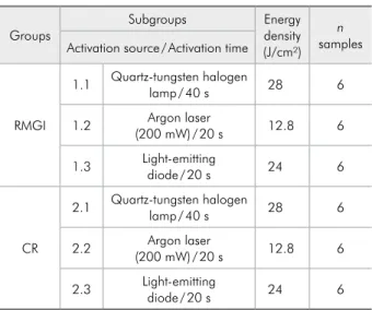
The experimental units consisted of 36 dental blocks (n = 6 per group) obtained from 18 unerupt-ed human third molars storunerupt-ed in 0.1% thymol solu-tion at 4°C. Blocks of 4 × 4 × 2 mm were sectioned from the third molars with double-faced diamond disks (#7020; KG Sorensen, Barueri, Brazil) at low speed (Kavo, Joinville, Brazil) under water irriga-tion. Cavities of approximately 1.6 mm in diameter and 1.6 mm in depth were prepared with #2292 dia-mond burs (KG Sorensen, Barueri, Brazil) under wa-ter spray. Teeth were distributed into two groups ac-cording to the restorative material, and each group was divided into three subgroups (Table 1).
Table 1 - Experimental groups.
Groups Subgroups Energy density
(J/cm²) n samples Activation source / Activation time
RMGI
1.1 Quartz-tungsten halogen lamp / 40 s 28 6
1.2 (200 mW) / 20 sArgon laser 12.8 6
1.3 Light-emitting diode / 20 s 24 6
CR
2.1 Quartz-tungsten halogen lamp / 40 s 28 6
2.2 (200 mW) / 20 sArgon laser 12.8 6
2.3 Light-emitting diode / 20 s 24 6
the Sof-lex (3M ESPE) polishing system for 15 s with each disk.
An 11-mm-diameter light tip was used with a quartz-tungsten-halogen (QTH)-based curing unit (Optilux 501; Demetron/Kerr, Danbury, USA), whereas the LED source (Radii Cal) had 1,200 mW/ cm² and an 8-mm-diameter light tip. The power density of both curing units was constantly mea-sured with a radiometer (Demetron/Kerr, Danbury, USA). The power selected for the AL (Accucure 3000; LaserMed, Salt Lake City, USA) was 200 mW for 20 s. A 6.3-mm-diameter spot size was mea-sured with the knife-edge method. The energy den-sity (J/cm²) of the light emitted by all curing units was calculated as the power density multiplied by the exposure time.
Both products were quantitatively evaluated by ive central indentations at 100-µm distance on the top surface (Figure 1). A microhardness tester (Pan-Tec; Panambra Ind. e Técnica SA, São Paulo, Brazil) was used with a 25-g load for 20 s, with a dwell time of 15 s. The specimens were thermocycled 1,000 times between distilled water baths held at 5°C and 55°C. The dwell time was 60 s and 5 s of transfer time (MSCT-3e; Elquip, Equipamentos para All cavities were prepared and restored by the
same calibrated operator. Table 2 shows composi-tion, lot number, and application instructions of the selected materials. A commercial RMGI (Vitremer; 3M ESPE, St. Paul, USA; A3 shade) was tested in this study. Prior to the RMGI application, Vitremer Primer (3M ESPE, St. Paul, USA) was applied for 30 s, dried for 5 s, and light-activated for 20 s. The RMGI was mixed according to the manufacturer’s instructions in a 1:1 proportion and inserted in a single increment by syringe (Centrix Inc., Shelton, USA). Groups 1.1, 1.2, and 1.3 were light-activated according to each activation source. After RMGI in-sertion, Vitremer Finishing Gloss was applied and light-activated for 20 s with a light-emitting diode (LED) (Radii Cal; SDI Limited, Bayswater, Austra-lia).
For all groups, the adhesive was light-activat-ed with a LED curing unit (Radii Cal; irradiance: 1,200 mW/cm²). After the bonding procedure, a nanoilled resin composite (Filtek Z350-OA3, 3M ESPE) was inserted in a single increment and acti-vated by the respective photoactivation source. Re-stored enamel blocks were Re-stored in 100% relative humidity at 37°C for 24 h and then polished with
Table 2 - Composition, lot number, and application mode of the selected materials.
Material Manufacturer Composition Directions for use
Adper Single Bond 2 (3M-ESPE,
Irvine, USA; Lot: 9XL) HEMA, Bis-GMA, DMAs, functional methacrylate, copolymer of polyacrylic and polyitaconic acids, water, ethanol, nanofilller, photoinitiator
Consecutively apply 2 coats, gently air-dry, and light-cure for 10 s
Conditioner (3M-ESPE, Irvine,
USA; Lot: 060808) Conditioner: etchant (37% phosphoric acid) Etch cavity for 15 s, wash and dry (do not desiccate) Z350-OA3 (3M-ESPE, Irvine, USA;
Lot: 7CN) Bis-GMA, Bis-EMA, UDMA and camphorquinone. Fillers: Zirconia-sílica Light-activate each increment for 40 s for QTH source Vitremer (3M-ESPE; St. Paul, USA;
Lot: 8HP; 8HH) Powder:potassium, Persulfate, ascorbic acid and pigments fluoroaluminosilicate glass, microencapsulated Liquid: aqueous solution of a polycarboxylic acid modified with pendant methacrylate groups, copolymer, water, HEMA and photoinitiators
Light-activate each increment for 40 s for QTH source with 2-mm maximum thickness
Vitremer Primer (3M-ESPE; St. Paul,
USA; Lot: 8CB) Vitrebond copolymer, HEMA, ethanol and photoinitiators Apply primer with a brush for 30 s to both dentin and enamel; air-dry and light-cure for 20 s
Vitremer Finishing Gloss (3M-ESPE;
Pesquisa Odontológica, São Carlos, Brazil).
The restored enamel blocks were covered with wax, except for the restoration area and 1 mm around this area, which remained exposed. The re-stored enamel blocks were submitted to pH challenge to induce caries-like lesions. The in vitro demineral-ization/remineralization dynamic model produced caries-like enamel lesions through a modiied Feath-erstone model18 to simulate the condition of high caries risk. Each restored block was placed in 15 mL of demineralization solution at 37°C (2.0 mmol/L of calcium, 2.0 mmol/L of phosphate in a buffer solu-tion of 74 mM of acetate, pH 4.3). The pH-cycling regimen was performed over 14 days, with 10 daily cycles of 6 h in demineralizing solution at 37°C. The remineralizing solution contained calcium and phos-phate at a known degree of saturation (50 mmol/L KCl, 1.5 mmol/L Ca, 0.9 mmol/L PO4, 20 mmol/L tri-hydroxymethil-aminomethan, pH 7.0) that was changed daily. The enamel blocks were washed with distilled water before an 18-h immersion in reminer-alizing solution. On the 6th, 7th, 13th, and 14th days of the cycle, the restored enamel blocks were kept only in the remineralizing solution.6
The quantitative KHN evaluation of the enamel caries-like lesions was performed by the PanTec mi-crohardness tester, with a 25-g load for 5 s. Four in-dentations located 100 µm from the bonded surface were created on the enamel surface in each enam-el block in the upper, lower, left, and right sides around the restorations (Figure 1).
Statistical analysis
Two-way ANOVA was performed to evaluate the inluence of the two variables tested: photoac-tivation source and material for supericial enamel and material evaluations. The means of the KHN values were compared by a Tukey post-hoc test (α = 0.05). The software used for statistical analysis in both evaluations was SANEST (EPAMIG, Belo Horizonte, Brazil). Power was calculated with the G power 3.1.2 software package (Heine, Universität Dusseldorf, Germany) (p = 1) for both material and enamel evaluation.
Results
ANOVA did not show differences for the “ma-terial” factor (p = 0.05), “photoactivation source” factor (p = 0.86), or interactions between factors (p = 0.25). Table 3 shows the KHN values and Tukey results of both materials exposed to the eval-uated photoactivation sources for material evalu-ation. In the evaluation of the supericial enamel Table 3 - Material evaluation of the Knoop microhardness values for the material and photoactivation source factors,
mean [standard deviation] and n samples.
Restorative
material QTH (28 J/cm²) AL (12.8 J/cm²) LED (24 J/cm²) RMGI (n = 6) 43.1 [12.6] 50.8 [11.6] 49.2 [13.3]
CR (n = 6) 65.3 [12.6] 55.5 [15.1] 52.5 [24.6] Figure 1 - Schematic
representation of the superficial enamel microhardness and indentation location on the restorative materials.
Restored cavity
Specimen
Delimited area with enamel demineralization
Indentation located 100 µm of restored margins
demineralization, ANOVA showed differences for the “material” factor (p < 0.05) but no differences for the “photoactivation source” factor or interac-tions between factors (both p = 0.7). Table 4 shows the KHN and Tukey results for the “material” and “photoactivation source” factors for the supericial enamel evaluation.
Discussion
According to the results of the present study, the irst null hypothesis (the supericial enamel KHN values around two restorative materials cured by different light-curing devices are not different) was rejected. Null hypothesis no. 2 (the KHN values of the two restorative materials are not different) was accepted. Although several studies have shown that AL irradiation provides reduced depth of caries-like lesions,10-14 and increased enamel microhardness12 compared with control groups, in the current study, the AL irradiation promoted no signiicant changes in enamel KHN values.
A previous study showed that the use of halo-gen or AL activation with 200 mW for 20 s resulted in similar degrees of conversion for the same resin composite, whereas decreased supericial KHN val-ues were obtained for AL activation with an expo-sure time of 10 s.19 Therefore, this protocol was used in the present study. The exposure time of 20 s al-lowed an energy density of 12.8 J/cm². This lower energy density of AL was suficient to promote high-er luoride retention for enamel, as demonstrated by Nammour et al.15 Turbino et al.20 recommended against the use of thickness increments of more than 1 mm for the AL with a power setting of 250 mW and an exposure time of 30 s.
The irst AL was cleared for marketing and clini-cal use in 1991 by the U.S. Food and Drug Admin-istration (FDA).21 Since that time, AL has been used
clinically to light-activate dental materials17 and has been shown to be capable of polymerizing com-pomers2 or CR.20,22 The use of AL in dentistry re-duces the chair time in dental ofices. The adhesion of orthodontic brackets previously showed favorable results in terms of the time required for the bonding procedure.23-25 There is evidence that topical luoride application decreases the depth of enamel primary caries lesions.10-14 The enamel resistance to caries was also demonstrated in vitro24,25 and in vivo23 af-ter AL irradiation. However, although AL was ef-fective at activating the restorative materials in the present study, laser technology is more expensive than QTH or LED.
The dynamic pH-cycling model has been shown to be adequate for studies of enamel caries-like le-sions.6,18 Various protocols (e.g., with 5 pH cycles17 or 14 days in demineralization solution16) have been used to simulate demineralization/remineralization phenomena in the oral environment with different restorative materials2 and activation sources.16 Nev-ertheless, other important clinical variables that are not addressed in the in vitro setting should also be considered, such as the cariogenicity and frequency of the patient’s diet or presence of saliva.6
Roberts et al.26 demonstrated that increments above 3 mm are not recommended for the same RMGI material evaluated in the present study. A 1.6-mm-deep standardized cavity preparation was used to ensure maximal setting from light activa-tion. Importantly, in this study, the P-value for the “material” factor was 0.05 when the microhardness of the CR or RMGI was evaluated (Table 3). This borderline signiicant result may be due to the small sample size of our study (n = 6), which is a limita-tion of this paper.
The preventive effect of the evaluated RMGI5-6 was evident once higher enamel KHN values were
Superficial
enamel QTH (28 J/cm²) AL (12.8 J/cm²) LED (24 J/cm²) Material factor RMGI (n = 6) 275.8 [8.5] 272.5 [17.4] 267.6 [29.0] 272.8 [19.9]A CR (n = 6) 115.4 [74.7] 82.7 [52.9] 80.7 [63.3] 93.3 [62.4]B Table 4 - Superficial enamel
evaluation of the enamel Knoop microhardness values for the material and photoactivation source factors, mean [standard
deviation] n samples and the results
of Tukey’s test. Means followed by different uppercase letters indicate significant differences (modified glass ionomer; CR: composite resin; QTH, quartz-tungsten-halogen; AL, argon laser; LED, p < 0.05). RMGI:
recorded around this restorative material com-pared to enamel KHN values around the resin composite (Table 4). This result was related to the luoride-release beneit of RMGI material.27 Fluo-ride release reduces demineralization on enamel around the restoration and along the cavity wall. The evaluated resin composite contains organic pigments. Because of its dark shade, the manufac-turer recommends an exposure time of 40 s, which is similar to the recommended exposure time for the RMGI when a halogen-based curing unit is used. The dark shade (OA3) of the CR promotes light attenuation.
Conclusion
The photoactivation source did not inluence enamel demineralization around dental restorations supericially. There was less development of enamel demineralization around RMGI restorations than around CR restorations.
Acknowledgements
The authors would like to thank the Special Lab-oratory of Lasers in Dentistry (LELO) of the Uni-versity of São Paulo, Brazil, for the use of their AL. This work was supported by FAPESP (São Paulo Re-search Foundation, grant no. 2009/02240-3).
References
1. Pereira PN, Inokoshi S, Tagami J. In vitro secondary caries in-hibition around fluoride releasing materials. J Dent. 1998 Jul-Aug;26(5-6):505-10.
2. Hicks MJ, Ellis R, Flaitz C, Werstermann G, Powell L. Res-toration-enamel interface with argon laser and visible light polymerization of compomer and composite resin restorations: a polarized light and scanning electron microscopic in vitro study. J Clin Pediatr Dent. 2003 Summer;27(4):353-8. 3. Hicks J, Flaitz C, Ellis R, Westerman G, Powell L. Primary
tooth enamel surface topography with in vitro argon laser irradiation alone and combined fluoride and argon laser treat-ment: scanning electron microscopic study. Pediatr Dent 2003 Sep-Oct;25(5):491-6.
4. Sarrett DC. Clinical challenges and the relevance of materi-als testing for posterior composite restorations. Dent Mater. 2005 Jan;21(1):9-20.
5. Dionysopoulos P, Kotsanos N, Koliniotou-Koubia E, Tolidis K. Inhibition of demineralization in vitro around fluoride releasing materials. J Oral Rehabil. 2003 Dec;30(12):1216-22. 6. Rodrigues JA, Marchi GM, Serra MC, Hara AT. Visual evalu-ation of in vitro cariostatic effect of restorative materials as-sociated with dentifrices. Braz Dent J. 2005;16(2):112-8. 7. Ellakuria J, Triana R, Mínguez N, Soler I, Ibaseta G, Maza
J, et al. Effect of one-year water storage on the surface micro-hardness of resin-modified versus conventional glass-ionomer cements. Dent Mater. 2003 Jun;19(4):286-9.
8. Swift EJ Jr, Pawlus MA, Vargas MA, Fortin D. Depth of cure of resin-modified glass ionomers. Dent Mater. 1995 May;11(3):196-200.
9. Mount GJ, Patel C, Makinson OF. Resin modified glass-ionomers: strength, cure depth and transucency. Aust Dent J. 2002 Dec;47(4):339-43.
10. Flaitz CM, Hicks MJ, Westerman GH, Berg JH, Blankenau RJ, Powell GL. Argon laser irradiation and acidulated phosphate
fluoride treatment in caries-like lesion formation in enamel: an in vitro study. Pediatr Dent. 1995 Jan-Feb;17(1):31-5. 11. Hicks MJ, Flaitz CM, Westerman GH, Blankenau RJ, Powell
GL, Berg JH. Enamel caries initiation and progression follow-ing low fluence (energy) argon laser and fluoride treatment. J Clin Pediatr Dent. 1995 Fall;20(1):9-13.
12. Westerman GH, Ellis RW, Latta MA, Powell GL. An in vitro study of enamel surface microhardness following argon laser irradiation and acidulated phosphate fluoride treatment. Pe-diatr Dent. 2003 Sep-Oct;25(5):497-500.
13. Westerman GH, Hicks MJ, Flaitz C, Powell GL. In vitro enamel caries formation: argon laser, light-emitting diode and APF treatment effect. Am J Dent. 2004 Dec;17(6):383-7. 14.Westerman GH, Hicks MJ, Flaitz CM, Ellis RW, Powell GL.
Argon laser irradiation and fluoride treatment effects on car-ies-like enamel lesion formation in primary teeth: an in vitro study. Am J Dent. 2004 Aug;17(4):241-4.
15. Nammour S, Rocca JP, Pireaux JJ, Powell GL, Morciaux Y, Demortier G. Increase of enamel fluoride retention by low fluence argon laser beam: a 6-month follow-up study in vivo. Lasers Surg Med. 2005 Mar;36(3):220-4.
16. Das UM, Prashanth ST. A comparative study to evaluate the effect of fluoride releasing sealant cured by visible light, argon lasers, and light emitting diode curing units: an in vitro study. J Indian Soc Pedod Prev Dent. 2009 Jul-Sep;27(3):139-44. 17. Magalhães CS, Hara AT, Turssi CP, Serra MC, Giannini M.
Microhardness evaluation around composite restorations us-ing fluoride-containus-ing adhesive systems. J Appl Oral Sci. 2005 Sep;13(3):259-64.
19. Cassoni A, Ferla JO, Shibli JA, Kawano Y. Knoop micro-hardness and FT-Raman Spectroscopy evaluation of a resin-based dental material light-cured by an argon íon laser and halogen lamp: an in vitro study. Photomed Laser Surg. 2008 Dec;26(6):531-9.
20. Turbino ML, Belan LC, Soprano V, Rode KM, Ramos PL, Youssef MY. Argon ion laser curing depth effect on a com-posite resin. Lasers Med Sci. 2011 Jul;26(4):421-5. Epub 2010 May 28.
21. Powell GL, Blankenau RJ. Laser curing of dental materials. Dent Clin North Am. 2000;Oct;44(4):923-30. Review. 22. Cassoni A, Ferla JO, Albino LG, Youssef MN, Shibli JA,
Rodrigues JA. Argon ion laser and halogen lamp activation of a dark and light resin composite: microhardness after long-term storage. Lasers Med Sci. 2010 Nov;25(6):829-34. Epub 2009 Jul 22.
23. Anderson AM, Kao E, Gladwin M, Benli O, Ngan P. The effects of argon laser irradiation on enamel decalcification:
an in vivo study. Am J Orthod Dentofacial Orthop. 2002 Sep;122(3):251-9.
24. Noel L, Rebellato J, Sheats RD. The effect of argon laser irradiation on demineralization resistance of human enamel adjacent to orthodontic brackets: an in vitro study. Angle Orthod. 2003 Jun;73(3):249-58.
25. Tavares JG, Eduardo CP, Burnett Jr LH, Boff TR, Freitas PM. Argon and Nd:YAG Lasers for Caries Prevention in Enamel. Photomed Laser Surg. 2012 Aug;30(8):433-7. Epub 2012 Jun 29.
26. Roberts HW, Berzins DW, Charlton DG. Hardness of three resin-modified glass-ionomer restorative materials as a func-tion of depth and time. J Esthet Restor Dent. 2009;21(4):262-72.