*Correspondence: C.H. Andrade/V. de Oliveira, Laboratório de Modelagem Molecular (LabMol)/Laboratório de Bioconversão (LaBioCon), Faculdade de Farmácia, Universidade Federal de Goiás, Avenida Universitária c/ 1ª Avenida, s/n, Setor Universitário, Caixa Postal 131, 74605-220 - Goiânia - GO, Brasil. E-mail: [email protected]; [email protected]
R
ev
Pharmaceutical Sciences vol. 47, n. 2, apr./jun., 2011
Twenty-Six Years of HIV science: an overview of anti-HIV drugs
metabolism
Carolina Horta Andrade
1,*, Lenis Medeiros de Freitas
2, Valéria de Oliveira
3,*
1Laboratório de Modelagem Molecular, Faculdade de Farmácia, Universidade Federal de Goiás, 2Programa de Pós-Graduação em Ciências Farmacêuticas, Faculdade de Farmácia, Universidade Federal de Goiás, 3Laboratório de
Bioconversão, Faculdade de Farmácia, Universidade Federal de Goiás
From the identiication of HIV as the agent causing AIDS, to the development of effective antiretroviral drugs, the scientiic achievements in HIV research over the past twenty-six years have been formidable. Currently, there are twenty-ive anti-HIV compounds which have been formally approved for clinical use in the treatment of AIDS. These compounds fall into six categories: nucleoside reverse transcriptase inhibitors (NRTIs), nucleotide reverse transcriptase inhibitors (NtRTIs), non-nucleoside reverse transcriptase inhibitors (NNRTIs), protease inhibitors (PIs), cell entry inhibitors or fusion inhibitors (FIs), co-receptor inhibitors (CRIs), and integrase inhibitors (INIs). Metabolism by the host organism is one of the most important determinants of the pharmacokinetic proile of a drug. Formation of active or toxic metabolites will also have an impact on the pharmacological and toxicological outcomes. Therefore, it is widely recognized that metabolism studies of a new chemical entity need to be addressed early in the drug discovery process. This paper describes an overview of the metabolism of currently available anti-HIV drugs.
Uniterms: AIDS/treatment. Drugs/anti-HIV. Drugs/metabolism. Antiretroviral drugs. Biotransformation.
Da identiicação do HIV como o agente causador da AIDS, ao desenvolvimento de fármacos antirretrovirais eicazes, os avanços cientíicos na pesquisa sobre o HIV nos últimos vinte e seis anos foram marcantes. Atualmente, existem vinte e cinco fármacos anti-HIV formalmente aprovados pelo FDA para utilização clínica no tratamento da AIDS. Estes compostos são divididos em seis classes: inibidores nucleosídeos de transcriptase reversa (INTR), inibidores nucleotídeos de transcriptase reversa (INtTR), inibidores não-nucleosídeos de transcriptase reversa (INNTR), inibidores de protease (IP), inibidores da entrada celular ou inibidores de fusão (IF), inibidores de co-receptores (ICR) e inibidores de integrase (INI). O metabolismo consiste em um dos maiores determinantes do peril farmacocinético de um fármaco. A formação de metabólitos ativos ou tóxicos terá impacto nas respostas farmacológicas ou toxicológicas do fármaco. Portanto, é amplamente reconhecido que estudos do metabolismo de uma nova entidade química devem ser realizados durante as fases iniciais do processo de desenvolvimento de fármacos. Este artigo descreve uma abordagem do metabolismo dos fármacos anti-HIV atualmente disponíveis na terapêutica.
Unitermos: AIDS/tratamento. Fármacos/anti-HIV. Fármacos/antirretrovirais. Fármacos/metabolismo. Biotransformação.
INTRODUCTION
The human immunodeficiency virus (HIV) has been established as the causative agent of the acquired immunodeiciency syndrome (AIDS) for over
more than 60 million people have been infected with the virus, and more than 25 million people have died as a result (Cohen et al., 2008).
According to the World Health Organization (WHO), globally there were an estimated 33 million [30 million–36 million] people living with HIV in 2007. Sub-Saharan Africa remains the region most heavily affected by HIV, accounting for 67% of all people living with HIV and for 75% of AIDS deaths in 2007 (UNAIDS/WHO, 2008). In addition, according to the same report, it was estimated that almost 3 mil lion HIV-infected people were receiving antiretroviral treatment in low- and middle-income countries, representing approximately 31% of the estimated 9.7 million people currently in need of treatment (UNAIDS/WHO, 2008).
Currently, there are twenty-five anti-HIV com-pounds that have been formally approved for clinical use in the treatment of AIDS. These compounds fall into six categories: nucleoside reverse transcriptase inhibi-tors (NRTIs), nucleotide reverse transcriptase inhibiinhibi-tors (NtRTIs), non-nucleoside reverse transcriptase inhibitors (NNRTIs), protease inhibitors (PIs), cell entry inhibitors or fusion inhibitors (FIs), co-receptor inhibitors (CRIs), and integrase inhibitors (INIs). Metabolism by the host organism is one of the most important determinants of the pharmacokinetic proile of a drug. Formation of active or toxic metabolites will have an impact on the pharmacolo-gical and toxicolopharmacolo-gical outcomes. Therefore, it is widely recognized that metabolism studies of a new chemical entity need to be addressed early in the drug discovery process. This paper describes an overview of the metabo-lism of currently available anti-HIV drugs.
ANTI-HIV DRUGS
The antiretroviral therapy advanced with the elu-cidation of the HIV cycle of life. Zidovudine (3’-azido-3’-deoxythymidine or AZT) (Retrovir®), a nucleoside
analogue, was the irst anti-HIV drug. AZT was originally synthesized as a potential anticancer drug by Horwitz in 1964 (Horwitz, Chua, Noel, 1964). In 1985, its anti-HIV activity was confirmed in vitro by a screening process using compounds that had previously been produced for others purposes (Fauci, 2003). AZT triphosphate acts by competitively inhibiting the utilization of thymidine tri-phosphate by reverse transcriptase (RT) of HIV-1 (Mitsuya
et al., 1985). In 1987, AZT was approved by the United States Food and Drug Administration (FDA) and supplied by Burroughs-Wellcome Laboratories.
In 2009, twenty-six years after the discovery of HIV as causative agent of AIDS, no less than twenty-ive
anti-HIV compounds have been formally approved for clinical use in the treatment of AIDS (Table I) (Adamson, Freed, 2009). These compounds fall into different categories, depending on the target within the HIV replicative cycle they interact with. In all, there are six categories: nucleo-side reverse transcriptase inhibitors (NRTIs: zidovudine, didanosine, zalcitabine, stavudine, lamivudine, abacavir, emtricitabine), nucleotide reverse transcriptase inhibitors (NtRTIs: tenofovir), non-nucleoside reverse transcriptase inhibitors (NNRTIs: nevirapine, delavirdine, efavirenz, etravirine), protease inhibitors (PIs: saquinavir, ritonavir, indinavir, nelinavir, amprenavir, lopinavir, atazanavir, fosamprenavir, tipranavir, darunavir), cell entry inhibitors or fusion inhibitors (FIs: enfuvirtide), co-receptor inhi-bitors (CRIs: maraviroc), and integrase inhiinhi-bitors (INIs: raltegravir) (Mehellou, De Clercq, 2009).
Although treatment with antiviral agents has proven to be a highly effective way to improve the health and survival of infected individuals, the epidemic will conti-nue to grow and there is an urgent need to develop new anti-HIV drugs.
METABOLISM STUDIES
Metabolism of a drug is one of the most important determinants of its pharmacokinetic properties. In most cases, metabolism leads to the inactivation of drugs. However, the metabolic transformation of a xenobiotic can also sometimes lead to the generation of an active metabolite, which can be solely or partially responsible for the pharmacological response. In some instances, me-tabolic transformation can also produce reactive or toxic intermediates or metabolites, with potential toxicological implications (Kumar, Surapaneni, 2001). Hence, a good understanding of the metabolism of a new chemical entity is needed early in the drug discovery process.
Mammalian metabolism of xenobiotics is traditio-nally subdivided into Phase I and Phase II processes. In Phase I, oxidation, reduction, or hydrolysis results in the introduction of new functional groups. Phase II involves conjugation reactions, where a highly hydrophilic moiety such as sulfate or glucuronide is attached to make the compounds more water-soluble and to prepare for excre-tion through urine or bile (Kumar, Surapaneni, 2001; Lee, Obach, Fisher, 2003).
N-oxidation, oxidative desalkylation and epoxide forma-tion (Omura, 1999).
Key Phase II enzymes include, for instance, uridine diphosphate-dependent glucuronosyltransferase (UGT), sulfotransferases and glutathione- S -transferase. Glucuro-nidation of small lipophilic molecules by UGTs is proba-bly the most important Phase II process for the clearance of drugs. Consistent with its broad substrate proile, UGT exists as an enzyme superfamily. So far, 18 UGT proteins, which can be divided into two families, UGT1 and UGT2, have been identiied (Mackenzie et al., 1997).
Prediction of drug metabolism in humans
In vitro and in vivo extrapolation
The ability to predict human pharmacokinetic from
in vitro and in vivo models with a reasonable degree of accuracy (2.0-fold variation) is an ongoing challenge with many different approaches yielding varying degrees of success (Prakash, Vaz, 2009). Traditionally, in vivo
drug metabolism studies have relied on the use of model systems to produce the expected human metabolites of drugs. Usually whole animal systems are used, especially
TABLE I - Approved antiretroviral drugs in the US and Europe
Generic Name Brand Name Manufacturer Date of FDA approval
Zidovudine Retrovir GlaxoSmithKline 19 March 1987
Didanosine Videx (tablet)
Videx EC (capsule) Bristol Myers-Squibb Bristol Myers-Squibb 31 October 20009 October 1991
Zalcitabine Hivid Hoffmann-La Roche 19 June 1992
Stavudine Zerit Bristol Myers-Squibb 24 June 1994
Lamivudine Epivir GlaxoSmithKline 17 November 1995
Saquinavir Invirase (hard gel capsule)
Fortovase (soft gel capsule) Hoffmann-La Roche Hoffmann-La Roche 7 November 19976 December 1995
Ritonavir Norvir Abbott Laboratories 1 March 1996
Indinavir Crixivan Merck 13 March 1996
Nevirapine Viramune Boehringer Ingelheim 21 June 1996
Nelinavir Viracept Agouron Pharmaceuticals 14 March 1997
Delavirdine Rescriptor Pizer 4 April 1997
Efavirenz Sustiva (US)
Stocrin (Europe) Bristol Myers-Squibb Merck 17 September 199817 September 1998
Abacavir Ziagen GlaxoSmithKline 17 December 1998
Amprenavir Agenerase GlaxoSmithKline 15 April 1999
Lopinavir + ritonavir Kaletra
Aluvia (developing world) Abbott Laboratories Abbott Laboratories 15 September 200015 September 2000
Tenofovir Viread Gilead Sciences 26 October 2001
Enfuvirtide Fuzeon Hoffmann-La Roche & Trimeris 13 March 2003
Atazanavir Reyataz Bristol-Myers Squibb 20 June 2003
Emtricitabine Emtriva Gilead Sciences 2 July 2003
Fosamprenavir Lexiva (US)
Telzir (Europe) GlaxoSmithKline GlaxoSmithKline 20 October 200320 October 2003
Tipranavir Aptivus Boehringer Ingelheim 22 June 2005
Darunavir Prezista Tibotec, Inc. 23 June 2006
Maraviroc Celsentri (Europe)
Selzentry (US) PizerPizer 18 September 200718 September 2007
Raltegravir Isentress Merck & Co., Inc. 12 October 2007
small laboratory animal models like rat, dog, cat, guinea pig, rabbit etc. However, interspecies differences must be considered since there is no “perfect” animal model destined for extrapolation to the human model. In vitro
studies are generally used to complement and specify the data obtained using perfused organs, tissue or cell cultures and microsomal preparations. Such methods suffer from a number of limitations, such as the cost of experimental animals, the ethical concerns and species variations (Asha, Vidyavathi, 2009; Collins, 2001).
Isolated perfused rat liver has the advantages of controlling blood and bile lows, ease of manipulation of the perfusion medium and the large number of perfusate samples that may be collected (Meijer, Swart, 1997). Inste-ad, microsomal preparations are derived from endoplasmic reticulum after homogenization and centrifugation and present CYPs and UGTs. They are developed to predict metabolic clearance of the compound and drug-drug inte-ractions, and are widely used for drug metabolism studies (Pelkonen et al., 2005).
Another important tool employed in drug metabo-lism is the application of human hepatocyte cultures, that present both Phase I and Phase II enzymes, representing a great model for biotransformation study mediated by P450, and allowing a good correlation with in vivo studies. On the other hand, the use of hepatocyte cultures is limi-ted by the restriclimi-ted availability of liver tissue (Li et al., 1997; Gomez-Lechon et al., 2004). Liver-derived HepG-2 and BC2 cell lines and lung-derived line A549 have the disadvantage of expressing few enzymes, restricting their metabolic capacity (Yoshitomi et al., 2001; Le Vee et al., 2006). However, the recent human hepatoma-derived cell line HepaRG has been shown to possess functions and morphological resemblance to normal human hepatocytes (Aninat et al., 2006).
The use of microorganisms as models of mammalian metabolism was introduced in the early 1970s by Smith and Rosazza (Smith, Rosazza, 1974, 1975). This method uses eukaryotic microorganisms to produce metabolites si-milar to mammals, since microbial transformation systems could closely mimic most of the Phase I transformations of drugs observed in mammals, mainly those catalyzed by cytochrome P450 (Azerad, 1999).
Compared with traditional methods, there are cle-arly a number of practical advantages in using microbial transformation as a model for drug metabolism, such as its chemo-, regio-, and stereoselectivity, impressive catalytic eficiency, and capability of accepting a wide range of complex molecules as substrates. In addition, it is conducive to easy scale up to produce suficient amounts of metabolites for unambiguous structural determination,
and to provide such metabolites as authentic standards (Zmijewski et al., 2006; Asha, Vidyavathi, 2009).
Therefore, it is a valuable tool in the production of molecules with improved, different or toxic activity originated by the fungal enzymatic biodiversity (Azerard, 1999; Asha, Vidyavathi, 2009). Recently, our laboratory has carried out a number of studies applying microbial mo-dels to study mammalian metabolism (Gomes et al., 2006; Pazzini et al., 2005, 2010; Carneiro et al., 2005, 2010; Dias et al., 2005; Costa et al., 2008; Braga et al., 2011).
In silico Computational Tools
In recent years, several in silico computational methods have become available for prediction of me-tabolism and pharmacokinetics of the NCEs and are increasingly becoming a part of the drug discovery process to select candidates with desirable ADME pro-perties (Canavan, 2007; Afzelius et al., 2007; De Graff, Vermeulen, Feenstra, 2005). Mechanism-based quantum chemical calculations on substrates and the enzyme, pharmacophore modeling of ligands, and protein homo-logy modeling in combination with automated docking and molecular dynamics simulations have been used to rationalize and predict ligand binding and formation of metabolites (Prakash, Vaz, 2009).
Moreover, the prediction of metabolites can be per-formed by means of large collections of transformation rules. A given compound is fragmented and then passed through all rules to identify putative metabolically labile sites. Expert systems and their databases, such as Meta-bolExpert (Darvas, 1988), META (Klopman, Dimayuga, Talafous, 1994), METEOR (Testa et al., 2005),MetaDrug (Ekins et al., 2006), and PK/DB (Moda et al., 2008), are examples of in silico methods used to predict drug bio-transformation pathways and possible metabolites, that provide a ranked list of most likely metabolites.
Metasite (Cruciani et al., 2005) is a computational program for metabolite formation prediction, which com-bines structural information, by matching the structural complementarity of the substrate and the protein, and rule-based and reactivity methods.
ANTI-HIV DRUGS METABOLISM
Since the discovery of antiretrovirals, metabolism studies of these drugs have been increasingly carried out, aiming to ensure their efficacy and safety and to explain their toxic effects (Li, Chan, 1999). This trend is graphically illustrated in Figure 1, which presents the number of publications about antiretrovirals metabolism, between 1987 and 2009, retrieved upon searching with the SciFinder engine.
Nucleoside Analogue Reverse Transcriptase Inhibitors (NRTIs)
The reverse transcriptase (RT) of HIV is actually the target for three classes of inhibitors: nucleoside RT inhibitors (NRTIs), nucleotide RT inhibitors (NtRTIs) and non-nucleoside RT inhibitors (NNRTIs). The NRTIs and NtRTIs interact with the catalytic site (that is the substrate-binding site) of the enzyme, whereas the NNRTIs interact with an allosteric site located a short distance from the catalytic site (De Clercq, 2007).
For the NRTIs and NtRTIs to interact with the subs-trate-binding site they need to be phosphorylated to the tri-phosphate and ditri-phosphate forms, respectively. There are at present, seven NRTIs that have been formally approved for the treatment of HIV infections: zidovudine (AZT), didanosine (ddI), zalcitabine (ddC), stavudine (d4T), lami-vudine (3TC), abacavir (ABC) and emtricitabine [(-)FTC]. All the NRTIs can be considered 2’,3’-dideoxynucleoside (ddN) analogues and act in a similar fashion. After they have been taken up as such by the cells, they are
phos-phorylated to their 5’-mono-, 5’-di- and 5’-triphosphate (MP, DP and TP, respectively) form following the same mechanism: ddN → ddNMP → ddNDP → ddNTP, before the latter will then act as a competitive inhibitor/alternate substrate of the normal substrate dNTP (either dATP, dTTP, dGTP or dCTP) (Furman et al., 1986; De Clercq, 2007). As competitive inhibitor of the normal substrate, the ddNTP will inhibit incorporation of this substrate into the growing DNA chain; as alternate substrate it will be incorporated into this chain (as ddNMP), thereby acting as a chain terminator (since the ddNMP is missing the 3’-hydroxyl group required for further chain elongation) (De Clercq, 2007).
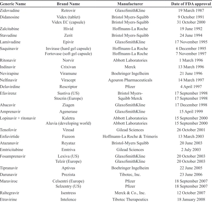
Among the NRTIs, metabolism studies have been focused on zidovudine (AZT). There are three main me-tabolic pathways of AZT: intracellular phosphorylation, glucuronidation and reduction (Figure 2). The intracellular metabolism, which involves the formation of zidovudine-5’-triphosphate (AZT-TP, 1), is limited in the last two steps of phosphorylation due to an ineficiency of the enzyme thymidine kinase to form zidovudine-5’-diphosphate (AZT-DP), leading to accumulation of zidovudine-5’-monophosfate (AZT-MP) within the cell, and serving as a deposit for the formation of AZT-TP. High concentrations of AZT-MP inhibit the thymidylate kinase, decreasing the intracellular concentration of deoxythymidine-5’-triphosphate and increasing the activity of antiretroviral. Furthermore, the accumulation of intracellular AZT-MP may be related to the toxicity observed during the use of AZT, mainly represented by hematological toxicity, likely caused by mitochondrial DNA polymerase γ inhibition (Veal, Back, 1995).
AZT undergoes fast irst-pass hepatic metabolism, producing zidovudine-5’-glucuronide (GAZT, 2), an inac-tive metabolite (Li, Chan, 1999). In studies using human microsomal preparations, it was observed that AZT’s glucuronidation is independent of sex or age, but the poly-morphism displayed by UGT2B7 isoform may contribute to the interindividual variations observed in plasma con-centration of AZT and its conjugate metabolite (Court et al., 2003). However, these variations may be involved in the conjugation catalyzed by other isoforms, as well as the stage of intracellular AZT’s phosphorylation, which implies a greater or lesser availability of substrate for hepatic meta-bolism. Also, according to studies on liver microsomes, the extent of metabolism of AZT by UGT can range from 60 to 75% in humans, while in rats it is only 10% (Naritomi et al., 2003) due to higher catalytic eficiency of these enzymes in the former (Trapnell et al., 1998).
The third metabolic pathway of AZT consists of its azido group reduction, probably mediated by both
FIGURE 1 - Graphical representation of the increasing trend for
cytochrome P450 isoenzymes, NADPH-cytochrome P450 reductase and cytochrome b5 reductase, resulting in 3’-amino-3’-deoxythymidine (AMT, 3). In studies using rat hepatocytes and liver microsomes of rats and humans, the participation of the subfamilies CYP2B, CYP3A and CYP4A was observed in this reaction, evidenced by an increased formation of AMT through the use of inducers of these enzymes. The CYP2C9 isoform also appears to be involved in this process. These studies have also indi-cated that the AMT is ive to seven times more toxic to the hematopoietic cells than AZT, conirming its role in the cytotoxicity of antiretroviral therapy observed in patients. In another similar study, the AMT glucuronide conjugate (GAMT), produced from the reduction of zidovudine-5’-glucuronide, was found to discard the AMT as a substrate for UGT (Cretton et al., 1991).
AZT metabolism studies were also performed using microbial models obtaining derivatives not previously reported in the literature (Figure 3). Kruszewska and co-workers(2003) studied the bioconversion of AZT with an environmental bacterium, Stenotrophomonas maltophilia
PCM 1942. After ten days of incubation, a hydroxylated metabolite at the C-2’ deoxyribose ring (4) was produced with a yield of about 97% (Kruszewska et al., 2003). In our laboratory, biotransformation of AZT was performed using Cunninghamella echinulata ATCC 9244 as a catalyst (Nunes, 2008). This ilamentous fungus strain is widely known for performing biotransformation reactions of a va-riety of compounds, similar to mammals, and is frequently employed in bioconversion processes. In our experiment,
C. echinulata produced a glycosylated metabolite of AZT (5) (Figure 3) after seven days of incubation, under mild conditions of temperature and pH, with a yield of about 46% (Nunes, 2008).
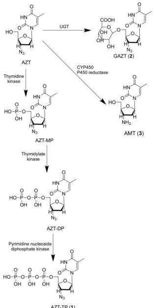
Didanosine (2’,3’-dideoxyinosine or ddI) (Figure 4) is first phosphorylated by 5’-nucleotidase to form didanosine-5’-monophosfate (ddI-MP) in human lym-phoid cells. Then, ddI-MP is converted to dideoxyade-nosine-5’-monophosfate (ddA-MP) by adenylosuccinate synthetase and lyase, with subsequent phosphorylation to dideoxyadenosine-5’-triphosphate (ddA-TP) by adenyla-te kinase (miokinase), producing active metaboliadenyla-te (6) responsible for inhibiting viral reverse transcriptase. In another metabolic pathway, ddI is hydrolyzed by purine nucleoside phosphorylase, to release the sugar deoxyri-bose (7) and hypoxanthine (8), which is degraded into
FIGURE 2 – Main metabolic routes of zidovudine in humans.
FIGURE 3 - Microbial metabolites of AZT: AZT-2’-hydroxyl
uric acid (9) and eliminated (Navé et al., 1994). In a study using beagle dogs, the following possible metabolites of didanosine were found in plasma and urine: allantoin, uric acid, hypoxanthine and xanthine (Kaul et al., 1993). Probably ddI is not metabolized by CYP450 isoforms, since studies using inducers of these enzymes showed no changed pharmacokinetic proile (Bruzzese et al., 1995).
Zalcitabine (2’,3’-dideoxycytidine or ddC), as well as other nucleoside analogues, must be phosphorylated to dideoxycitidine-5’-triphosphate (ddC-TP) in the intra-cellular environment to become active (10) (Figure 5). The
irst stage of phosphorylation, with production of ddC-MP, seems to be ineficient due to a low afinity between ddC and the enzyme deoxycytidine kinase. In addition to the mono-, di- and triphosphate, the only reported metabolite of zalci-tabine is dideoxyuridine, found in urine (Li, Chan, 1999).
Human metabolism of stavudine (2’,3’-didehydro-2’,3’-dideoxythymidine or d4T) (Figure 6) has yet to be completely elucidated. Intracellularly, it is sequentially phosphorylated to produce its active form, stavudine-5’-triphosphate (d4T-TP) (11). In a study using monkeys and isolated rat hepatocytes, the degradation of stavudine was detected in thymine (12) and the d4T-TP derivative, with subsequent production of beta aminoisobutyric acid (BAI-BA) (13) (Cretton et al., 1993). The glucuronide metabolite (14) of stavudine was found in the urine of monkeys but has remained controversial (Schinazi, et al., 1990).
A recent study reported by our group (Freitas, 2009) described the metabolic proile of stavudine, applying in vivo, in vitro and in silico approaches. The bioconversion experiments with nine strains of ilamentous fungi, detect-ed one major metabolite, which was further identiidetect-ed as thymine (12). The in vivo study with Balb/c mice detected the same metabolite (thymine, 12), highlighting the strains used in this study as microbial models of stavudine metab-olism (Figure 7). Unlike in vivo and in vitro experiments, the in silico study based upon the docking and molecular dynamics simulations of stavudine in complex with the active site of cytochrome P450 CYP3A4 structure, identi-ied that the methyl group is the most probable site for the oxidation reaction mediated by CYP450 enzymes. It is likely that the in vivo and in vitro results described in this study must be mediated by enzymes other than CYP-450.
Lamivudine (3TC) (Figure 8) is another NRTI, whose intracellular phosphorylation occurs eficiently to produce its active metabolite, lamivudine-5’-triphosphate (3TC-TP) (15). There was no difference in the
concen-FIGURE 4 – Proposed metabolic pathways of didanosine.
tration of 3TC and its pharmacokinetic proile in healthy individuals and patients with moderate to severe hepatic dysfunction, suggesting that lamivudine metabolism by liver enzymes is minimal (Johnson, Horak, Breuel, 1998).
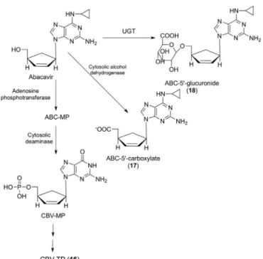
Abacavir (ABC) (Figure 9), an analogue of 2’-deo-xyguanosine, undergoes fast intracellular phosphorylation
catalyzed by the enzyme adenosine phosphotransferase, producing its monophosphate form, which is then con-verted into the monophosphate form of carbovir (CBV-MP) by a cytosolic deaminase. Subsequently, CBV-MP is converted into the di- and triphosphate form (16), the latter is another analogue of deoxyguanosine with potent inhibitory activity of viral reverse transcriptase (Faletto
et al., 1997).
In addition, it also undergoes hepatic biotransforma-tion in humans, mice and monkeys, producing two major metabolites, identiied as 5’-carboxylate (17) and 5’-glu-curonide derivatives (18), formed by cytosolic alcohol dehydrogenase (ADH) and UGT, respectively (Good et al., 1995; McDowell et al., 1999). In in vitro experiments with human liver microsomes, it was demonstrated that CYP450 enzymes appear to be involved in the metabolism of abacavir (Ravitch et al., 1998).
Nucleotide Reverse Transcriptase Inhibitors (NtRTIS)
NtRTIs should be clearly distinguished from the NRTIs as they are nucleotide analogues (not nucleoside analogues), which means they only need two (not three) phosphorylation steps to be converted to their active form. Most importantly, they contain a phosphonate group that
FIGURE 6 – Proposed metabolic routes of stavudine.
FIGURE 7 – Metabolism studies of stavudine by C. elegans and
mice, producing the metabolite thymine (Freitas, 2009).
cannot be cleaved by hydrolases (esterases), which would make it more dificult to cleave off these compounds once incorporated at the 3’-terminal end, as compared to their regular nucleotide counterparts (i.e. AZTMP, ddAMP, ddCMP, etc.). The prototype of the NtRTIs, (R)-9-(2- phos-phonomethoxypropyl) adenine (tenofovir) (Figure 10) was irst described in 1993 (Balzarini et al., 1993) and is the only NtRTIs currently approved for use in patients suffe-ring from HIV. Due to its limited oral bioavailability, teno-fovir has been converted to its oral prodrug form, tenoteno-fovir disoproxil fumarate (TDF, Viread®) and has become one of the most frequently prescribed drugs for the treatment of HIV infections (De Clercq, 2003). Since 2008, it has also been approved for the treatment of chronic hepatitis B virus (HBV) infections. Tenofovir is metabolized intra-cellularly to its active metabolite, tenofovir diphosphate (19), which is a competitive inhibitor of HIV-1 RT that
terminates the growing DNA chain. Phosphorilation to its diphosphate form can be achieved in one or two steps with the aid of 5-phosphoribosyl-1-pyrophosphonate (PRPP) synthetase (Balzarini et al., 1995) or AMP kinase (Merta
et al., 1992), respectively (Figure 10).
Non-nucleoside Reverse Transcriptase Inhibitors (NNRTIs)
These compounds are noncompetitive inhibitors of HIV-1 RT, because they act differently compared with nucleoside analogues. They are mainly metabolized by the CYP3A subfamily of cytochrome P450 enzymes. Nevirapine (NVP) (Figure 11), for example, is capable of inducing its own metabolism mediated by CYP450 with production of several hydroxylated metabolites, with sub-sequent glucuronidation. These derivatives were found in urine and feces of human and animals (dog, mouse, rat, chimpanzee, rabbit and monkey), after oral administration of nevirapine, and identiied as 2-hydroxy-nevirepine (20), 3-hydroxy-nevirapine (21), 8-hydroxy-nevirapine (22), 12-hydroxy-nevirapine (23), their respective glucuronide conjugates (24-27), and 4-carboxy-nevirapine (28), for-med by secondary oxidation of 23 (Riska et al., 1999a, b). In experiments with human liver microsomes, it was observed that CYP3A and CYP2B6 are responsible for 2- and 3-hydroxy-nevirapine production, respectively, while the formation of 8- and 12-hydroxi-nevirapine is mediated by several enzymes, primarily CYP3A4 and CYP2D6 (Erickson et al., 1999).
Delavirdine (Figure 12) is another NNRTI, whi-ch produces various metabolites in rats (Chang et al.,
1997a), mice (Chang et al., 1997b) and monkeys (Chang
et al., 1997c) through oxidation reactions catalyzed mainly by CYP450. An N-desalkylation reaction pro-duces the desalkyl delavirdine metabolite (29), with subsequent sulfation (30), while hydroxylation at C-6’ position of the pyrimidinic ring produces 6’-hydroxy-delavirdine (31), followed by glucuronidation to form the
FIGURE 9 – Main metabolic pathways of abacavir in humans.
6’-O-glucuronide conjugate (32). The cleavage of pyrimi-dinic ring of 6’-hydroxy-delavirdine was also observed, resulting in depyridinyl delavirdine, and cleavage of the amide bond forming indole carboxylic acid (33) and N -isopropylpyridinepiperazine (34). Moreover, 6’-hydroxy delavirdine is sulfated (35) in rats, depyridinyl delavirdine is conjugated in rats and mice, and desalkyl delavirdine is hydroxylated at positions C-4’ (36) and C-6’ (37) of the pyrimidinic ring in rats and monkeys, with subsequent sulfation (38 and 39).
In mice, N-desalkylation of N -isopropylpyridine-piperazine produces N-deisopropylpyridinepiperazine, while the indole carboxylic acid undergoes conjugation. In monkeys, three additional metabolites were observed, identiied as conjugation of desalkyl delavirdine with N -acetylglucosamine, hydroxylation of delavirdine at the C-4 position of the indole ring to form 4-hydroxy delavirdine, followed by glucuronidation of 4-O-glucuronide, and
N-glucuronidation of the indole ring of delavirdine. The
N-desalkylation was the major metabolic fate in rats and monkeys, while in mice it was the cleavage of the amide bond (Chang et al., 1997a, b, c).
Experiments with human liver microsomes sho-wed that delavirdine is an inhibitor of the isoforms CYP2C9, CYP2C19, CYP2D6 and CYP3A4, and that the N-desalkylation reaction is mediated by CYP2D6 and CYP3A4, while hydroxylation at position C-6’ of
pyrimi-dinic ring is catalyzed mainly by CYP3A4 (Voorman et al., 1998a, b; Voorman et al., 2001).
Efavirenz (EFV) (Figure 13) is extensively metabo-lized and small or insigniicant amounts of the unchanged drug were found in urine of several species, including humans (Mutlib et al., 1999). The major metabolite isolated is the O-glucuronide (40) conjugate of the me-tabolite 8-hydroxy-efavirenz (41). EFV also undergoes glucuronidation to form the N-glucuronide conjugate (42). The sulfation of 8-hydroxy-efavirenz (43) occurs in all species, except humans, while EFV hydroxylation to form 7-hydroxy-efavirenz (44), with subsequent glu-curonidation (45) or sulfation (46), is not only observed in rats. Other reactions include the hydroxylation of the cyclopropane ring (C-14) of 8-hydroxy-efavirenz, produ-cing 8,14-dihydroxy-efavirenz (47), and hydroxylation of its sulfated conjugate (48), which can also be formed from the sulfation of 47, followed by glucuronidation (49) or conjugation with glutathione (GSH), with subsequent degradation, forming the adducts cysteinylglicine and cysteine. The hydroxylation of 44 is also observed, follo-wed by glucuronidation (50) or sulfation (51) in position C-7 whereas 57 can still undergo glucuronidation (52) in position C-8 (Mutlib et al., 1999).
In vitro experiments using human liver microsomes have shown that 8-hydroxy-efavirenz was obtained as the major metabolite, as well as during in in vivo studies, while the metabolites 8,14-dihydroxy-efavirenz and 7-hydroxy-efavirenz were also isolated. It was further demonstrated
FIGURE 11 – Main metabolic routes of nevirapine.
that CYP2B6 is the major isoform responsible for the production of 8-hydroxy-efavirenz and 8,14-dihydroxy-efavirenz, and that other isoforms (CYPs 1A2, 3A4 and 3A5) seem to participate in a minority of this process (Ward et al., 2003). Rodriguez-Novoa and co-workers (2005) reported that CYP2B6 genetic polymorphisms contribute to variations in plasma concentration of EFV, proving the involvement of this isoform in its metabolism. In addition to being metabolized by CYP2B6, efavi-renz is also its inhibitor, as well as its major metabolite, 8-hydroxy-efavirenz (Bumpus et al., 2006), which is also an inducer of the isoform CYP3A4 (Mouly et al., 2002).
Protease Inhibitors (PIs)
At present, there are ten protease inhibitors (PIs) licensed for clinical use in the treatment of HIV infections. With the exception of tipranavir (which is based on a cou-marine scaffold), all these PIs are based on the “peptidomi-metic” principle, that is they contain an hydroxyethylene scaffold which mimics the normal peptide linkage (cleaved by the HIV protease) but which can itself not be cleaved. They thus prevent the HIV protease from carrying out its normal function, which is the proteolytic processing of precursor viral proteins into mature viral proteins. The ten PIs presently available for the treatment of HIV infections are saquinavir, ritonavir, indinavir, nelinavir, amprenavir, lopinavir, atazanavir, fosamprenavir, tipranavir and daru-navir (Madruga et al., 2007; Lazzarin et al., 2007).
Akin to NNRTIs, PIs are usually metabolized by oxidation mediated by CYP450. Saquinavir (SQV)
(Fi-gure 14) has low oral bioavailability due to its extensive irst-pass metabolism, which occurs mainly by CYP3A4 in small intestine, but also in liver, producing the same meta-bolites mono- and dihydroxy. In incubations of saquinavir with human small intestine and liver microsomes, two ma-jor metabolites (53 and 54) were isolated, produced from mono-hydroxylations of octahydro-2-(1H)-isoquinolynil and (1,1-dimethylethyl)amino, respectively. Another ive minority metabolites were found (Flitzsimmons, Collins, 1997). One of the major metabolites was subsequently identiied as 6-equatorial-hydroxy-saquinavir (Eaglings
et al., 2002).
Biotransformation of indinavir (IDV) (Figure 14) takes place primarily in liver by CYP3A4 (Chiba et al.,
1996; Lin et al., 1996), and can also occur in the small intestine. Oxidative metabolites were found in both in vivo
and in vitro experiments using liver and intestinal microso-mes of humans and rats (Chiba, Hensleigh, Lin, 1997). In a study with healthy volunteers, seven major metabolites were found in urine, whose metabolic pathways were identiied as: nitrogen pyridine glucuronidation (55); N-oxidation of pyridine (56); p-hydroxylation of the phenylmethyl radical (57); 3’-hydroxylation of indan (58 and 59); and N-depyrido methylation (Balani et al., 1995). In experiments with liver microsomes, the production of the metabolite cis-(indan) hydroxylated by isoforms of the CYP2D subfamily was observed in Rhesus and Cynomolgus monkeys, while
trans-(indan) hydroxylation and N-desalkylation were also observed in chimpanzees and humans, mediated by CYP3A4. The extent of metabolism in humans was the lowest among the four species studied (Chiba et al., 2000).
Anari and co-workers (2004) described the meta-bolic proile of indinavir, combining data obtained after analysis of human liver post-mitochondrial preparations in liquid chromatography coupled to mass spectroscopy (LC-MS/MS), and metabolic predictions generated by research based on structural similarity using the MDL Metabolite Database software. Two desalkylated, eleven monoxydated, three deoxygenated and two desalkylated/ monoxydated metabolites were identiied, and the major derivatives resulted from the oxidation of pyridine, in-danyl and fenylmethyl groups, and from N-desalkylation of piperazinil (Anari et al., 2004). Similar metabolites were found previously in another experiment employing LC-MS/MS analysis of the metabolites obtained from in vitro study (Yu, Cui, Davis, 1999).
Ritonavir (Figure 14) is extensively metabolized by rat, dog and human livers, through a variety of oxidative pathways and conjugation with glucuronic acid (60). The latter was only observed in dogs. The major metabolites identiied in urine and feces of these three species were
produced by loss of the thiazolyl carbamate, oxidation and loss of isopropyl-thiazolyl (61). Moreover, there was formation of N-hydroxymethyl urea (62), oxidation of the thiazolyl heteroatom (63) and a second oxidation of isopropyl of 61 (64), in rats only, as well as oxidation of the methyl thiazolyl from the metabolite that lost the carbamate thiazolyl, in dogs, loss of formaldehyde from
62 in rats and dogs, producing N-demethyl urea followed by oxidation to produce N-hydroxy urea. Glucuronidation of 62 was also observed in dogs (65). In vitro experiments demonstrated that the metabolite 61 and the metabolite that lost the isopropyl-thiazolyl group have antiretroviral activity (Denissen et al., 1997). A study using human liver microsomes showed that the production of 61 is mediated by enzymes of the CYP3A subfamily and by the isoform CYP2D6 (Kumar et al., 1996). In addition to being meta-bolized by CYP3A, ritonavir is also a potent inhibitor of this subfamily (Kumar et al., 1999).
Nelinavir (NFV) (Figure 14) is biotransformed in the liver into three major metabolites, identiied as nelina-vir-hydroxy-t-butyl-amide (66), 3,4-dihydroxy-nelinavir (67) and 3-methoxy-4-hydroxy-nelinavir (68) (Zhang et al., 1998). Compound 66 shows antiretroviral activity in vitro comparable to that of NFV, contributing to its oral activity in anti-HIV therapy, while 68 showed low activity (Zhang et al., 2001; Hirani, Raucy, Lasker, 2004). It has been shown that CYP3A4 is involved in the formation of metabolites 67 and 68, while 66 is produced by CYP2C19 (Wu et al., 1998; Lillibridge et al., 1998; Khaliq et al., 2000).66 can also be metabolized by CYP3A4. Nelinavir is also an inhibitor of CYP3A4(Lillibridge et al., 1998) and CYP2B6(Hesse et al., 2001) isoforms.
Amprenavir (Figure 14) is primarily oxidized in the liver by enzymes CYP450 (Decker et al., 1998). Experiments with human liver microsomes showed the production of ive metabolites, the majority obtained by
oxidation of the tetrahydrofuran ring (THF) (69). The others were formed by N-desalkylation (70), oxidation of the aliphatic chain (71), dehydration of the THF ring (72) and mono-oxidation of the aniline ring (73). Enzymes of CYP3A subfamily appear to be involved in the formation of 69, 71 and 73, while CYP2C9 contributes to the achie-vement of 71. It was also observed that the metabolism of amprenavir is reduced in neonates due to non-maturation of CYPs 3A4 and 3A5 (Tréluyer et al., 2003).
Fusion Inhibitors (FIs)
Enfuvirtide is the only fusion inhibitor (FI) appro-ved (March 2003) by the US FDA, and to be licensed for the treatment of HIV-1 infection in the US, Australia and Europe (Fletcher, 2003). This compound is a synthetic polypeptide of 36 amino acids that is homologous to, and engages in a coil-coil interaction, with the heptad repeat (HR) regions of the viral glycoprotein gp41 (Matthews et al., 2004). As a consequence of this interaction, the fusion of the virus particle with the outer cell membrane is blo-cked. The FI enfuvirtide is the only anti-HIV compound that has a polymeric (i.e. polypeptidic) structure (structure not disclosed), and hence, is not orally bioavailable: it has to be injected parenterally (subcutaneously) twice daily. This makes the long-term use of enfuvirtide cum-bersome and problematic. Enfuvirtide is primarily used in salvage therapy as part of drug combination regimens. Because enfuvirtide is a peptide, it is expected to undergo catabolism rather than metabolism by cytochrome P450 enzymes (Ferraiolo, Mohler, Gloff, 1992). Enfuvirtide did not inhibit the activities of CYP1A2, CYP2C19, CYP2D6, CYP2E1 or CYP3A4 in an open label, crossover trial in 12 HIV-infected adults (Hammer et al., 2006).
Co-Receptor Inhibitors (CRIs)
Co-receptor inhibitors (CRIs) interact with the co-receptor CCR5 or CXCR4 used by M (macrophage)-tropic and T (lymphocyte)-tropic HIV strains, respectively (now generally termed R5 and X4 strains, respectively), to enter the target cells. Within the whole virus-cell entry process, the interaction of the viral glycoprotein gp120 and the co-receptor falls between the interaction of the viral glyco-protein gp120 and the CD4 receptor, and the fusion of the viral glycoprotein gp41 with the outer cell membrane (Westby, van der Ryst, 2005). There is, at present, only one CRI available (licensed in 2007 for clinical use), namely, the CCR5 antagonist maraviroc (Perros, 2007). The major problem with CCR5 antagonists is that they are only active against R5 HIV strains, and that from a mixed population
of X4/R5 HIV strains, they stimulate the selection of X4 strains. Ideally, a CCR5 antagonist should be combined with a CXCR4 antagonist so as to block both X4 and R5 HIV strains (Door et al., 2005).
Maraviroc is rapidly absorbed and extensively meta-bolized, although unchanged drug is the major circulating component in plasma and is the major excreted component after oral dosing. The metabolite proiling of plasma, urine and fecal samples showed that maraviroc is extensively metabolized with the major pathways involving oxidation and N-dealkylation reactions (Figure 15) (Walker et al.,
2005; Abel et al., 2008). The most abundant metabolite detected in human plasma was a secondary amine product of N-dealkylation (74, Figure 15). Moreover, Hyland and co-workers (2008) have demonstrated that maraviroc is predominantly metabolized by CYP3A4, and is not itself an inhibitor of CYP enzymes.
Integrase Inhibitors (INIs)
Although the integrase has been pursued for many years as a potential target for the development of new anti-HIV compounds, the irst integrase inhibitor (INI) licensed to treat HIV-1 infection, raltegravir, has only recently (in late 2007) been approved. The HIV integrase has essen-tially two important catalytic functions (3’-processing and strand transfer) (Chiu, Davies, 2004). Raltegravir (Figure 16) is targeted at the strand transfer reaction, as is elvitegravir (Figure 16), which is now entering phase III clinical trials. Elvitegravir is intended for once daily dosing (orally), while raltegravir has to be administered twice daily. It has proven highly effective in reducing viral loads in HIV-infected patients (Grinsztejn et al., 2007; Steigbigel et al., 2008; Cooper et al., 2008).
In preclinical studies, raltegravir was metabolized predominantly in the liver via the UDP-glucuronosyltrans-ferase (UGT)-mediated pathway. In vivo and in vitro studies using isoform selective chemical inhibitors and cDNA-ex-pressed UGTs showed that UGT1A1 is the primary enzyme responsible for the formation of raltegravir-glucuronide (87) (Merck & Co, 2007; Kassahun et al., 2007). In vitro studies demonstrate that raltegravir is not a substrate for cytochro-me P450 (CYP) enzycytochro-mes and does not inhibit CYP1A2, 2B6, 2C8, 2C9, 2C19, 2D6, or 3A or induce 3A4 (Iwamoto
et al., 2008). Similarly, raltegravir is not an inhibitor of UGTs (UGT1A1 and UGT2B7) or P-glycoprotein (P-gp)-mediated transport (Merck & Co, 2007).
into urine). The only metabolite detected in humans was the phenolic hydroxyl glucuronide (87), and this metabolite does not have antiviral activity (Kassahun
et al., 2007) (Figure 16). In a human ADME study, ∼51
and 32% of a radiolabeled raltegravir dose (parent and metabolite) was excreted in the feces and urine, res-pectively. The major circulating entity in plasma was the parent compound (69% of the total drug-related material in plasma), whereas most of the drug-related material in urine was accounted for by the glucuronide derivative (72% of the drug-related material in urine). In feces, only parent compound was detected, but it is likely that a good proportion of the raltegravir detected in feces is derived from hydrolysis of the glucuronide derivative secreted in bile as observed in preclinical species (Kassahun et al., 2007).
Elvitegravir undergoes metabolism primarily by CYP3A and also by glucuronidation via UGT1A1/3, which is the major pathway of elimination. Metabolites formed by these pathways are markedly less potent (5- to
38-fold) than the parent drug and are present in plasma at lower concentrations; thus, the metabolites do not contri-bute to the antiviral activity of elvitegravir (Ramanathan
et al., 2007a).
FIGURE 15 – Proposed metabolic pathways for maraviroc.
FIGURE 16 – Structures of raltegravir, its glucuronide
In a human ADME study with radiolabeled elvi-tegravir and boosting doses of ritonavir, >90% of the circulating plasma was present as the parent compound. Radiolabeled compound was excreted primarily in the feces (94.8%), with a minor amount in the urine (6.7%). Fecal radioactivity consisted primarily of parent com-pound (36%) and an oxidative metabolite (46%). Urine contained low levels of various metabolites (Ramanathan
et al., 2007b).
FINAL CONSIDERATIONS
Despite the signiicant advances made in understan-ding the mechanism of HIV infection and in identifying effective treatment approaches, the search for optimum treatment strategies for AIDS remains a major challenge.
This review has demonstrated that the cytochrome P450 family of enzymes is responsible for the metabolism of the majority of antiretroviral drugs, but for some of the nucleoside reverse transcriptase inhibitors (NRTIs), hepatic conjugation via UDP-glucuronosyltransferases as well as via alcohol dehydrogenase is important. NRTIs are the most studied class of anti-HIV agents in terms of metabolism. In contrast, limited published data are cur-rently available on the other classes of anti-HIV agents and much of the available information is obtained from manufacturers and the clinical studies sponsored by the manufacturers. More systematic studies on metabolism, such as metabolite formation, the presence of active me-tabolites and mechanisms of chemical toxicity, are needed regarding antiretroviral therapy.
ACKNOWLEDGEMENTS
The authors gratefully acknowledge inancial sup-port from CNPq (The National Council for Scientiic and Technological Development), CAPES (The Coordenação de Aperfeicoamento de Pessoal de Nível Superior) and FAPEG (The State of Goias Research Foundation), Brazil.
REFERENCES
ABEL, S.; RUSSELL, D.; WHITLOCK, L.A.; RIDGEWAY, C.E.; NEDDERMAN, A.N.R.; WALKER, D.K. Assessment of the absorption, metabolism, and absolute bioavailability
of maraviroc in healthy male subjects. Br. J. Clin.
Pharmacol., v.65, suppl.1, p.60-67, 2008.
ADAMSON, C.S.; FREED, E.O. Anti-HIV-1 Therapeutics: from FDA-approved drugs to hypothetical future targets.
Mol. Interv., v.9, p.70-74, 2009.
AFZELIUS, L.; ARNBY, C.H.; BROO, A.; CARLSSON, L.; ISAKSSON, C.; ULRIK JURVA, U.; KJELLANDER, B.; KOLMODIN, K.; NILSSON, K.; RAUBACHER, F.; WEIDOLF , L. State of the art tools for computational site of metabolism predictions: comparative analysis, mechanistically insights, and future applications. Drug Metab. Rev., v.39, p.61-86, 2007.
ANARI, M.R.; SANCHEZ, R.I.; BAKHTIAR, R.; FRANKLIN, R.B.; BAILLIE, T.A. Integration of knowledge-based metabolic predictions with liquid chromatography data-dependent tandem mass spectrometry for drug metabolism studies: application to studies on the biotransformation of indinavir. Anal. Chem., v.76, p.823-832, 2004.
ANINAT, C.; PITON, A.; GLAISE, D.; LE CHARPENTIER, T.; LANGOUET, S.; MOREL, F.; GUGUEN-GUILLOUZO, C.; GUILLOUZO, A. Expression of cytochromes p450, conjugating enzymes and nuclear receptors in human hepatoma heparg cells. Drug Metab. Dispos., v.34, p.75-83, 2006.
ASHA S.; VIDYAVATHI, M. Cunninghamella – A microbial model for drug metabolism studies – A review. Biotechnol. Adv., v.27, p.16-29, 2009.
AZERAD, R. Microbial models for drug metabolism. Adv.
Biochem. Eng. Biotechnol., v.63, p.163-218, 1999.
BALANI, S.K.; ARISON, B.H.; MATHAI, L.; KAUFFMAN, L.R.; MILLER, R.R.; STEARNS, R.A.; CHEN, I.W.; LIN, J.H. Metabolites of L-735, 524, a potent HIV 1-protease inhibitor, in human urine. Drug Metab. Dispos., v.23, p.266-270, 1995.
BALZARINI, J.; HOLÝ, A.; JINDRICH, J.; NAESENS, L.; SNOECK, R.; SCHOLS, D.; DE CLERCQ, E. Differential antiherpesvirus and antiretrovirus effects of the (S) and (R) enantiomers of acyclic nucleoside phosphonates: potent and selective in vitro and in vivo antiretrovirus activities of (R)-9-(2-phosphonomethoxypropyl)- 2,6-diaminopurine.
Antimicrob. Agents Chemother., v.37, p.332-338, 1993.
BARRÉ-SINOUSSI, F.; CHERMANN, J.C.; REY, F.; NUGEYRE, M.T.; CHAMARET, S.; GRUEST, J.; DAUGUET, C.; AXLER-BLIN, C.; VÉZINET-BRUN, F.; ROUZIOUX, C.; ROZENBAUM, W.; MONTAGNIER, L. Isolation of a T-lymphotropic retrovirus from a patient at risk for acquired immune deiciency syndrome (AIDS).
Science, v.220, p.868-871, 1983.
BRAGA, R.C.; TORRES, A.C.B.; PERSIANO, C.B.; ALVES, R.O.; FRAGA, C.A.M.; BARREIRO, E.J.; DE OLIVEIRA, V. Determination of the cardioactive prototype LASSBio-294 and its metabolites in dog plasma by LC-MS/MS: Application for a pharmacokinetic study.
J. Pharm. Biomed. Anal., in press, 2011, DOI: 10.1016/j. jpba.2011.02.031.
BRUZZESE, V.L.; GILLUM, J.G.; ISRAEL, D.S.; JOHNSON, G.L.; KAPLOWITZ, L. G.; POLK, R.E. Effect of luconazole on pharmacokinetics of 2’,3’-dideoxyinosine in persons seropositive for human immunodeiciency virus.
Antimicrob. Agents Chemother., v.39, p.1050-1053, 1995.
BUMPUS, N.N.; KENT, U.M.; HOLLENBERG, P.F. Metabolism of efavirenz and 8-hydroxyefavirenz by P450 2B6 leads to inactivation by two distinct mechanisms. J. Pharmacol. Exp. Ther., v.318, p.345-351, 2006.
CANAVAN, N. FDA and drug companies alike want ADME-tox testing performed earlier and earlier in a drug’s life cycle.
Drug Discov. Dev., v.10, p.34-36, 2007.
CARNEIRO, E.O.; MENEGATTI, R.; FRAGA, C.A.M.; BARREIRO, E.J.; DE OLIVEIRA, V. Microbial models of animal metabolism: application to a study of the metabolism of LASSBio-873. Braz. J. Pharm. Sci., v.41, p.392-392, 2005.
CARNEIRO, E.O.; ANDRADE, C.H.; BRAGA, R.C.; TÔRRES, A.C.B., ALVES, R.O.; LIÃO, L.M.; FRAGA, C.A.M.; BARREIRO, E.J.; DE OLIVEIRA, V. Structure-based prediction and biosynthesis of the major mammalian metabolite of the cardioactive prototype LASSBio-294.
Bioorg. Med. Chem. Lett., v.20, p.3734–3736, 2010.
CHANG, M.; SOOD, V.K.; KLOOSTERMAN, D.A.; HAUER, M.J.; FAGERNESS, P. E.; SANDERS, P.E.; VRBANAC, J.J. Identiication of the metabolites of the HIV-1 reverse transcriptase inhibitor delavirdine in monkeys. Drug Metab. Dispos., v.25, p.814-827, 1997a.
CHANG, M.; SOOD, V.K.; WILSON, G.J.; KLOOSTERMAN, D.A.; SANDERS, P.E.; HAUER, M.J.; ZHANG, W.; BRANSTETTER, D.G. Metabolism of the HIV-1 Reverse transcriptase inhibitor delavirdine in mice. Drug Metab. Dispos., v.25, p.828-839, 1997b.
CHANG, M.; SOOD, V.K.; WILSON, G.J.; KLOOSTERMAN, D.A.; SANDERS, P. E.; HAUER, M.J.; FAGERNESS, P.E. Metabolism of the human immunodeiciency virus type 1 reverse transcriptase inhibitor delavirdine in rats. Drug Metab. Dispos., v.25, p.228-242, 1997c.
CHIBA, M.; HENSLEIGH, M.; LIN, J.H. Hepatic and intestinal metabolism of indinavir, an HIV protease inhibitor, in rat
and human microsomes: Major role of CYP3A. Biochem.
Pharmacol., v.53, p.1187-1195, 1997.
CHIBA, M.; HENSLEIGH, M.; NISHIME, J.A.; BALANI, S.K.; LIN, J.H. Role of cytochrome P450 3A4 in human metabolism of MK-639, a potent human immunodeiciency virus protease inhibitor. Drug Metab. Dispos., v.24, p.307-314, 1996.
CHIBA, M.; NISHIME, J.A.; NEWAY, W.; LIN, Y.; LIN, J.H. Comparative in vitro metabolism of indinavir in primates - a unique stereoselective hydroxylation in monkey.
Xenobiotica, v.30, p.117-129, 2000.
CHIU, T.K.; DAVIES, D.R. Structure and function of HIV-1 integrase. Curr.Top. Med. Chem., v.4, p.965-77, 2004.
COHEN M.S.; HELLMANN, N.; LEVY, J.A.; DECOCK, K.; LANGE, J. The spread, treatment, and prevention of HIV-1: evolution of a global pandemic. J. Clin. Invest., v.118, p.1244-1254, 2008.
COLLINS, J.M. Inter-species differences in drug properties.
Chem. Biol. Interact., v.134, p.237-242, 2001.
COSTA, E.M.M.B.; PIMENTA, F.C.; LUZ, W.C.; DE OLIVEIRA V. Selection of filamentous fungi of the Beauveria genus able to metabolize quercetin like mammalian cells. Braz. J. Microbiol., v.39, p.405-408, 2008.
COURT, M.H.; KRISHNASWAMY, S.; HAO, Q.; DUAN, S.X.; PATTEN, C.J.; VON MOLTKE, L.L.; GREENBLATT, D.J. Evaluation of 3_-azido-3_-deoxythymidine, morphine and codeine as probe substrates for UDP-glucuronosyltransferase 2B7 (UGT2B7) in human liver microsomes: speciicity and
inluence of the UGT2B7*2 polymorphism. Drug Metab.
Dispos., v.31, p.1125-1133, 2003.
CRETTON, E.M.; SCHINAZI, R.F.; MCCLURE, H.M.; ANDERSON, D.C.; SOMMADOSSI, J.P. Pharmacokinetics of 3’-azido-3’-deoxythymidine and its catabolites and interactions with probenecid in rhesus monkeys.
Antimicrob. Agents Chemother., v.35, p.801-807, 1991.
CRETTON, E.M.; ZHOU, Z.; KIDD, L.B.; MCCLURE, H.M.; KAUL, S.; HITCHCOCK, M.J.; SOMMADOSSI, J.P. In vitro and in vivo disposition and metabolism of 3’-deoxy-2’,3’-didehydrothymidine. Antimicrob. Agents Chemother., v.37, p.1816-1825, 1993.
CRUCIANI, G.; CAROSATI, E.; DE BOECK, B.; ETHIRAJULU, K.; MACKIE, C.; HOWE, T.; VIANELLO, R. MetaSite: Understanding metabolism in human cytochromes from the perspective of the chemist. J. Med. Chem., v.48, p.6970-6979, 2005.
DARVAS F. Predicting metabolic pathways by logic programming. J. Mol. Graph., v.6, p.80-86, 1988.
DE CLERCQ, E. Anti-HIV drugs. Verh. K. Acad. Geneesk.
Belg., v.64, p.81-104, 2007.
DE CLERCQ, E. Potential of acyclic nucleoside phosphonates in the treatment of DNA virus and retrovirus infections.
Expert Rev. Anti-Infect. Ther., v.1, p.21-43, 2003.
DE GRAFF, C.; VERMEULEN, N.P.E.; FEENSTRA, K.A. Cytochrome P450 in silico: an integrative modeling approach. J. Med. Chem., v.48, p.2725-2755, 2005.
DECKER, C.J.; LAITINEN, L.M.; BRIDSON, G.W.; RAYBUCK, S.A.; TUNG, R.D.; CHATURVEDI, P.R. Metabolism of amprenavir in liver microsomes: role of CYP3A4 inhibition for drug interactions. J. Pharm. Sci.,
v.87, 803-807, 1998.
DENISSEN, J.F.; GRABOWSKI, B.A.; JOHNSON, M.K.; BUKO, A.M.; KEMPF, D. J.; THOMAS, S.B.; SURBER, B.W. Metabolism and disposition of the HIV-1 protease inhibitor ritonavir (ABT-538) in rats, dogs, and humans.
Drug Metab. Dispos., v.25, p.489-501, 1997.
DIAS, L.E.S.; ANDRADE, C.H.; PAZINI, F.; DE OLIVEIRA, V. Preparation of new metabolites from lamivudine by ilamentous fungi bioconversion. Braz. J. Pharm. Sci., v.41, p.133-133, 2005.
DORR, P.; WESTBY, M.; DOBBS, S.; GRIFFIN, P.; IRVINE, B.; MACARTNEY, M.; MORI, J.; RICKETT, G.; SMITH-BURCHNELL, C.; NAPIER, C.; WEBSTER, R.; ARMOUR, D.; PRICE, D.; STAMMEN, B.; WOOD, A.; PERROS, M. Maraviroc (UK-427 857), a potent, orally bioavailable, and selective small-molecule inhibitor of chemokine receptor CCR5 with broad-spectrum anti-human immunodeiciency virus type 1 activity. Antimicrob. Agents Chemother., v.49, p.4721-4732, 2005.
EAGLINGS, V.A.; WILTSHIRE, H.; WHITCOMBE, I.W.A.; BACK, D.J. CYP3A4-mediated hepatic metabolism of the HIV-1 protease inhibitor saquinavir in vitro. Xenobiotica, v.32, p.1-17, 2002.
EKINS, S.; ANDREYEV, S.; RYABOV, A.; KIRILLOV, E.; RAKHMATULIN, E.A.; SOROKINA, S.; BUGRIM, A.; NIKOLSKAYA, T. A combined approach to drug metabolism and toxicity assessment. Drug Metab. Dispos., v.34, p.495-503, 2006.
ERICKSON, D.A.; MATHER, G.; TRAGER, W.F.; LEVY, R.H.; KEIRNS, J.J. Characterization of the in vitro biotransformation of the HIV-1 reverse transcriptase inhibitor nevirapine by human hepatic cytochromes P-450.
Drug Metab. Dispos., v.27, p.1488-1495, 1999.
FALETTO, M.B.; MILLER, W.H.; GARVEY, E.P.; ST. CLAIRE, M.H.; DALUGE, S. M.; GOOD, S.S. Unique intracellular activation of the potent anti-human immunodeficiency virus agent 1592U89. Antimicrob. Agents Chemother., v.41, p.1099-1107, 1997.
FAUCI, A.S. HIV and AIDS: 20 years of science. Nat. Med.,
v.9, p.839-843, 2003.
FITZSIMMONS, M.E.; COLLINS, J.M. Selective biotransformation of the human immunodeficiency virus protease inhibitor saquinavir by human small intestinal cytochrome P450 3A4: Potential contribution to high irst-pass metabolism. Drug Metab. Dispos., v.25, p.256-266, 1997.
FLETCHER, C.V. Enfuvirtide, a new drug for HIV infection.
Lancet, v.361, p.1577-78, 2003.
FREITAS, L.M. Avaliação do peril metabólico da estavudina através do emprego da bioconversão e da modelagem molecular do citocromo P-450 CYP3A4. Goiânia, 2009. 173 p. [Dissertação de Mestrado. Faculdade de Farmácia. Universidade Federal de Goiás].
FURMAN, P.A.; FYFE, J.A.; ST. CLAIR, M.H.; WEINHOLD, K.; RIDEOUT, J.L.; FREEMAN, G.A.; NUSINOFF-LEHRMAN, S.; BOLOGNESI, D.P.; BRODER, S.; MITSUYA, H.; BARRY, D.W. Phosphorylation of 3’-azido-3’-deoxythymidine and selective interaction of the 5’-triphosphate with human immunodeiciency virus reverse transcriptase. Proc. Natl. Acad. Sci. USA, v.83, p.8333-8337, 1986.
GOMES, T.C.F.; ANDRADE, C.H.; CIRILO, H.N.C.; RIOS, D.P.; DIAS, L.E.S.; PAZINI, F.; DE OLIVEIRA, V. Bioconversão de antiretrovirais por Cunninghamella echinulata ATCC 9244. In: ENCUENTRO REGIONAL BIOCATÁLISIS Y BIOTRANSFORMACIONES, 2., 2006, São Paulo. Resumos. São Paulo: Instituto de Química da Universidade de São Paulo, 2006. p.51-51.
GOMEZ-LECHON, M.J.; DONATO, M.T.; CASTELL, J.V.; JOVER, R. Human hepatocytes in primary culture: the choice to investigate drug metabolism in man. Curr. Drug Metab., v.5, p.443-462, 2004.
GOOD, S.S.; DALUGE, S.M.; CHING, S.V.; AYERS, M.M.; MAHONY, W.B.; FALETTO, M.B.; DOMIN, B.A.; OWENS, B.S.; DORNSIFE, R.E., MCDOWELL, J.A.; LAFON, S.W.; SYMONDS, W.T. 1592U89 succinate-preclinical toxicological and disposition studies and preliminary clinical pharmacokinetics. Antiviral Res., v.26, p.A229, 1995.
GRINSZTEJN, B.; NGUYEN, B.Y.; KATLAMA, C.; GATELL, J.M.; LAZZARIN, A.; VITTECOQ, D.; GONZALEZ, C.J.; CHEN, J.; HARVEY, C.M.; ISAACS, R.D. Safety and eficacy of the HIV-1 integrase inhibitor raltegravir (MK-0518) in treatment-experienced patients with multidrug-resistant virus: a phase II randomised controlled trial.
Lancet, v.369, p.1261-1269, 2007.
GUENGERICH, F.P. Cytochrome P450s and other enzymes in drug metabolism and toxicity. AAPS Journal, v.8, p.E101-111, 2006.
HAMMER, S.M.; SAAG, M.S.; SCHECHTER, M.; MONTANER, J.S.; SCHOOLEY, R.T.; JACOBSEN, D.M.; THOMPSON, M.A.; CARPENTER, C.C.; FISCHL, M.A.; GAZZARD, B.G.; GATELL, J.M.; HIRSCH, M.S.; KATZENSTEIN, D.A.; RICHMAN, D.D.; VELLA, S.; YENI, P.G.; VOLBERDING, P.A. Treatment for adult HIV infection: 2006 recommendations of the International AIDS Society--USA panel. JAMA, v.296, p.827-843, 2006.
HESSE, L.M.; VON MOLTKE, L.L.; SHADER, R.I.; GREENBLATT, D.J. Ritonavir, efavirenz, and nelinavir inhibit CYP2B6 activity in vitro: potential drug interactions with bupropion. Drug Metab. Dispos., v.29, p.100-102, 2001.
HIRANI, V N.; RAUCY, J.L.; LASKER, J.M. Conversion of the HIV protease inhibitor nelinavir to a bioactive metabolite
by human liver CYP2C19. Drug Metab. Dispos., v.32,
p.1462-1467, 2004.
HORWITZ, J.P.; CHUA, J.; NOEL, M. Nucleosides V. Monomethanesulfonates of 1-(2-deoxy-b-D-threo-pentofuranosyl)thymine. J. Org. Chem., v.29, 2076-2078, 1964.
HYLAND, R.; DICKINS, M.; COLLINS, C.; JONES H.; JONES, B. Maraviroc: in vitro assessment of drug-drug interaction potential. Br. J. Clin. Pharmacol., v.66, suppl.4, p.498-507, 2008.
IWAMOTO, M.; KASSAHUN, K.; TROYER, M.D.; HANLEY, W.D.; LU, P.; RHOTON, A.; PETRY, A.S.; GHOSH, K.; MANGIN, E.; DENOIA, E.P.; WENNING, L.A.; STONE, J.A.; GOTTESDIENER, K.M.; WAGNER, J.A. Lack of a pharmacokinetic effect of raltegravir on midazolam: in vitro/ in vivo correlation. J. Clin. Pharmacol., v.48, p.209-214, 2008.
JOHNSON, M.A.; HORAK, J.; BREUEL, P. The pharmacokinetics of iamivudine in patients with impaired hepatic function. Eur. J. Clin. Pharmacol., v.54, p.363-366, 1998.
KAUL, S.; SHYU, W.C.; SHUKLA, U.A.; DANDEKAR, K.A.; BARBHAIYA, R.H. Absorption, disposition, and metabolism of [14C]didanosine in the beagle dog. Drug Metab. Dispos., v.21, p.447-453, 1993.
KHALIQ, Y.; GALLICANO, K.; SEGUIN, I.; FYKE, K.; CARIGNAN, G.; BULMAN, D.; BADLEY, A.; CAMERON, D.W. Single and multiple dose pharmacokinetics of nelinavir and CYP2C19 activity in human immunodeficiency virus-infected patients with chronic liver disease. Br. J. Clin. Pharmacol., v.50, p.108-115, 2000.
KLOPMAN, G.; DIMAYUGA, M.; TALAFOUS, J. META A program for the evaluation of metabolic transformation of chemicals. J. Chem. Inf. Comput. Sci., v.34, p.1320-1325, 1994.
KRUSZEWSKA, H.; CHMIELOWIEC, U.; BEDNAREK, E.; WITOWSKA-JAROSZ, J.; DOBROWOLSKI, J.C.; MISICKA, A. Spectroscopic identiication of AZT derivative obtained from biotransformation of AZT by Stenotrophomonas maltophilia. J. Mol. Struct., v.651-653, p.645-650, 2003.
KUMAR, G.N.; DYKSTRA, J.; ROBERTS, E.M.; JAYANTI, V.K.; HICKMAN, D.; UCHIC, J.; YAO, Y.; SURBER, B.; THOMAS, S.; GRANNEMAN, G.R. Potent Inhibition of the cytochrome P-450 3A-mediated human liver microsomal metabolism of a novel HIV protease inhibitor by ritonavir: a positive drug-drug interaction. Drug Metab. Dispos., v.27, p.902-908, 1999.
KUMAR, G.N.; RODRIGUES, A.D.; BUKO, A.M.; DENISSEN, J.F. Cytochrome P450-mediated metabolism of the HIV-1 protease inhibitor ritonavir (ABT-538) in human liver microsomes. J. Pharmacol. Exp. Ther., v.277, p.423-431, 1996.
KUMAR, G.N.; SURAPANENI, S. Role of drug metabolism in drug discovery and development. Med. Res. Rev., v.21, p.397-411, 2001.
LAZZARIN, A.; CAMPBELL, T.; CLOTET, B.; JOHNSON, M.; KATLAMA, C.; MOLL, A.; TOWNER, W.; TROTTIER, B.; PEETERS, M.; VINGERHOETS, J.; DE SMEDT, G.; BAETEN, B.; BEETS, G.; SINHA, R.; WOODFALL, B. Eficacy and safety of TMC125 (etravirine) in treatment-experienced HIV-1-infected patients in DUET-2: 24-week results from a randomised, double-blind, placebo-controlled trial. Lancet, v.370, p.39-48, 2007.
LE VEE, M.; JIGOREL, E.; GLAISE, D.; GRIPON, P.; GUGUEN-GUILLOUZO, C.; FARDEL, O. Functional expression of sinusoidal and canalicular hepatic drug transporters in the differentiated human hepatoma HepaRG cell line. Eur. J. Pharm. Sci., v.28, p.109-117, 2006.
LEE, J.S.; OBACH, R.S.; FISHER, M.B. Drug metabolizing
enzymes. Cytochrome P450 and other enzymes in drug discovery and development. New York: Marcel Dekker, Inc., 2003. 587 p.
LI, A.P.; MAUREL, P.; GOMEZ-LECHON, M.J.; CHENG, L.C.; JURIMA-ROMET, M. Preclinical evaluation of drug-drug interaction potential: present status of the application of primary human hepatocytes in the evaluation of cytochrome P450 induction. Chem. Biol. Interact., v.107, p.5-16, 1997.
LI, X.; CHAN, W.K. Transport, metabolism and elimination mechanisms of anti-HIV agents. Adv. Drug Delivery Rev.,
v.39, p.81-103, 1999.
LILLIBRIDGE, J.H.; LIANG, B.H.; KERR, B.M.; WEBBER, S.; QUART, B.; SHETTY, B.V.; LEE, C.A. Characterization of the selectivity and mechanism of human cytochrome P450 Inhibition by the human immunodeiciency virus-protease inhibitor nelinavir mesylate. Drug Metab. Dispos., v.26, p.609-616, 1998.
LIN, J.H.; CHIBA, M.; BALANI, S.K.; CHEN, I. W.; KWEI, G.Y.; VASTAG, K.J.; NISHIME, J.A. Species differences in the pharmacokinetics and metabolism of indinavir, a potent HIV protease inhibitor. Drug Metab. Dispos., v.24, p.1111-1120, 1996.
MACKENZIE, P.I.; OWENS, I.S.; BURCHELL, B.; BOCK, K.W.; BAIROCH, A.; BÉLANGER, A.; FOURNEL-GIGLEUX, S.; GREEN, M.; HUM, D.W.; IYANAGI, T.; LANCET, D.; LOUISOT, P.; MAGDALOU, J.; CHOWDHURY, J.R.; RITTER, J.K.; SCHACHTER, H.; TEPHLY, T.R.; TIPTON, K.F.; NEBERT, D.W. The UDP glycosyltransferase gene superfamily: recommended nomenclature update based on evolutionary divergence.
Pharmacogenet., v.7, p.255-269, 1997.
MATTHEWS, T.; SALGO, M.; GREENBERG, M.; CHUNG, J.; DEMASI, R.; BOLOGNESI, D. Enfuvirtide: the irst therapy to inhibit the entry of HIV-1 into host CD4 lymphocytes. Nat. Rev. Drug Discov., v.3, p.215-225, 2004.
MCDOWELL, J.A.; CHITTICK, G.E.; RAVITCH, J.R.; POLK, R.E.; KERKERING, T.M.; STEIN, D.S. Pharmacokinetics of [14C]Abacavir, a human immunodeiciency Virus Type 1 (HIV-1) reverse transcriptase inhibitor, administered in a single oral dose to HIV-1-infected adults: a mass balance study. Antimicrob. Agents Chemother., v.43, p.2855-2861, 1999.
MEHELLOU, W.; DE CLERCQ, E. Twenty-six years of anti-HIV drug discovery: where do we stand and where do we go? J. Med. Chem., v.53, p.521-538, 2010.
MEIJER, D.K.F.; SWART, P.J. Isolated perfused liver as a tool to study the disposition of peptides, liver irst-pass effects, and cell-speciic drug delivery. J. Controlled Release, v.46, p.139-156, 1997.
MERCK & CO., Inc. Raltegravir tablets FDA Advis. Comm. Meet. Briefing Document (Background Package), Whitehouse Station, NJ: Merck & Co, Inc; 2007. p.19.
MERTA, A.; VOTRUBA, I.; JINDRICH, J.; HOLÝ, A.; CIHLÁR, T.; ROSENBERG, I.; OTMAR, M.; HERVE, T.Y. Phosphorylation of 9-(2-phosphonomethoxyethyl) adenine and 9-(S)-(3-hydroxy-2-phosphonomethoxypropyl)adenine
by AMP (dAMP) kinase from L1210 cells. Biochem.
Pharmacol., v.44, p.2067-2077, 1992.
MITSUYA, H.; WEINHOLD, K.J.; FURMAN, P.A.; ST CLAIR, M.H.; LEHRMAN, S.N.; GALLO, R.C.; BOLOGNESI, D.; BARRY, D.W.; BRODER, S. 3’-Azido-3’-deoxythymidine (BW A509U): an antiviral agent that inhibits the infectivity and cytopathic effect of human T-lymphotropic virus type III/lymphadenopathy associated virus in vitro. Proc. Natl. Acad. Sci. USA, v.82, p.7096-7100, 1985.
MODA, T.L.; TORRES, L.G.; CARRARA, A.E.; A N D R I C O P U L O , A . D . P K / D B : d a t a b a s e f o r pharmacokinetic properties and predictive in silico ADME models. Bioinformatics, v.24, p.2270-2271, 2008.
MOULY, S.; LOWN, K.S.; KORNHAUSER, D.; JOSEPH, J.L.; FISKE, W.D.; BENEDEK, I.H.; WATKINS, P.B. Hepatic but not intestinal CYP3A4 displays dose-dependent
induction by efavirenz in humans. Clin. Pharmacol.
Therap., v.72, p.1-9, 2002.
MUTLIB, A.E.; CHEN, H.; NEMETH, G.A.; MARWALDER, J.A.; SEITZ, S.P.; GAN, L.S.; CHRIST, D.D. Identiication and characterization of efavirenz metabolites by LC/MS and high ield NMR. Species differences in the metabolism of efavirenz. Drug Metab. Dispos., v.27, p.1319-1333, 1999.
NARITOMI, Y.; TERASHITA, S.; KAGAYAMA, A.; SUGIYAMA, Y. Utility of hepatocytes in predicting drug metabolism: comparison of hepatic intrinsic clearance in rats and humans in vivo and in vitro. Drug Metab. Dispos., v.31, p.580-588, 2003.
NAVÉ, J.F.; ESCHBACH, A.; WOLFF-KUGEL, D.; HALAZY, S.; BALZARINI, J. Enzymatic phosphorylation and pyrophosphorylation of 2’,3’-dideoxyadenosine-5’-monophosphate, a key metabolite in the pathway for activation of the anti-HIV (human immunodeiciency virus) agent 2’,3’-dideoxyinosine. Biochem. Pharmacol., v.48, p.1105-1112, 1994.
NUNES, E.S. Aplicação da bioconversão da zidovudina por
Cunninghamella echinulata ATCC 9244 na síntese de
derivados funcionalizados por carboidratos. Goiânia, 2008. 146 p. [Dissertação de Mestrado. Faculdade de Farmácia. Universidade Federal de Goiás].
OMURA, T. Forty years of cytochrome P450. Biochem.
Biophys. Res. Commun., v.266, p.690-698, 1999.
PAZINI, F.; DE OLIVEIRA, V.; MENEGATTI, R.; FIGUEIRA, A.P.; FRAGA, C.A.M.; BARREIRO, E.J. Preparação de metabólitos fase I e II do derivado N-fenilpiperazínico (LASSBio-581) via bioconversão por Cunninghamella echinulata ATCC 9244 e Mortierella isabelina NRRL 1757.
Rev. Eletr. Farm., v.2, supl.2, p.157-160, 2005.
PAZINI, F.; MENEGATTI, R.; SABINO, J.R.; ANDRADE, C.H.; NEVES, G.; RATES, S. M.K.; NOËL, F.; FRAGA, C.A.M.; BARREIRO, E.J.; DE OLIVEIRA, V. Design of new dopamine D2 receptor ligands: Biosynthesis and pharmacological evaluation of the hydroxylated metabolite of LASSBio-581. Bioorg. Med. Chem. Lett., v.20, p.2888-2891, 2010.
PELKONEN, O.; TURPEINEN, M.; UUSITALO, J.; RAUTIO, A.; RAUNIO, H. Prediction of drug metabolism and interactions on the basis of in vitro investigations. Basic Clin. Pharmacol. Toxicol., v.96, p.167-175, 2005.