Influence of different light curing units
on the bond strength of indirect resin
composite restorations
Abstract: The aim of this study was to evaluate the inluence of different light sources on the bond strength of indirect resin composite restora-tions cemented with a dual-cure resin cement. The supericial dentin of human third molars was exposed and acid-etched and an adhesive sys-tem was applied (Single Bond 2). Four-mm-thick indirect resin compos-ite restorations (Gradia) were fabricated and cemented using a dual-cure resin cement (Rely X). Four light sources were used to polymerize the cement: QTH – Optilux 401; LED1 – L.E.Demetron 1; LED2 – Opti-light CL; and LED3 – Ultralume 5. The teeth were stored for 24 h and then sectioned, yielding stick-shaped specimens for each group with a bonded area of 1.0 mm². The specimens were then tested in a universal testing machine, at a crosshead speed of 1 mm/min. Data were analyzed using ANOVA. Bond strength mean values were: QTH: 22.5 (± 8.4); LED1: 22.7 (± 9.4); LED2: 21.4 (± 10.2); and LED3: 27.3 (± 13.8). No statistically signiicant difference was observed among the experimental groups. The bond strength values when the cement was polymerized us-ing different LED lights were equivalent to the values when the QTH light was used. It can be concluded that the variety of light sources used in the present study did not inluence the bond strength of indirect resin composite restorations cemented with a dual-cure resin cement.
Descriptors: Resin cements; Composite resins/standards. Veridiana Camilotti(a)
Patricia Grau Grullón(a) Márcio José Mendonça(b)
Paulo Henrique Perlatti D’Alpino(c) João Carlos Gomes(d)
(a) MSc Students in Operative Dentistry; (c)Assistant Professor; (d)Professor and
Section Director – Department of Restorative Dentistry, School of Dentistry, State University of Ponta Grossa, PR, Brazil.
(b) Assistant Professor, Department of
Prosthodontics, School of Dentistry, Oeste do Paraná University, PR, Brazil.
Corresponding author:
Dr. João Carlos Gomes
Universidade Estadual de Ponta Grossa – PR Departamento de Odontologia
Campus Uvaranas - Bloco M - 3° Piso Avenida General Carlos Cavalcanti, 4748 Ponta Grossa - PR - Brazil
CEP: 84030-900
E-mail: [email protected]
Introduction
Advances in the ield of light-curing have been remarkable, mainly after the development of blue light-emitting diodes (LED) lights for the photoac-tivation of resin composites.1 These new light
cur-ing devices are very compact, promise unlimited life, work at reduced voltage, do not require ilters to limit the wavelength range and the light emit-ted is very speciic for the camphorquinone/amine system.2 These devices are composed of solid-state
LEDs that use junctions of doped semiconductors based on gallium nitride to directly emit light in the blue region of the spectrum, without excessive heat-ing.3 These devices have improved and are now
clas-siied into generations.1
The irst generation of LED lights was very limited4 due to a low power density (around
150 mW/cm²), and had a worse performance than that of conventional quartz-tungsten-halogen (QTH) lights.1 The second-generation LED lights provided
superior results, delivering a greater power output.5
These light sources have a large area chip, which allows higher power operation; thus they are ca-pable of achieving a polymerization degree similar to that produced by QTH lights with the same ex-posure time.2 An increase in temperature may
oc-cur; however it is dissipated quite quickly. The large area chip and a special thermal management, which prevents overheating, allow high power operation without thermal damage of the curing unit. These factors allow a higher light output and shorter ex-posure times.1,2,5 The spectral distribution of both
irst- and second-generation LED lights is narrower than that provided by QTH lights.6 However, these
units are only able to effectively polymerize cam-phorquinone-amine-based composites. Also, LED light sources are less effective in light curing darker composite shades as these materials do not achieve a high degree of conversion when photoactivated with this type of units.4 An increase in the power density
and spectral distribution delivered would overcome the aforementioned drawbacks.4
A third generation of LED lights was then de-veloped.1,6 In this case, there is an association with
one or more low power density chips that emit light wavelengths in the violet color area of the
electro-magnetic spectrum (400 nm).1 The once narrow
bandwidth and photo-initiator-speciic nature of irst- and second-generation lights was eliminated. The inclusion of short wave violet light may allow curing of alternate photoinitiators found in some speciic types of resin-based restorative materials. Thus, LED lights can now be classiied as “broad-banded” with respect to their output range. As the heating of these devices is directly correlated with their power density and the spectral distribution de-livered, the LED elements must be cooled, or other-wise these components may burn out.1
The use of LED lights to polymerize direct com-posite restorations has been widely assessed in the dental literature.5,7 Therefore, the inluence of LED
devices on the microtensile bond strength of indi-rect composite restorations has, up to now, not been evaluated. The purpose of the present study is to investigate the inluence of a variety of generations of blue light-emitting-diode lights to polymerize a dual-cure resin cement when an indirect resin com-posite is used to restore dental specimens. The null hypothesis to be tested is that there will be no dif-ference in the bond strength values when laboratory processed resin restorations are cemented using a dual-cure resin cement. A selection of commercial LED lights was used to polymerize this restorative material and the results were compared to those of a conventional QTH light.
Material and Methods
Twenty sound human third molars were selected in the present study. Teeth were obtained and used in accordance with a protocol approved by the Hu-man Ethical Committee (#660/05, State University of Ponta Grossa, PR, Brasil). The teeth were stored in saline solution at 4°C and used within 6 months after extraction.8 A lat dentin surface was exposed
on each tooth after wet grinding the occlusal enam-el on a #180-grit silicon-carbide paper.9 In the event
of pulp exposure, the specimen was discarded. The exposed dentin surfaces were further polished on a wet #600-grit SiC paper for 60 s to standardize the smear layer.9 After specimen preparation, the
facilitate the posterior adaptation of the indirect res-torations during the luting procedures, a groove was prepared in the mesial or distal aspect of the teeth using of a diamond bur (3031 KG, Sorensen, Baru-eri, SP, Brazil). This area took part of the restored area and guided the cementation of the indirect res-torations.
During restoration fabrication, the teeth were kept moist in saline solution at room temperature. A cylindrical-shaped restoration was constructed us-ing a second generation laboratory processed resin (shade A2, Gradia, GC America, Alsip, IL, USA). The protocol to obtain the indirect restorations fol-lowed the application of the resin in four 1-mm-thick consecutive increments. All increments were polymerized for 90 s in a xenon-stroboscopic device (Xenon Pulse Curing System, Kulzer, Belo Horizon-te, MG, Brazil). The inal composite increment was polymerized for 180 s.
After restoration fabrication, the internal surfac-es of the indirect rsurfac-estorations were sandblasted with 50 µm aluminum oxide glass spheres (Sandblaster Micro Etcher, Syosset, NY, USA) for 10 s. The den-tin surfaces were then acid-etched (37% phosphoric acid gel) for 15 s, and then water-rinsed for 30 s. Two coats of the adhesive system Adper Single Bond 2 (3M/ESPE, St. Paul, MN, USA) were applied fol-lowing the manufacturer’s directions, and then a blowing air was applied for solvent evaporation (5 s).10 The dentin surface was kept slightly shiny
af-ter adhesive application in all specimens. The light exposure time to polymerize the dentin-bonding agent was 10 s for all groups using a conventional quartz-tungsten-halogen light (Optilux 401 Dem-etron, Sybron, Newport Beach, CA, USA).
The dual-cure resin cement (3M/ESPE Rely X ARC, St. Paul, MN, USA) was proportioned accord-ing to the manufacturer’s directions.11 Thereafter,
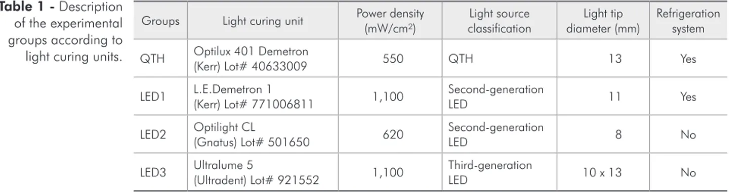
the resin cement was applied to the internal surface of the indirect restorations and then placed accord-ing to their respective guides. Cement excesses were removed and then polymerized for 60 s in all tooth surfaces: buccal, lingual, mesial, distal and occlusal. Different light sources were used in the groups, as described in Table 1.
The power density was assessed using a con-ventional hand-held radiometer for the QTH light (Demetron Radiometer) and an L.E.D. Demetron Radiometer for the LED lights (Sybron, Newport Beach, CA, USA). All specimens were then stored for 24 h in a dark environment at room temperature (37°C) and 100% relative humidity. After the stor-age time, the teeth were longitudinally sectioned in both the “x” and “y” directions across the bonded interface with a diamond saw mounted in a Labcut 1010 machine (Extec, Enield, CT, USA), under wa-ter-cooling at 300 rpm. Bonded stick-shaped speci-mens were obtained with a cross-sectional area of 1.0 (± 0.2 mm²). The stick area was measured with a digital caliper after testing (Digimatic, Mitutoyo, Tokyo, Japan). Individual bonded sticks were posi-tioned in a Universal Testing Machine (DL10000 EMIC, São José dos Pinhais, PR, Brazil) by means of cyanoacrylate-based cement and then subjected to tensile forces at a cross-head speed of 1.0 mm/ min until failure. The results were recorded, and the debond stress values, converted into MPa. Data were submitted to one-way ANOVA and Tukey’s
post-hoc test, at a pre-set alpha of 0.05. The
distri-bution of failure mode of the tested specimen was
Groups Light curing unit Power density
(mW/cm²)
Light source classification
Light tip diameter (mm)
Refrigeration system
QTH Optilux 401 Demetron
(Kerr) Lot# 40633009 550 QTH 13 Yes
LED1 L.E.Demetron 1
(Kerr) Lot# 771006811 1,100
Second-generation
LED 11 Yes
LED2 Optilight CL
(Gnatus) Lot# 501650 620
Second-generation
LED 8 No
LED3 Ultralume 5
(Ultradent) Lot# 921552 1,100
Third-generation
LED 10 x 13 No
also evaluated at 40 X magniication using a dis-secting microscope (Lambda LEB-3, São Paulo, SP, Brasil) and classiied as:
Cohesive failure (failure exclusively within the laboratory resin [CR] or dentin [CD]);
Adhesive failure (fracture at the composite/den-tin interface);
Adhesive/Mixed failure (failure at the composite/ dentin/resin cement interface including cohesive failures in the neighboring substrates).
Results
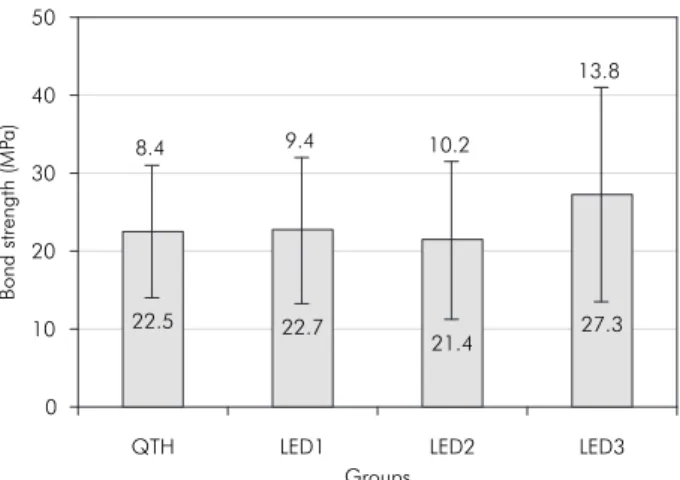
The microtensile bond strength values for the ex-perimental groups are shown in Graph 1.
The highest bond strength mean value was seen when the LED3 light (Ultralume 5) was used (27.3 ± 13.8 MPa). The second-generation LED2 light (Optilight CL) presented the lowest bond strength mean value (21.4 ± 10.2 MPa). The control group (QTH light) bond strength mean value was similar to that obtained when the LED1 light was used (22.5 ± 8.4 and 22.7 ± 9.4 MPa, respectively). One-way ANOVA revealed that all the results were statistically equivalent (p > 0.05).
The failure modes of the fractured specimens, after microtensile testing, are presented in Table 2. This table also shows the percentage of failure in the experimental groups. The great majority of fail-ures observed were adhesive (from 88% to 100%). The adhesive/mixed mode was the next most
fre-1.
2.
3.
quently observed failure mode (from 0% to 8%). The cohesive failure mode in resin (CR) was ob-served the least in all the experimental groups (0% to 4%). The cohesive failure mode in dentin (CD) was not detected in any of the microscopic analy-ses. The overall fracture mode results proved that the predominant failure mode was the adhesive fracture mode (94%).
Discussion
The null hypothesis, that there would be no dif-ference in bond strength when different lights were used to polymerize a dual-cure resin cement in in-direct composite restorations, was validated. Sta-tistical analysis revealed no signiicant difference among the groups (p > 0.05). The reason to explain the values when different commercial light curing sources were used to polymerize the resin cement seems to be related neither to their power density output nor to their spectral irradiance. Equivalent bond strength values were observed when the QTH and the LED lights were used to polymerize the res-in composite. The second-generation LED2 and the QTH light provided almost the same power density, whereas both the second- (LED1) and third-genera-tion (LED3) LED lights showed an enormous differ-ence in power density, but comparable dentin adhe-sion values. In addition, in terms of irradiance, the “broad-banded” QTH and LED3 lights provided similar bond strength values compared to the “nar-row-banded” LED1 and LED2.
The evolving technology of curing light sources led to the advent of broad-banded LED lights in
or-Graph 1 - Mean and standard deviation of bond strength values according to experimental groups. All values were statistically equivalent (p > 0.05).
0 10 20 30 40 50
QTH LED1 LED2 LED3
Groups
B
o
nd
strength
(MP
a)
22.5 8.4
22.7 9.4
21.4 10.2
27.3 13.8
Table 2 - Percentage of failure modes after microtensile bond strength testing for all experimental groups.
Groups A % AM % CD % CR %
QTH 88 8 0 4
L.E.Demetron 1 (LED1) 100 0 0 0
Optilight CL (LED2) 100 0 0 0
Ultralume 5 (LED3) 88 8 0 4
Overall percentage of
failure mode 94 4 0 2
der to allow the photoactivation of all types of res-in-based materials, irrespective of the photoinitiator added.1 An increase in power density in conjunction
with a wide spectral distribution seen in the third-generation LED lights produces heating as a conse-quence.12,13 Despite the absence of signiicant
differ-ences, the heating produced by the third-generation LED light (LED3) might have led to an additional polymerization, which in turn, can explain why a higher bond strength mean value was observed when this light source was used.
It has been claimed that the resin cement polym-erization should be optimized in order to resist de-terioration of mechanical and chemical properties, among them strength, hardness, stiffness, and wear resistance.14 Studies have shown that dual-curing
resin cements depend on photoactivation to achieve a high degree of conversion, and thus, a better per-formance.15 There is no agreement about the ideal
power density needed to obtain optimal energy den-sity, nor is there agreement about the irradiance of the light source, and the exposure time needed to cure resin cements suficiently.16 These parameters
are of particular interest since, in practice, they are under the control of the clinician.17,18 On the other
hand, the results obtained in the present study do not corroborate these assumptions. In fact, these indings can be explained because a dual resin lut-ing cement was used in the present study. The speci-mens were evaluated with the microtensile bond strength test after 24 h, time after which the resin cement may have chemically completed its polym-erization.19 Irrespective of the light source used, the
results proved that the bond strength was signii-cantly equivalent.
Dual-curing resin cements have been used for luting indirect esthetic restorations and most recent-ly metal castings such as crowns and ixed partial dentures, as an alternative to zinc phosphate and glass ionomer cements.20 Some advantages of this
type of resin cement are low solubility, adequate consistency and ilm thickness, superior mechani-cal properties, optimal bonding to dental struc-tures and restoring materials by adhesive systems
and reduced microleakage.21,22 Since the
introduc-tion of LED sources by Millis in 1995, several con-cerns about their eficiency for the light curing of resin-based materials have arisen. Regarding resin composites, several studies have demonstrated that LED devices are effective.23,24 On the other hand,
as regards resin cements, few studies25-27 have only
reported no signiicant differences using shear bond strength testing when QTH devices were compared to high-intensity LED light sources to polymerize a dual-cure resin cement through a 3-mm-thick ceramic restoration. It has been claimed that LED light units with a relatively low power output re-quire a higher exposure time to perform as well as QTH lights.12
The indings observed in the present investiga-tion suggest that, although high-intensity LED lights were also used to activate the resin cement, the po-lymerization of this restorative material was effec-tive. The bond strength values seen in the experi-mental groups were equivalent. Clinically, the use of a light source when cementing an indirect restora-tion is important to allow immediate polymerizarestora-tion of the marginal cement, avoiding premature crown removal. Polymerization completion in the areas not reached by the curing light energy occurs through chemical reaction, which takes about 24 h,11 and it
is essential for achieving adequate bond strength to the dental tissues. Also, despite the evolving tech-nology, the conventional broad-banded QTH lights are effective for light curing of resin-based materials and can still be considered a control light for being able to polymerize all resin-based materials, irre-spective of the photoinitiator.
Conclusion
References
1. Rueggeberg FA, Blalock JS, Callan RS. LED curing lights – what’s new? Compend Contin Educ Dent. 2005;26(8):586-91. 2. Uhl A, Sigusch BW, Jandt KD. Second generation LEDs
for the polymerization of oral biomaterials. Dent Mater. 2004;20(1):80-7.
3. Kurachi C, Tuboy AM, Magalhaes DV, Bagnato VS. Hardness evaluation of a dental composite polymerized with experimen-tal LED-based devices. Dent Mater. 2001;17(4):309-15. 4. Caughman WF, Rueggeberg FA. Shedding new light on
com-posite polymerization. Oper Dent. 2002;27(6):636-8. 5. Price RB, Felix CA, Andreou P. Evaluation of a
second-genera-tion LED curing light. J Can Dent Assoc. 2003;69(10):666. 6. Price RB, Felix CA, Andreou P. Evaluation of a dual peak
third generation LED curing light. Compend Contin Educ Dent. 2005;26(5):331-8.
7. Park YJ, Chae KH, Rawls HR. Development of a new photo-initiation system for dental light-cure composite resins. Dent Mater. 1999;15(2):120-7.
8. Pashley DH, Carvalho RM, Sano H, Nakajima M, Yoshiyama M, Shono Y et al. The microtensile bond test: a review. J Adhes Dent. 1999;1(4):299-309.
9. Reis A, Loguercio AD, Azevedo CL, de Carvalho RM, da Julio Singer M, Grande RH. Moisture spectrum of demineralized dentin for adhesive systems with different solvent bases. J Adhes Dent. 2003;5(3):183-92.
10. Coelho Santos MJ, Navarro MF, Tam L, McComb D. The effect of dentin adhesive and cure mode on film thickness and microtensile bond strength to dentin in indirect restorations. Oper Dent. 2005;30(1):50-7.
11. 3M ESPE. Rely X ARC Adhesive resin cement system. Techni-cal product profile. St. Paul: 3M Center; 1998.
12. Nomoto R, McCabe JF, Hirano S. Comparison of halogen, plasma and LED curing units. Oper Dent. 2004;29(3):287-94. 13. Trujillo M, Newman SM, Stansbury JW. Use of near-IR to
monitor the influence of external heating on dental composite photopolymerization. Dent Mater. 2004;20(8):766-77. 14. Davidson-Kaban SS, Davidson CL, Feilzer AJ, de Gee AJ,
Erdilek N. The effect of curing light variations on bulk curing and wall-to-wall quality of two types and various shades of resin composites. Dent Mater. 1997;13(6):344-52.
15. Papazoglou E, Rahiotis C, Kakaboura A, Loukidis M. Curing efficiency of a photo- and dual-cured resin cement
polymer-ized through 2 ceramics and a resin composite. Int J Prostho-dont. 2006;19(1):34-6.
16. Ernst CP. Clinical aspects of photopolymerization. In: Scien-tific insights into dental ceramics and photopolymer networks; 2004. Geneva: Academy of Dental Materials 2004. p. 105-17.
17. Daronch M, Rueggeberg F, de Goes MF. Monomer conversion of pre-heated composite. J Dent Res. 2005;85(7):663-7. 18. Halvorson RH, Erickson RL, Davidson CL. Energy
depend-ent polymerization of resin-based composite. Ddepend-ent Mater. 2002;18(6):463-9.
19. Venhoven BA, de Gee AJ, Davidson CL. Light initiation of dental resins: dynamics of the polymerization. Biomaterials. 1996;17(24):2313-8.
20. Fonseca RG, Santos JG, Adabo GL. Influence of activation modes on diametral tensile strength of dual-curing resin ce-ments. Braz Oral Res. 2005;19(4):267-71.
21. Van Meerbeek B, Inokoshi S, Davidson CL, De Gee AJ, Lam-brechts P, Braem M et al. Dual cure luting composites – Part II: Clinically related properties. J Oral Rehabil. 1994;21(1):57-66.
22. Yoshida K, Tanagawa M, Atsuta M. In vitro solubility of three types of resin and conventional luting cements. J Oral Rehabil. 1998;25(4):285-91.
23. Bennett AW, Watts DC. Performance of two blue light-emit-ting-diode dental light curing units with distance and irradia-tion-time. Dent Mater. 2004;20(1):72-9.
24. Teshima W, Nomura Y, Tanaka N, Urabe H, Okazaki M, Nahara Y. ESR study of camphorquinone/amine photoinitia-tor systems using blue light-emitting diodes. Biomaterials. 2003;24(12):2097-103.
25. Foxton RM, Pereira PN, Nakajima M, Tagami J, Miura H. Durability of the dual-cure resin cement/ceramic bond with different curing strategies. J Adhes Dent. 2002;4(1):49-59. 26. Nalcaci A, Kucukesmen C, Uludag B. Effect of high-powered
LED polymerization on the shear bond strength of a light-po-lymerized resin luting agent to ceramic and dentin. J Prosthet Dent. 2005;94(2):140-5.