HISTOMORPHOMETRICAL STUDY OF THE ELASTIC FIBER
HISTOMORPHOMETRICAL STUDY OF THE ELASTIC FIBER
HISTOMORPHOMETRICAL STUDY OF THE ELASTIC FIBER
HISTOMORPHOMETRICAL STUDY OF THE ELASTIC FIBER
HISTOMORPHOMETRICAL STUDY OF THE ELASTIC FIBER
SYSTEM IN THE ANTERIOR CEREBR
SYSTEM IN THE ANTERIOR CEREBR
SYSTEM IN THE ANTERIOR CEREBR
SYSTEM IN THE ANTERIOR CEREBR
SYSTEM IN THE ANTERIOR CEREBRAL AR
AL AR
AL AR
AL AR
AL ARTERY OF MAN
TERY OF MAN
TERY OF MAN
TERY OF MAN
TERY OF MAN
RENATO PAULO CHOPARD* , RENÊ GERHARD* *
ABSTRACT - The aim of the present study was to quantify the distribution of the elastic fiber system within the wall of the anterior cerebral artery. The study is based on the works of Glynn (1940) and Stehbens (1989) concerning the incidence and origin of brain aneurysms and recent studies of the elastic fibers. The anterior cerebral artery was divided into three segments, S1, S2 and S3: S1 corresponds to the origin of the anterior cerebral artery, S2 is located at the junction of the anterior cerebral artery with the anterior communicating artery, and S3 at the junction of the rostrum and genu of the corpus callosum,which were submitted to routine histological procedures. A histomorphometrical study was undertaken using an estimation of the linear density (Ld) of the components of the fibrous elastic system which evaluates their full length in each segment. Data were analyzed using first order linear regression methods. The results show a decreasing quantity of elastic fibers in the three segments (S1>S2>S3). Study of the elastic fiber system may originate new concepts regarding the genesis of cerebral artery aneurysm.
KEY WORDS: elastic fibers, cerebral arteries, anterior cerebral artery, aneurysm.
Estudo histomorfométrico do sistema de fibras elásticas da artéria cerebral anterior no homem
RESUMO - O objetivo deste estudo foi o de quantificar a distribuição dos sistemas de fibras elásticas encontrados na parede da artéria cerebral anterior. O estudo foi baseado nos trabalhos de Glynn (1940) e Stehbens (1989) referentes a incidência e origem dos aneurismas cerebrais e estudos recentes sobre fibras elásticas. A artéria cerebral anterior foi dividida em três segmentos S1, S2 e S3; S1 corresponde ao segmento de origem da artéria cerebral anterior, S2 localizado entre a junção da artéria com o ramo comunicante anterior, e S3 ao segmento arterial ao nível da junção do rostro e joelho do corpo caloso; todos estes foram submetidos a procedimentos histológicos rotineiros . Foi realizado estudo histomorfométrico calculando-se a densidade linear dos componentes do sistema de fibras elásticas , que avalia o comprimento total de cada tipo de fibra em cada segmento analisado. Os dados obtidos foram calculados e as duas variáveis idade e densidade linear foram tratadas e correlacionadas estatisticamente através de métodos de regressão linear. Os resultados mostram de maneira geral um aumento da quantidade de fibras do sistema elástico com a idade e uma distribuição não homogênea destes sistemas na túnica média da artéria cerebral anterior. As fibras elaunínicas no entanto diminuem com a idade, sugerindo menor elasticidade do sistema, principalmente no segmento intermediário.
PALAVRAS-CHAVE: fibras elásticas, artérias cerebrais, artéria cerebral anterior, aneurisma.
Human cerebral arteries have been studied extensively both in terms of their macroscopic anatomy and with regard to the diseases that affect the structure of their walls, in particular cerebral aneurysm. The present study quantifies the distribution of the elastic fiber system in the wall of the human anterior cerebral artery at ages ranging from 39 to 74 years.
The elastic component plays an important role in maintaining the dynamic balance of the blood vessels in general. It also has a function in the quantitative and qualitative modifications
Departamento de Anatomia, Instituto de Ciências Biomédicas, Universidade de São Paulo (USP): *PhD; **MD. Aceite: 31 de agosto 2000.
which take place after birth. In adults, these changes are of interest in the study of vascular diseases1-3.
According to Stehbens 4, the frequency of defects in the media layer of the cerebral arteries of man
increases with age; these failures thus cannot be considered congenital in origin. While the direct causes of the defects themselves are unknown, they probably result from mechanical factors.There is no evidence available to suggest that such defects represent areas of decreased wall resistance that lead to the development of cerebral aneurysm. Studies have underscored the importance of the elastic, collagen and muscle elements in the pathophysiology of the cerebral arteries 5- 7. In the
cerebral arteries the incidence of aneurysm is high. In a review of the literature by McDonald & Korb 8 about 8.8% of cerebral aneurysms in the circle of Willis occur in the anterior cerebral artery.
The high incidence of aneurysm in the cerebral arteries, when compared to other arteries, is the result of differences in the distribution of the elastic tissue within the walls of these vessels. In the cerebral vessels most of the elastic tissue is concentrated in the internal elastica. Such a characteristic distribution of the elastic tissue presumably renders these arteries more susceptible to injury and degradation than if the elastic tissue where more widely distributed in the media and adventitia, as seen in other arteries1.
Ladzinski et al. 9, consider that the anatomical structure of intracranial arteries may favour
the development of aneurysms because of poorly developed esternal elastic lamina and adventitia, gaps in the muscular layer and great number of collagenous fibres; however, the haemodynamic factor has the principal significance in initiation , growth and rupture of the aneurysms. Various studies have been undertaken on the system of elastic fibers with regard to their composition: oxytalanic, elauninic and elastic fibers exhibit specific morpho-functional characteristics 10-12.
According to Pasquali-Ronchetti et al.13, the elastic fibers seem to be composed of several matrix
constituents,which are different in different organs and change with age and in patological conditions. Apparently a continous evolutionary process takes place within the elastic fiber system starting with the oxytalanic fiber and evolving to the elastic fiber 14. The elastogenesis follows the sequence
oxytalanic, elauninic and elastic fibers, but the process only completes its maturation in arterial walls, thus leading to the internal elastic lamina 15.
METHOD METHOD METHOD METHOD METHOD
A) Sample collection
A quantitative study of the elastic fiber system of the anterior cerebral artery was undertaken in 19 cadavers,seven were female and 12 were male subjects who had died from various causes (Table 1). No significant macroscopic or microscopic alterations were seen in any of the cases examined.
B) Histological preparations
The anterior cerebral artery was divided into three segments (S1,S2 and S3) which correspond in part to the division of the anterior cerebral artery as proposed by Fischer16. Segment S1 corresponds to the origin of the anterior cerebral artery, segment S2 is located at the junction of the anterior cerebral artery with the anterior communicating artery, and segment S3 at the junction of the rostrum and genu of the corpus callosum.
The segments were fixed in 10% neutral formalin at room temperature for 5 days and embedded in paraffin, transverse serial sections, 5 micrometers thick. Of these, five sections were chosen using a table of random numbers, thus sampling the serial sections, which were then stained according to the following methods: ferric hematoxylin, which stains elastic fibers (Verhoeff) , resorcin-fuchsin, which stains elastic and elauninic fibers (Weigert ) and resorcin-fuchsin, after oxidation with 1% aqueous oxone solution (Weigert-Oxone) which stains elastic, elauninic and oxytalanic fibers.
C) Histomorphometrical study
The histomorphometrical study was performed using a linear density (Ld) estimation of the elastic system. The estimated elastic fiber length was derived from the formula
L=2Q x EV
Table 1. Subjects and segments studied, with age, sex, and causes of death.
Case Segments Age Sex Cause of death
1 S1 S2 S3 39 M Pulmonary edema
2 S1 S2 S3 42 F Bronchopneumonia
3 S2 S3 45 M Congestive heart failure
4 S1 S2 S3 45 M Indeterminate
5 S1 S3 46 F Pulmonary edema
6 S1 S2 S3 46 F Indeterminate
7 S2 S3 47 F Intra-abdominal hemorrage
8 S2 S3 47 F Bronchopneumonia
9 S1 S2 S3 50 F Pulmonary embolysm
10 S2 52 M Indeterminate
11 S1 S2 53 M Pulmonary edema
12 S1 S2 S3 55 M Indeterminate
13 S1 57 M Pulmonary edema
14 S1 S2 57 F Indeterminate
15 S1 S2 S3 58 M Hemopericardium
16 S2 S3 60 M Indeterminate
17 S1 S3 70 M Cachexia
18 S3 73 M Heart failure
19 S1 S2 S3 74 M Indeterminate
The fiber length per unit volume is directly proportional to the number of fiber intersections within the unit section area 17,18. Histological sections of the anterior cerebral artery were analyzed using a light microscope equipped with a 100 X oil immersion objective and a 10X18 Kf compensating eyepiece containing a 400 point integrating graticule and 20 parallel lines.The distance (L) between the points was 5 um and between the lines (D) 4.94 um. An integrating graticule containing 100 points was also employed. In this case L was 12.5 um and D was 12.36 um.
The Ld of the elastic fiber system in the wall of the anterior cerebral artery evaluates the total length of the elastic fibers in this tissue. This is the fundamental basis of histomorphometrical analysis as applied to fibrous systems 18.
Estimation of Ld of the elastic fiber system was perfomed,as follows:
At = L x D x P x m ; f = Pu/P x m ; A = At x f ; Ld =2 x C/ A
where: At is the total area of the test-system ; A is the total area examined in m fields; L is length of the line segment in the test system ; D is the distance between lines of the test-system ; P is the number of points in the test-system; m is the number of fields examined ; f is a correction factor for non-homogenous samples.That is calculated by dividing the number of incident dots (Pi) on the tunica media by the total number of dots utilized (Pu) which corresponds to the total number of points in the test-system that coincide with the elastic fiber system; C corresponds to the number of intersections of the elastic fibers with the parallel straight lines of the test-system.
the elastic fiber system (Pi) and of the total number of points lying over the other fiber systems as a whole (Pu). The Volume Density (VVi) occupied by the elastic fiber system in the histological sections transverse to the anterior cerebral artery is given by the equation: Vvi = Pi/Pu. The measurement of the volume density (VVi) thus provide a correction factor (f) that allows, the linear density analysis in non-homogeneous histological sections like those of the anterior cerebral artery. For an estimated error of 0.05 , twenty five fields (m=25) were examined in each segment (S1, S2 and S3) using two test-systems (100 and 400 points).
D) Statistical analysis
The numerical values of the linear densities were initially analyzed using the non-parametric test 19 which does not consider the values as such but rather their ranks. Mathematical relationships were then established between the linear densities and the age classes. Since Kendall’s test assumes a first order correlation, a simple linear regression model was used in those cases where the hypothesis was accepted (p<0.05).First order linear regressions were obtained by means of specif computer program (EXCEL V 7.0 ,1997).
RESUL RESUL RESUL RESUL RESULTSTSTSTSTS
A) Histological preparations
Histological preparations were obtained using the staining methods of Verhoeff, Weigert and Weigert-Oxone.These techniques respectively show elastic fibers; elastic and elauninic fibers; and elastic, elauninic and oxytalanic fibers. In all cases there was a low density of elastic fibers in the media of the anterior cerebral artery .
B) Histomorphometrical study
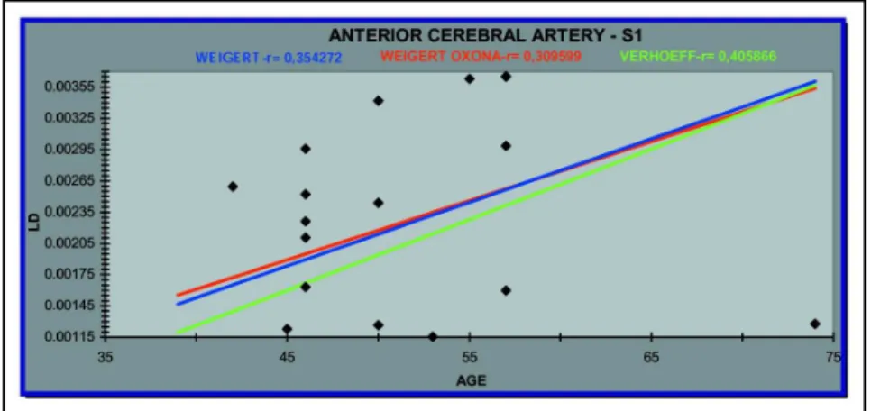
Linear correlations for the three segments (S1, S2, S3) of the anterior cerebral artery were obtained for each specific stain. The corresponding regression lines are given in Figures 1, 2 and 3. In S1 segment, it is observed a great increase of elastic fibre while elauninics and oxitalanics decreases. In S2 segment, it is verified the increase of elastic and oxitalanic fibres in lower degree but the elauninics decreases considerably. In S3 the opposite is verified: the elastic fibres decreases and the elauninics increases. The oxitalanics decreases very little.
DISCUSSION DISCUSSIONDISCUSSION DISCUSSIONDISCUSSION
The tables comparing the linear densities of the elastic fiber system in the three segments, and the plots of the average linear density for each segment show that the density is greater in segment S1 and lower in segment S3, while S2 occupies an intermediate position.
The elastic fiber system is not distributed in a homogeneous form in the walls of the anterior cerebral artery.The number of elastic fibers also varies significantly from one field to the other within the same case. For example in case 19 (segment S2 at elastics fibers) the minimum number of fibers was two and the maximum was 56; in this same case the variance was 134.
The difference in distribution of the elastic tissue within the wall of the cerebral arteries has been emphasized by Glynn 1. This author stressed that the elastic tissue was predominantly distributed
in the internal elastic lamina, located between the internal tunica (intima) and the tunica media. The histomorphometrical study quantitatively demonstrated the heterogeneous distribution of the elastic tissue in the media of the anterior cerebral artery, validating the observations of Chopard et al.20 in
the basilar artery.
With regard to the linear regression analysis, different results were obtained for the three segments (S1, S2, S3) of the anterior cerebral artery.
Fig 1. Linear regression analysis between linear density (Ld) of the segment one (S1), in the different stains, and age. Coefficient of correlation are mentioned for each different stains.
Fig 2. Linear regression analysis between linear density (Ld) of the segment one (S2), in the different stains, and age. Coefficient of correlation are mentioned for each different stains.
system are similar (Fig 2). However, the linear density of the elastic fibers and elauninic elastic fibers decreases with age and the linear correlation coefficients are negative (Fig 2) The elastic fibre, comparatively, are the ones with most increased number by ageing. In segment S3, the linear density of the elastic fibers decreases with age, presenting a negative linear correlation coefficient. However, the linear densities of the elastic and elauninic fibers and of the elastic, elauninic and oxytalanic fibers increase with age. Linear correlation coefficients of similar value are found (Fig 3). Also in this segment ,the elauninics increases and the elastics decreases by ageing, what comes to show a higher possibility of ruining structure in elderly people. According to some authors 2,9, the
frequency of failures in the media of human cerebral arteries increases with age. Hemodynamic stress is responsible for the degeneration and loss of the elastic tissue.
This pathological process may explain the phenomenon observed in segments S2 and S3 of the anterior cerebral artery; that is, the decrease in the linear density of the elastic and elauninic fibers and of the elastic fibers, respectively, with age. However, this same phenomenon is not seen in segment S1, where the linear density of all components of the elastic system increases progressively with age, being the wall less susceptible to injure by ageing. The same observation is valid for the elastic fibers and elastic, elauninic and oxytalanic fibers in segment S2 (Fig 2); this is also the case for the elastic and elauninic fibers and elastic, elauninic and oxytalanic fibers in segment S3 (Fig 3). In the studied segments is a progressive increase of oxytalanic fibre what makes the wall higher resistance to traction. Differential behavior of the cerebral artery has been shown through study of the proximal segment of the basilar artery. In this artery the linear density of all components of the elastic system decreases progressively with age 20. A certain similarity between the behavior of the
elastic and elauninic fibers (Weigert staining) and of the elastic, elauninic and oxytalanic fibers (Weigert-Oxone staining) can be seen in segment S1 of the anterior cerebral artery (Fig 1). Such similarity is also present in segment S3 of the same artery (Fig 3), and in the proximal segment of the basilar artery, where both systems exhibit similar linear correlation coefficients. Segment S2 of the anterior cerebral artery shows the opposite behavior (Fig 2). This finding appears to contradict the use of Weigert staining while may have led to bias in the fiber counts for this segment. The fibers of the elastic system undergo a continuous process of maturation which begins with the oxytalanic fiber, proceeds to the elauninic fiber and continues until elastic fiber develops. Elastic fibers may be subject to degeneration and loss with the passing of the years, although this is probably not the case for the oxytalanic and elauninic fibers. Such degeneration and loss of the elastic fibers apparently takes place in segment S3 of the anterior cerebral artery (Fig 3) and in the proximal segment of the basilar artery. However, no loss of elastic fibers was found in segments S1 and S2 of the anterior cerebral artery. On the contrary, the linear density of this component of the elastic system increases with age in both segments (Figs 1 and 2). It is clear that the fibres of the elastic system are distributed in the arterial wall depending on the intensity of blood flux It is also clear that the places with less quantity of this connective tissue elements have higher probability of aneurysm.
Conclusions
The linear density of the elastic fiber system is greatest in segment S1 and lowest in segment S3, with an intermediate position in segment S2: S1>S2>S3.
REFERENCES REFERENCES REFERENCES REFERENCES REFERENCES
1. Glynn LE. Medial defects in the circle of Willis and their relation to aneurysm formation. J Pathol Bacteriol 1940;51:213-222.
2. Stehbens WE. Medial defects of the cerebral arteries of some mammals. Nature 1957;179:327-328. 3. Stehbens WE. Medial defects of the cerebral arteries of man. J Pathol Bacteriol 1959;78:179-185. 4. Stehbens WE. Etiology of intracranial berry aneurysms. J Neurosurg 1989;70:823-831.
5. Pope FM, Narcisi P, Neil-Dwyer G. Some patients with cerebral aneurysms are deficient in type III collagen. Lancet 1081;1:973-975.
6. Pope FM, Limburg M, Schievink WI. Familial cerebral aneurysms and type III collagen deficiency. J Neurosurg 1990;72:156-158.
7. Leblanc R, Lozano AM, Van Der Rest M. Absence of collagen deficiency in familial cerebral aneurysms. J Neurosurg 1989;70:837-840.
8. McDonald CA, Korb M. Intracranial aneurysms. Arch Neurol Psychiat 1938;32:16-25.
9. Ladzinki P, Koper R, Maliszewski M, Majchzark H. Views on the etiology and pathogenesis of intracranial saccular aneurysms. Neurol Neurochir Pol 1996;30:649-657.
10. Bittencourt-Sampaio S, Cotta-Pereira G. Distribuição das fibras elásticas, elaunínicas e oxitalânicas na derme superior em pele humana. An Bras Derm 1971;46:333-347.
11. Cotta-Pereira G, Rodrigo FG, Bittencourt-Sampaio S. Ultrastructural study of elaunin fibers in the secretory coil of human exocrine sweat glands. Br J Derm 1975;93:623-629.
12. Keith DA, Paz MA, Gallop PM. Elastic tissue histochemistry. Adv Exp Med Biol 1977;79:57-60.
13. Pasqualli-Ronchetti I, Fornieri C, Baccarini-Contri M, Quaglino D.Ultrastructure of elastin.Ciba Found Symp 1995;192:31-50.
14. Montes GS. Structural biology of the fibres of the collagenous and elastic systems. Cell Biol Int 1996;20:15-27. 15. Monte A, Costa A, Porto LC. Distribution of elastic fibres in the humam fetal liver. J Anat 1966;188(III):645-650. 16. Fischer E. Die Lageabweichungen der vorderen Hirnarterie im Gefassbild. Zentralbl Neurochir 1938;3:300-312. 17. Weibel ER. Principles and methods for the morphometric study of the lung and other organs. Lab Invest 1963;12:131-151. 18. Niewoehner DE, Kleinerman J. Morphometric study of elastic fibers in normal and emphysematous human lungs. Am Rev
Resp Dis 1977;115:15-21.
19. Kendall W. In Braley JV. Distribution free statistical tests. New Jersey: Englewood Cliffs, 1968: 284-288.