Letters to the Editor
Radiol Bras. 2016 Jul/Ago;49(4):267–276
274
In most cases are asymptomatic patients, and the diagnosis is established by examination of cardiac imaging performed for other purposes. The symptoms, when present, correspond to pal-pitations, dyspnea, and syncope(11). The prognosis of caseous de-generation of the mitral annulus is good, especially in patients who are asymptomatic, although some patients develop severe symptomatic valvular dysfunction; in the latter group of patients, the prognosis is poor and surgery should be considered(9,12).
On the CCT scans, we noted a hyperintense crescent-shaped mass or a well-defined oval-shaped mass with peripheral calcifica-tion, usually along the posterior mitral annulus, which was not enhanced after contrast administration(13). The heterogeneity of the content of the mass was confirmed by the variation in its den-sity, which can range from negative Hounsfield units, suggest-ing fatty degeneration, to elevated Hounsfield units, suggestsuggest-ing a high protein content and structural calcification(14). The cen-tral hypointensity was secondary to liquefaction of the calcium that fills the center of mass(11,13,15).
In this context of our findings in the case presented here, we can conclude that CCT helps confirm the diagnosis, allows the degree of mitral valve stenosis to be evaluated, and offers mea-sures to improve treatment strategies, especially those involving transcatheter or percutaneous transapical mitral valve implanta-tion. Therefore, CCT is considered an excellent tool for the diag-nosis of caseous degeneration of the mitral annulus.
REFERENCES
1. Neves PO, Andrade J, Monção H. Coronary anomalies: what the radiolo-gist should know. Radiol Bras. 2015;48:233–41.
2. Araújo Neto CA, Oliveira Andrade AC, Badaró R. Intima-media com-plex in the investigation of atherosclerosis in HIV-infected patients [Let-ter]. Radiol Bras 2014;47(1):x.
3. Brasileiro Junior VL, Luna AHB, Sales MAO, et al. Reliability of digital panoramic radiography in the diagnosis of carotid artery calcifications. Radiol Bras. 2014;47:28–32.
4. Ramos SMO, Glavam AP, Kubo TTA, et al. Optimization of a protocol for myocardial perfusion scintigraphy by using an anthropomorphic phantom. Radiol Bras. 2014;47:217–22.
Fernanda Boldrini Assunção1, Diogo Costa Leandro
de Oliveira1, Alair Augusto Sarmet Moreira Damas
dos Santos2, Marcelo Souto Nacif2
1. Complexo Hospitalar de Niterói (CHN), Niterói, RJ, Brazil. 2. Univer-sidade Federal Fluminense (UFF), Niterói, RJ, Brazil. Mailing address: Dra. Fernanda Boldrini Assunção. Hospital Universitário Antonio Pedro – Radiologia. Rua Marquês do Paraná, 303, 2º andar, Centro. Niterói, RJ, Brazil, 24030-900. E-mail: [email protected]. 5. Barranhas AD, Santos AASMD, Coelho-Filho OR, et al. Cardiac
mag-netic resonance imaging in clinical practice. Radiol Bras. 2014;47:1– 8.
6. Stamou SC, Braverman AC, Kouchoukos NT. Caseous calcification of the anterior mitral valve annulus presenting as intracardiac mass. J Thorac Cardiovasc Surg. 2010;140:e9–e10.
7. França LA, Rodrigues ACT, Vieira MLC, et al. Calcificação caseosa do anel mitral: relato de caso. Einstein. 2013;11:370–2.
8. Sequeira A, Morris L, Patel, B, et al. Calcific mitral stenosis in the he-modialysis patient. Hemodial Int. 2014;18:212–4.
9. Mozenska O, Sypula S, Celinska-Spoder M, et al. Mitral annulus caseous calcification mimicking cardiac mass in asymptomatic patient – multimodality imaging approach to incidental echocardiographic find-ing. Pol J Radiol. 2014;79:88–90.
10. Stone E, Cohn D, Deal C, et al. Calcific atrial mass in end-stage renal failure. Nephrol Dial Transplant. 1997;12:807–10.
11. Elgendy IY, Conti CR. Caseous calcification of the mitral annulus: a review. Clin Cardiol. 2013;36:E27–31.
12. Mallat N, Limeme M, Zaghouani H, et al. Caseous calcification of the mitral annulus on MDCT: a rare intracardiac mass. Acta Radiol Short Rep. 2013;2:2047981613502177.
13. Vanovermeire OM, Duerinckx AJ, Duncan DA, et al. Caseous calcifica-tion of the mitral annulus imaged with 64-slice multidetector CT and magnetic resonance imaging. Int J Cardiovasc Imaging. 2006;22:553– 9.
14. Harpaz D, Auerbach I, Vered Z, et al. Caseous calcification of the mitral annulus: a neglected, unrecognized diagnosis. J Am Soc Echocardiogr. 2001;14:825–31.
15. Ribeiro S, Salgado A, Salomé N, et al. Caseous calcification of the mitral annulus: a multi-modality imaging perspective. Rev Port Cardiol. 2012; 31:313–6.
http://dx.doi.org/10.1590/0100-3984.2015.0096
Aortic arch anomaly in an adult patient: a case of right aortic arch with aberrant left subclavian artery and Kommerell’s diverticulum
Dear Editor,
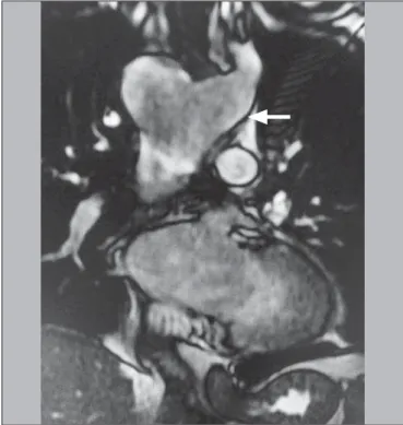
We report the case of a 54-year-old male presenting with vague symptoms of discomfort when swallowing. The patient un-derwent magnetic resonance imaging of the chest. The exami-nation showed right aortic arch with an aberrant left subclavian artery and Kommerell’s diverticulum (Figures 1 and 2).
Thoracic diseases of vascular origin have been the subject of a number of recent publications in the radiology literature of Bra-zil(1–5). First described by Fioratti et al., right aortic arch is an un-common birth defect, of unknown cause, occurring in 0.05% of the general population. It is often asymptomatic but can be ac-companied by dysphagia and complications arising from the for-mation of an aneurysm. Such an aneurysm generally occurs at the origin of the left subclavian artery and is known as Kommerell’s aneurysm or Kommerell’s diverticulum, which can cause com-pression of mediastinal structures or can rupture spontaneously(6– 13). In children, the symptoms can also be associated with
exist-ing congenital cardiac abnormalities(7).
Various systems for classifying right aortic arch have been proposed. The most widely used classification system is that de-vised by Edwards, who described three main types of right aortic
arch: type I, with mirror-image branching of the major arteries; type II, with an aberrant subclavian artery; and type III, with an isolated subclavian artery (connected to the pulmonary artery via the ductus arteriosus)(8). In the case presented here, the variant was classified as an Edwards type II right aortic arch, which ac-counts for approximately 40% of all cases(7).
In an autopsy study cited by Faucz et al.(7), 50% of cases of right aortic arch were associated with an aberrant left subclavian artery, which can be located behind the esophagus (in 80%), be-tween the trachea and the esophagus (in 15%), or anterior to the trachea (in 5%). In some cases, right aortic arch is associated with a congenital heart defect(7,9,10).
The treatment of right aortic arch is generally surgical and is quite complex. Preoperative imaging tests are extremely impor-tant for the surgical planning, which relies heavily on knowledge of the anatomical distribution of the local structures, as well as of the size and extent of the aneurysm. Although outpatient treat-ment is an option, endovascular repair has been performed suc-cessfully(7,11).
Letters to the Editor
Radiol Bras. 2016 Jul/Ago;49(4):267–276
275
REFERENCES
1. Neves PO, Andrade J, Monção H. Coronary anomalies: what the radiolo-gist should know. Radiol Bras. 2015;48:233–41.
2. Herrero Lara JA, de Araújo Martins-Romêo D, Caparrós Escudero C, et al. Hybrid treatment of penetrating aortic ulcer. Radiol Bras. 2015;48: 192–4.
3. Batista MN, Barreto MM, Cavaguti RF, et al. Pulmonary artery sar-coma mimicking chronic pulmonary thromboembolism [Letter]. Radiol Bras. 2015;48:333–4.
4. Amaral RH, Souza VVS, Nin CS, et al. Aortic lesion simulating pulmo-nary disease: a case report. Radiol Bras. 2014;47:320–2.
5. Araújo Neto CA, Andrade ACO, Badaró R. Intima-media complex in the investigation of atherosclerosis in HIV-infected patients [Letter]. Radiol Bras. 2014;47(1):x.
6. Sprong DH, Cutler NL. A case of human right aorta. Anat Rec. 1930; 45:365–75.
7. Faucz RA, Furlan S, Barros AS, et al. Arco aórtico direito com artéria subclávia esquerda aberrante e divertículo de Kommerell. Radiol Bras. 2005;38:381–4.
8. Edwards JE. Anomalies of the derivatives of the aortic arch system. Med Clin North Am. 1948;32:925–49.
9. Costa RN, Andrade IS, Reyes RO, et al. Arco aórtico direito com diver-tículo de Kommerell. Rev Bras Cardiol Invas. 2009;17:279–80. 10. Kimura-Hayama ET, Meléndez G, Mendizábal AL, et al. Uncommon
congenital and acquired aortic diseases: role of multidetector CT an-giography. Radiographics. 2010;30:79–98.
11. Tanaka A, Milner R, Ota T. Kommerell’s diverticulum in the current era: a comprehensive review. Gen Thorac Cardiovasc Surg. 2015;63: 245–59.
12. Barranhas AD, Indiani JM, Marchiori E, et al. Atypical presentation of Kommerell’s diverticulum. Arq Bras Cardiol. 2009;93:e88–90, e101– 3.
13. Barranhas AD, Santos AASMD, Coelho-Filho OR, et al. Cardiac mag-netic resonance imaging in clinical practice. Radiol Bras. 2014;47:1– 8.
Alexandre Ferreira Silva1, José Antônio dos Santos2
1. Ecotomo S/C Ltda., Belém, PA, Brazil. 2. Dimagem – Diagnóstico por Imagem, Belém, PA, Brazil. Mailing address: Dr. Alexandre Ferreira Silva. Rua Bernal do Couto, 93/1202, Umarizal. Belém, PA, Brazil, 06055-080. E-mail: [email protected].
http://dx.doi.org/10.1590/0100-3984.2015.0087
Rosai-Dorfman disease affecting the nasal cavities and paranasal sinuses
Dear Editor,
Here, we report the case of a 17-year-old male who presented with a three-month history of nasal obstruction, asthenia, and febrile episodes. Physical examination revealed bilateral enlarge-ment of cervical and axillary lymph nodes, all of which were pain-less on palpation. Laboratory tests showed mild leukocytosis, an elevated increased C-reactive protein level, and a high erythrocyte sedimentation rate. The venereal disease research laboratory test and monospot test were both negative, as was serology for HIV, toxoplasmosis, and cytomegalovirus. Computed tomography (CT) of the sinuses showed multiple, homogeneous, hypointense, rounded polypoid masses, which effectively narrowed the nasal
passages, together with opacification of the ethmoid cells and sphenoid sinuses, with no evidence of bone erosion (Figure 1). Biopsies of a cervical lymph node and nasal lesions were negative for neoplasia and acid-fast bacilli, showing diffuse lymphoplas-macytic infiltration, foamy histiocytes, and emperipolesis. Immu-nohistochemistry showed positivity for S-100 protein, positivity for CD68, and negativity for CD1a. A diagnosis of Rosai-Dorfman disease was made, and corticosteroid therapy was started, result-ing in slow, progressive improvement.
Recent studies in the radiology literature of Brazil have stressed the importance of CT and magnetic resonance imaging (MRI) in improving the diagnosis of head and neck masses(1–5). Rosai-Dorfman disease, also known as sinus histiocytosis with massive lymphadenopathy, is a rare, benign lymphoproliferative, usually self-limiting, condition characterized by bilateral, painless cervical Figure 1. A,B: Axial T2-weighted spin-echo magnetic resonance imaging
show-ing right aortic arch (arrow).
A
B
Figure 2. Coronal T2-weighted spin-echo magnetic resonance imaging showing Kommerell’s diverticulum (arrow).