Case Report
Key words
Aorta, thoracic; adult; heart failure,congestive.
The interrupted aortic arch (IAA) is a rare cardiopathy, with high morbimortality when treatment is delayed. This study is a case of IAA with atypical clinical behavior in a 19-year-old patient. The history and the clinical assessment were compatible with recent heart failure, associated to syncope episodes and reduction of pulses in the lower limbs. The electrocardiogram showed biventricular and biatrial overload. The echocardiogram showed severe ventricular dysfunction and altered blood flow in the descending aorta. The angiotomography disclosed occlusion of the thoracic aorta after the left subclavian artery. The attained diagnosis was interrupted aortic arch and surgical treatment was indicated.
Interrupted Aortic Arch with Cardiac Heart Failure in Young Adult
Rogério Fortes Lobato, Luis Augusto R. Saliba, Carlos Regenga Ferreiro, Fernando Bacal
Hospital do Coração, Associação do Sanatório Sírio, Instituto do Coração- INCOR, São Paulo, SP - Brazil
Mailing address: Rogério Fortes Lobato •
Rua Borges Lagoa, 933/104 - Vila Clementino - 04038- 000, São Paulo, SP - Brazil
E-mail: [email protected]
Manuscript received June 02, 2007; revised manuscript received July 02, 2007; accepted October 08, 2007.
preceding six months. A few days before the first clinical visit, he had presented 3 syncope episodes and worsening of the dyspnea as well as of the edema. Prior to symptom onset, he was asymptomatic with good weight and height development and denied personal or family morbid antecedents. At the physical examination, he presented clinical signs that were suggestive of heart failure (LLLL edema, enlarged liver and jugular turgescence), with no other alterations at the ectoscopy. The pulses were normal in the upper limbs (UULL), BP was 140/90 mmHg in both limbs, whereas the LLLL presented reduced amplitude, with BP = 90/50 mmHg in the right lower limb (RLL) and 100/60mmHg in the left lower limb (LLL).
The 12-derivation ECG and the chest x-ray showed overload of the four chambers and increased cardiac area of 2+/4+ on account of the left ventricle (LV). Myocardial dysfunction was confirmed by the Doppler echocardiogram, with a LV ejection fraction (LVEF) of 22% (Simpson’s biplane) and slight dilatation; the right ventricle showed moderate dilatation with diffuse severe hypokinesis, with a pulmonary systolic pressure of 76 mmHg in addition to suggestive signs of aortic obstruction after the left subclavian artery.
An angiotomography of the aorta was then requested (Figures 1 and 2), which disclosed aortic occlusion after the origin of the left subclavian artery, with an extension of approximately 3 cm, associated to an extensive collateral network that retrogradely filled the descending and iliac aortas, which had normal diameters, consistent with the patient’s age. The clinical and radiological findings pointed to a diagnosis of aortic arch interruption type A, according to the classification of Celoria and Patton4, with severe myocardial dysfunction.
The patient was submitted to the surgical correction on April 19. A lateral thoracotomy was performed, with the insertion of an 18-mm polytetrafluoroethylene tube between the left subclavian artery and the descending aorta, with the need for extracorporeal circulation.
The echocardiogram performed on the 4th postoperative (PO) day showed a LVEF of 40% with slight hypertrophy and dilatation; the maximum systolic gradient between the left subclavian artery and the descending aorta was 8 mmHg and the pulmonary systolic pressure estimated by the tricuspid insufficiency was 58 mmHg. The patient was discharged from the hospital with good clinical condition on the 9th PO day and was referred to outpatient follow-up.
Discussion
Some diseases present high mortality whereas others are extremely rare; when aortic arch interruption in an adult
Introduction
The interruption of the aortic arch (IAA) is a congenital cardiopathy that has devastating consequences, with a 75% mortality rate at 10 days and 90% at 12 months of life. With an incidence of 3 cases per million live births1, it represents 1.3% of the severe congenital cardiopathies2 and only eventually it is not associated to other anomalies such as persistence of the arterial canal, bicuspid aortic valve, subaortic stenosis, truncus arteriosus communis and transposition of the great arteries3.
The IAA is part of the differential diagnosis of cardiac heart failure in the newborn; however, this entity is extremely unusual in the adult patient.
Case Report
A 19-year old single Caucasian male patient, who was a student from the city of Sao Paulo and had recently arrived from the United States of America, was evaluated. The diagnostic investigation was initiated in March 2007; the patient complained of fatigue following moderate exertion and incipient edema of the lower limbs (LLLL) in the
Case Report
Lobato et al Interrupted aortic arch in adult
Arq Bras Cardiol 2007; 91(1) : e4-e6
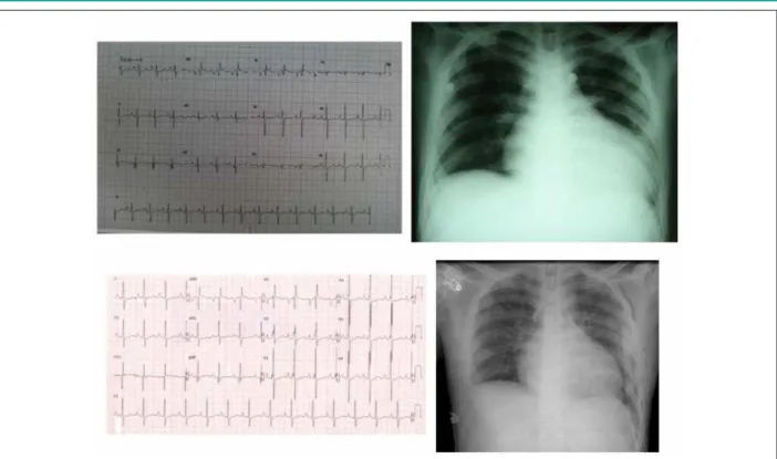
Figure 1 -Above - Preoperative ECG and chest x-Ray; below - postoperative ECG and chest x-Ray.
Figure 2 - A - The interrupted aortic arch can be observed; B - Subsequent correction with tube; C - Extensive collateral network; D - The arrow indicates the Adamkiewicz artery9, implicated in cases of paraplegia after aortic surgeries.
Case Report
Lobato et al
Interrupted aortic arch in adult
Arq Bras Cardiol 2007; 91(1) : e4-e6
patient is reported, a disease that joins these two factors is under discussion5-7.
This disease displays the absence of communication between the two segments of the thoracic aorta and, consequently, of the blood flow; thus, most cases are expected to be fatal. However, even under this extreme condition, sometimes the body develops mechanisms that are capable to maintaining long-term hemodynamic stability and absence of symptoms.
The common characteristic among the survivors is the presence of an extensive collateral network, which is necessary for the maintenance of the distal flow and the consequent organ viability. Another determinant factor is the presence of a large-caliber arterial canal with gradual closing, allowing the development of collateral circulation. It is also thought that in some cases the initial affection would be a coarctation of the aorta, which evolved to the progressive closing of the lumen8.
The few reports in literature show a higher incidence in the male sex, with diagnosis being made in patients up to the eight decade of life9. All of the patients were submitted to
surgical treatment with extra-anatomical tube interposition, with a reasonable mid- and long-term prognosis.
Conclusion
We conclude that the interrupted aortic arch must be recalled as a heart failure etiology in the adult, although it is a very rare entity; it is important to remember that an accurate physical examination can result in the identification of this entity with high sensitivity, thus allowing diagnosis attainment.
Potential Conflict of Interest
No potential conflict of interest relevant to this article was reported.
Sources of Funding
There were no external funding sources for this study.
Study Association
This study is not associated with any graduation program.
References
1. Irwin ED, Braulin EA, Folker JE. Staged repair of interrupted aortic arch and ventricular septal defect in infancy. Ann Thorac Surg. 1991; 52: 632-9. 2. Sturm JT, VanWeeckeren DW, Borkst G. Surgical treatment of interupted
aortic arch in infancy with expanded polytetrafluoroethylene grafts. J Thorac Cardiovasc Surg. 1981; 81: 245-50.
3. Bove EL, Minich LI, Pridjian AK, Lupinetti FM, Snider AR, Dick M 2nd. The management of severe subaortic stenosis, ventricular septal defect and aortic arch obstruction in the neonate. J Thorac Cardiovasc Surg. 1993; 105 (2): 289-96.
4. Celoria GC, Patton RB. Congenital absence of the aortic arch. Am Heart J. 1959; 58: 407-13.
5. Pillsbury RC, Lower RR, Shumway NE. Atresia of the aortic arch. Circulation.
1964; 30: 749-54.
6. Dische WP, Tsai M, Balthaze HA. Solitary interruption of the aortic arch: clinicopathologic review of eight cases. Am J Cardiol. 1971; 27: 271-7. 7. Fowler BN, Lucas SK, Razook JD, Thompson WM, Williams GR, Elkins RD.
lnterruption of the aortic arch: experience in 17 infants. Ann Thorac Surg. 1984; 37: 25-32.
8. Higgins CB, French JW, Silverman JF, Wexler L. lnterruption of aortic arch: preoperative and postoperative clinical, hemodynamic and angiographic features. Am J Cardiol. 1977; 39: 563-71.
9. Messner G, Reul GJ, Flamm SD, Gregoric ID, Opfermann UT. Interrupted aortic arch in an adult single-stage extra-anatomic repair. Tex Heart Inst J. 2002; 29 (2): 118-21.