Aortopulmonary Window - Impact of Associated Lesions on Surgical
Results
Cinthia Siqueira Gangana, Ana Flávia de Araújo Malheiros, Elizabete Vilar Alves, Maurício Amir de Azevedo,
Renata Moll Bernardes, Luiz Carlos Simões
Instituto Nacional de Cardiologia Laranjeiras – INCL-MS - Rio de Janeiro, RJ - Brazil
Summary
Objectives: The aortopulmonary window (APW) is a communication between the pulmonary artery (PA) and the ascending aorta in the presence of two separate semilunar valves. This review describes the natural history of the APW and the impact of associated defects on surgical results in patients treated at our institution.
Methods: Retrospective longitudinal study, based on the review of medical files of patients diagnosed between 1995 and 2005.
Results: Of nine patients diagnosed as having APW, six had associated lesions. Seven patients were submitted to surgical treatment with two deaths. One patient was not submitted to surgery due to pulmonary hypertension and another one died before the surgery due to a respiratory infection complication.
Conclusion: The surgical results are satisfactory when the APW presents as an isolated defect and when surgery is performed early, preventing the development of irreversible arterial pulmonary hypertension (APH). The presence of associated complex congenital heart disease is a bad prognostic factor in our series.
Key words: Aortopulmonary septal defect / diagnosis; heart disease, congenital; child; infant, newborn; adolescent.
Mailing address: Cinthia Siqueira Gangana •
Av. Epitácio Pessoa, 2566/301 - 22471-003 – Rio de Janeiro, RJ - Brazil E-mail: [email protected]
Manuscript received January 18, 2006; revised manuscript received June 30, 2006; accepted October 9, 2006.
Introduction
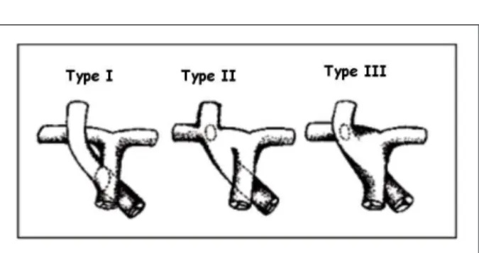
The aortopulmonary window (APW) is a congenital malformation characterized by a communication between the ascending aorta and the pulmonary artery in the presence of separate semilunar valves. It is a rare malformation and represents 0.15 to 0.60% of the congenital heart diseases, being associated with other cardiac defects in ¼ to ½ of the cases. The clinical impact depends on the size of the window and the presence of associated defects and the evolution to pulmonary arterial hypertension1. The most accepted classification is that by Richardson2 (Fig. 1):
Type I: the defect is located between the origin of the main pulmonary artery (MPA) and the ascending aorta immediately above the sinus of Valsalva (embryologically, it is due to the deficient septation of the aortopulmonary trunk);
Type II: it is more distal, between the ascending aorta and the origin of the right pulmonary artery (it is due to the abnormal migration of the sixth aortic arch);
Type III: consists in the anomalous origin of the right pulmonary artery of the aorta (results from the uneven septation of the truncus arteriosus).
The diagnosis is achieved through imaging assessment
(Doppler Echocardiogram with Color Flow and magnetic angioresonance), but in some cases, the hemodynamic study might be necessary, mainly for the study of pulmonary vascular reactivity and associated complex defects, especially the vascular ones3.
The treatment is surgical and must be carried out immediately after the diagnosis is established in order to prevent complications such as pulmonary obstructive vascular disease and heart failure4,5.
The prognosis depends on the presence of other associated congenital heart malformations3.
Considering that this pathology is a rare one and the importance of an early diagnosis, the present study carried out a review of APW cases, correlating clinical manifestations, diagnostic methods, surgical treatment and impact of the associated lesions on prognosis.
diagnosis varied from 2.7 to 34.6 Kg (median of 5.0 Kg). The clinical picture of pulmonary hyperflow with dyspnea and fatigue on exertion was observed in all cases. Two patients presented repeated respiratory infection and two others had congestive heart failure of difficult control.
In 7 cases, the diagnosis was achieved through echocardiography (Figs. 2 and 3) and in 2 cases, by cardiac catheterism (Figs. 4A and 4B).
Associated lesions - Six cases presented associated congenital heart defects: type B aortic arch interruption (1), anomalous origin of the right coronary artery of the pulmonary artery (1), interatrial communication (IAC) with left superior vena cava persistence (1), single ventricle with single atrioventricular (AV) valve (1), interventricular communication (IVC) associated to pulmonary arterial hypertension (1) and
Methods
A retrospective longitudinal study was carried out, with the review of the medical files of 9 patients with APW, diagnosed during January 1995 to September 2005 at the Instituto Nacional de Cardiologia Laranjeiras, Rio de Janeiro, Brazil. Age varied from two days to thirteen years; six patients were males and three were females. Diagnosis was confirmed by the review of echocardiograms, hemodynamic studies and in surgical cases, by the surgical findings. The following variables were obtained and analyzed: gender, age, clinical presentation, associated defects and APW classification.
Results
Patients - Nine patients were identified with a diagnosis of APW, 6 males and 3 females. Of the 9 patients, eight were less than a year old, and one was an adolescent at the time of the diagnosis (median of 4 months). Weight at the time of
Fig. 2 - A - Echocardiography at parasternal view, showing the aortopulmonary window (arrow) connecting the aorta (AO) to the main pulmonary artery (MPA). B - Echocardiography at parasternal view, showing color flow (in red) through the distal APW; RPA - right pulmonary artery; LPA - left pulmonary artery.
As for the surgical technique used, five patients underwent defect repair with a bovine pericardial patch, used to close the AP window; in one patient the defect was closed with a PTFE prosthesis and in another by dissection and ligature. The transpulmonary surgical approach was used in five patients and the transaortic approach was used in one patient. Of the seven patients submitted to surgery, two died. Of the two surgical deaths, one occurred in the OR, with the APW being associated with the interruption of aortic arch and broad interventricular communication, and the second occurred in the immediate postoperative period as a consequence of pulmonary arterial hypertension (the patient had interatrial communication (IAC) and left superior vena cava persistence.
Hospital stay duration ranged from 4 days to 3 months. Two patients developed complications during this period, one with PAH and another with systemic infection and acute respiratory distress syndrome (ARDS); a third patient presented a late complication with surgical wound infection and was readmitted at the hospital (the three cases had associated heart defects).
Of the 5 patients in whom the procedure was successful, three presented complete closure of the APW. Of the two patients who evolved with a residual shunt, one was successfully treated by the surgical repair of the associated defect (tetralogy of Fallot) and the other remained with a small residual shunt.
Four of the patients submitted to surgery are being followed at the outpatient clinic of our Institution, with good clinical evolution throughout a period of 15 months to 6 years (mean of 18.7 months). The patient submitted to the repair of the tetralogy of Fallot at another institution also shows good clinical evolution.
Discussion
The aortopulmonary window (APW) was first described by Elliotson in 18306.
Patients with APW present a physiology of left-right shunt and therefore, when this malformation is isolated and large-sized, the clinical presentation is due to the increased pulmonary flow, with congestive heart failure, pulmonary hypertension and early development of pulmonary obstructive vascular disease.
Smaller defects may not present a clinical manifestation and go undetected at image assessments. When other malformations are associated, the clinical findings are variable and this can result in atypical clinical manifestations, which are more related to the associated defect1,3. In our series, 8 patients (88.9%) had clinical manifestations caused by the pulmonary hyperflow and only one remained asymptomatic until 7 years of age.
The image diagnosis (Doppler echocardiogram with color flow and magnetic angioresonance) allows the exact assessment of the defect location and size. Sometimes it may be difficult to differentiate the defect from the common
truncus arteriosus, the anomalous origin of the pulmonary aortic artery (which, according to Richardson, corresponds to the type III of the APW) and even the short-extension
Fig. 4 - A and B: Hemodynamic angiographic study of patient with aortopulmonary window (APW), showing the course of the catheter through the ascending aorta (AO) reaching the main pulmonary artery (MPA) through the APW (arrow).
tetralogy of Fallot (1).
Morphology of the aortopulmonary window - In seven patients, the APW was of the proximal type (type I, according to the classification by Richardson) (Figs. 2A and 3A). In 2 patients, the APW was of the distal type (Figs. 2B and 3B), involving the main pulmonary artery and the right pulmonary artery (type II of Richardson). The size of the APW varied from 0.5 to 1.2 mm.
arterial canal. In these cases, the cardiac catheterism becomes important in the differential diagnosis, by also providing additional information on arterial pressure and pulmonary vascular resistance3. In the present study, two cases were diagnosed through the hemodynamic study and the other seven by the Doppler echocardiogram.
The APW can present different sizes and locations. According to studies such as the one by Tkebuchava et al3, the most frequent form is the type I of Richardson, which is in agreement with our series, where 90% of the cases presented this type of window.
The prognosis depends, in a large number of cases, on the presence of other associated congenital heart malformations1. In our study, 66% of the cases presented associated defects, which is in agreement with the literature that shows an incidence of 47 to 77%1,4. The most common associated
defect is the interruption of the aortic arch (15 to 20%)4; other defects are the anomalous origin of the coronary artery, aortic arch to the right, interventricular communication, bicuspid aortic valve, tetralogy of Fallot with or without agenesis of the pulmonary valve, discordant ventricle-arterial connection, double aortic arch and tricuspid atresia3,7. Dissimilarly from the data found in literature, our series did not show a predominance of any associated heart defect, with only one case of aortic arch interruption (Table 1).
The APW does not close spontaneously and does not decrease in size with age. The prevention of pulmonary obstructive vascular disease is the first indication for surgical closure and this procedure must be carried out immediately after the diagnosis is established, regardless of the patient’s age8.
The surgical repair consists in the closing of the APW window using a patch, normally of bovine pericardium. Since
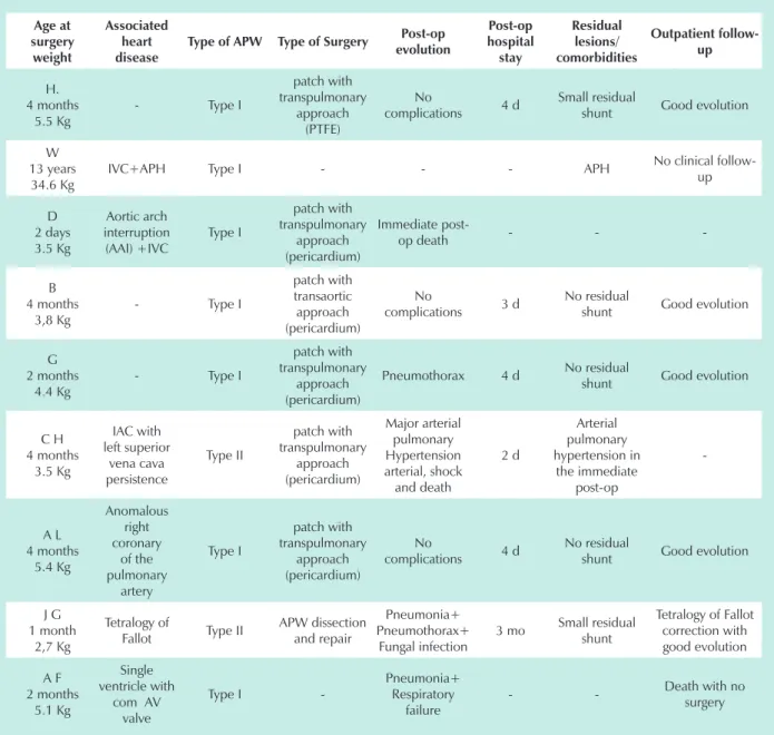
Table 1 – Anatomical, surgical and evolution characteristics of patients with APW
Age at surgery weight Associated heart disease
Type of APW Type of Surgery Post-op evolution Post-op hospital stay Residual lesions/ comorbidities Outpatient follow-up H. 4 months 5.5 Kg
- Type I
patch with transpulmonary
approach (PTFE)
No
complications 4 d
Small residual
shunt Good evolution
W 13 years 34.6 Kg
IVC+APH Type I - - - APH No clinical follow-up D 2 days 3.5 Kg Aortic arch interruption (AAI) +IVC Type I patch with transpulmonary approach (pericardium) Immediate
post-op death - -
-B 4 months
3,8 Kg
- Type I
patch with transaortic approach (pericardium)
No
complications 3 d
No residual
shunt Good evolution
G 2 months
4.4 Kg
- Type I
patch with transpulmonary
approach (pericardium)
Pneumothorax 4 d No residual
shunt Good evolution
C H 4 months 3.5 Kg IAC with left superior vena cava persistence Type II patch with transpulmonary approach (pericardium) Major arterial pulmonary Hypertension arterial, shock and death 2 d Arterial pulmonary hypertension in the immediate post-op -A L 4 months 5.4 Kg Anomalous right coronary of the pulmonary artery Type I patch with transpulmonary approach (pericardium) No
complications 4 d
No residual
shunt Good evolution
J G 1 month
2,7 Kg
Tetralogy of
Fallot Type II
APW dissection and repair
Pneumonia+ Pneumothorax+
Fungal infection
3 mo Small residual shunt
Tetralogy of Fallot correction with good evolution A F 2 months 5.1 Kg Single ventricle with
com AV valve
Type I
-Pneumonia+ Respiratory
failure
References
1. Bagtharia R, Trivedi KR, Burkhart HM. Outcomes for patients with an aortopulmonary window, and the impact of associated cardiovascular lesions. Cardiol Young. 2004; 14: 473-80.
2. Erez E, Dagan O, Georghiou G, Gelber O, Vidne B, Birk E. Surgical management of aortopulmonary window and associatted lesions. Ann Thorac Surg. 2004; 77: 484-7.
3. Tkebuchava T, Segesser LK, Vogt PR, Bauersfeld U, Jenni R, Kunzli A, et al. Congenital aortopulmonary window: diagnosis, surgical technique and long-term results. Eur J Cardiothorac Surg. 1997; 11: 293-7.
4. Tirado AM, Soto JS, Montero JG, Camacho JLG, Madrid AA, Fournier MG, et al. Ventana aortopulmonar: valoración clínica y resultados quirúrgicos. Rev Esp Cardiol. 2002; 55: 266-70.
5. Unlo Y, Ceviz N, Erkut B, Velioglo Y, Koçak H. The large aortopulmonary window without pulmonary vascular disease. Turk J Med Sci. 2001; 31: 451-3.
6. Di Bella I, Gladstone JD. Surgical manangement of aortopulmonary window. Ann Thorac Surg. 1998; 65: 768-70.
7. Botura EM, Piazzalunga M, Barutta F Jr, Grion DS, Neves Fº M, Ueda R. Janela
aortopulmonar e duplo arco aórtico: uma rara associação. Arq Bras Cardiol. 2001; 77: 487-9.
8. Ventemiglia RA, Oglietti J, Izquierdo J, Muasher I, Frazier OH, Cooley DA. The surgical treatment of aortopulmonary window. Tex Heart Inst. 1983; 10 (1): 31-7.
9. Atiq M, Rashid N, Kazmi NA, Qureshi SA. Closure of aortopulmonary window with Amplatzer duct occluder device. Pediatr Cardiol. 2003; 24: 298-9.
10. Rohit M, Nandakurmar S, Bahl A, Kubba S, Talwar KK. Transcatheter closure of aortopulmonary window. Indian Heart J. 2005; 57: 161-3.
11. Mert M, Paker T, Akcevin A, Cetin G, Ozkara A, Salti KL, et al. Diagnosis, management, and results of treatment for aortopulmonary window. Cardiol Young. 2004; 14: 506-11.
12. Backer CL, Mavroudis C. Surgical management of aortopulmonary window: a 40-year experience. Eur J Cardiothorac Surg. 2002; 21: 773-9.
13. Soares AM, Atik E, Cortez TM, Albuquerque AM, Castro CP, Barbero Marcial M, et al. Aortopulmonary window: clinical and surgical assessment of 18 cases. Arq Bras Cardiol. 1999; 73: 67-74.
the first repair made by Gross in 1952, through a thoracotomy, several techniques have been described, using the transaortic or the transpulmonary approaches. Most Centers recommend that the closing be made through the transaortic patch9-11, in contrast with our experience, where five patients (71.4%) underwent repair through the transpulmonary approach, 1 patient (14.2%) through the transaortic approach and in another case (14.2%), the closing was accomplished through dissection and repair.
The closing through hemodynamic intervention is also reported for small defects and to close any residual defect11,12. According to Soares and cols., for this procedure, the type of APW must be characterized and it must be type I (proximal), of a small dimension (3 to 4 mm) and not associated with anomalous origin of the coronary arteries. Therefore, the percutaneous closing is restricted to special situations13.
The surgical prognosis depends on the existence of of pulmonary arterial hypertension as well as of the associated malformations3. In our series, the surgery was contraindicated
for one patient who already presented APH with pulmonary hyperresistance at the moment of diagnosis. Of the patients with associated heart defect, two died: one had aortic arch interruption and another developed severe APH and shock in the immediate postoperative period. Among the patients with and without associated heart defect, mortality was 50% and 0%, respectively, which confirms the literature data.
We conclude that, when APW presents as an isolated defect, the surgical result is a good one, with low morbimortality. However, in the presence of an associated complex defect, the surgical result is worse. The surgery must be carried out as early as possible in order to prevent the development of the most feared complication, the irreversible pulmonary arterial hypertension.
Potential Conflict of Interest