ISSN 0100-879X
BIOMEDICAL SCIENCES
AND
CLINICAL INVESTIGATION
www.bjournal.com.br
www.bjournal.com.br
Volume 44 (12) 1194-1298 December 2011
Institutional Sponsors
The Brazilian Journal of Medical and Biological Research is partially financed by
Faculdade de Medicina de Ribeirão Preto Campus
Ribeirão Preto
Ex plor e H igh - Pe r for m a n ce M S Or bit r a p Te ch n ology I n Pr ot e om ics & M e t a bolom ics
analit icaw eb.com .br S C I E N T I F I C
Braz J Med Biol Res, December 2011, Volume 44(12) 1251-1255
doi: 10.1590/S0100-879X2011007500144
Interaction between human cytomegalovirus UL136 protein and
ATP1B1 protein
Interaction between human cytomegalovirus
UL136 protein and ATP1B1 protein
Xin Cui*, Zheng-Rong Sun*, Gao-Wei Ren, Gui-Li Wang, Ying Qi,
Yan-Ping Ma and Qiang Ruan
Virus Laboratory, the Affiliated Shengjing Hospital, China Medical University, Shenyang, Liaoning, China
Abstract
Interplay between the host and human cytomegalovirus (HCMV) has a pivotal role in the outcome of infection. A region (re
-ferred to as UL/b’) present in the Toledo strain of HCMV and low passage clinical isolates contains 19 additional genes, which
are absent in the highly passaged laboratory strain AD169. Products of the UL/b’ genes may determine the manifestations
of HCMV infection in vivo. However, little is known about the host factors, which interact with UL/b’ proteins. This study was
conducted to investigate the function of the HCMV UL136 protein. By yeast two-hybrid screening, the β1 subunit of the host
Na+/K+-ATPase (ATP1B1) was identified to be a candidate protein, which interacts with the HCMV UL136 protein. The inter -action was further evaluated both in vitro by pull-down assay and in vivo by immunofluorescent co-localization. The results showed that the UL136 protein can interact with ATP1B1 in vitro. Co-localization of UL136-EGFP and ATP1B1-DsRed in cell
membranes suggests that ATP1B1 was a partner of the UL136 protein. It can be proposed that the HCMV UL136 protein may
have important roles in processes such as cell-to-cell spread, and in maintaining cell osmotic pressure and intracellular ion
homeostasis during HCMV infection.
Key words: HCMV; UL136; ATPase; Yeast two hybridization
Introduction
Correspondence: Zheng-Rong Sun or Qiang Ruan, Virus Laboratory, the Affiliated Shengjing Hospital, China Medical University,
Shenyang, Liaoning 110004, China. Fax: +86-024-2389-2617. E-mail: ruanq@sj-hospital.org (Qiang Ruan), or sunzr@sj-hospital.org (Zheng-Rong Sun).
*These authors contributed equally to this study.
Received April 1, 2011. Accepted October 13, 2011. Available online October 28, 2011. Published November 28, 2011. Human cytomegalovirus (HCMV), a member of the
Betaherpesvirinae subfamily, is widely distributed in human populations. It can cause a minor or asymptomatic infec-tion in immunocompetent individuals, and cause severe disease in neonates and immunosuppressed individuals such as allograft transplant recipients and AIDS patients
(1). Although the exact mechanisms are not known, a
majority of researchers believe that a substantial portion
of the HCMV-encoded proteins have the potential to affect
virulence through cell tropism, immune evasion, molecular
mimicry, or interference with host chemokines (2,3). The HCMV genome comprises 236 kb dsDNA, and
many of its genes are nonessential for viral replication in
fibroblasts (4). Since 1996, it has been recognized that a 15-kb unique region (UL/b’) contains at least 19 open
reading frames (ORFs). These genes are found in clinical
HCMV isolates but not in the extensively passaged labo -ratory AD169 strains, which are dispensable for growth
in vitro (5). The retention of these ORFs in clinical HCMV
isolates, and the fact that the virus can replicate in SCID mice implanted with human tissues suggest that the UL/b’
region is required for viral infection in vivo (6). The
func-tions of several UL/b’ region genes, such as UL132, UL141, UL142, UL144, UL146, and UL147 have been previously investigated. The gpUL132 was observed to co-localize
with markers for the trans-Golgi network (7) and with other viral envelope glycoproteins such as gB and gH, serving
as mediators for attachment and fusion with the target
cells. The gpUL141 specifically acts to downregulate cell surface expression of the natural killer (NK) cell-activating
receptor CD155 (8). Soluble tagged gpUL141 inhibits the binding of the monoclonal antibody D171 to CD155, sug-gesting a direct interaction between CD155 and gpUL141. The UL142 gene product is capable of modulating natural
killer NK cell recognition when it is expressed in isolation
or in the context of viral infection and inhibiting NK
cell-mediated lysis (9). The UL144gene encodes a structural
1252 Xin Cui et al.
NFκB-dependent transcription through a mechanism involv -ing TNFR-activated factor 6 (TRAF6) (10). The products
of the UL146 and UL147genes have limited homology to
CXC chemokines. The pUL146protein induces neutrophil chemotaxis and calcium mobilization (11).
The UL136 ORF is 723 bp in size, and has the potential to encode a 241-amino acid protein. The UL136 protein is
a weakly alkaline protein with a molecular mass of ~27.3 kDa; however, little is known about the function of the
UL136 gene product. In the present study, ATPase, a Na/K
transporting beta 1 polypeptide was identified as one of the
binding candidates of the UL136 putative protein.
Material and Methods
Yeast two-hybrid screen
The sequence of UL136 ORF was amplified by the polymerase chain reaction (PCR) using HCMV H strain DNA (GenBank No. GQ981646) as a template with primers 5′-CCGGAATTCTTGTCGACCTCAGTCAAGGGCGTGG
AGATG-3′ (forward, EcoRI included) and 5′-CGCGGAT CCGGTACCTTACGTAGCGGGAGATACGG-3′ (reverse, BamHI included), and then cloned into the C-terminal domain of the shuttle plasmid pGBKT7 (Clontech, USA)
vector via EcoRI and BamHI restriction sites. The construct
of pGBKT7-UL136 was used as the bait in yeast two-hybrid experiments.
The yeast two-hybrid experiments were performed
ac-cording to manufacturer instructions (Matchmaker GAL4 Two-Hybrid System 3; Clontech). Saccharomyces cerevisae
AH109 was co-transformed with plasmids of both
pGBKT7-UL136 and the human fetus brain cDNA Library, pACT2-cDNA. Positive clones were selected on synthetic dropout
medium in the absence of 4 nutrients (Leu/Trp/Ade/His) and the selected clones were then confirmed by detection of α-galactosidase activity. Colonies turning blue were kept, and the positive results were confirmed by repeat assays.
The recombinant plasmids isolated from the positive clones were co-transformed into yeast with the pGBKT7-UL136
for further confirmation of the interaction. Sequencing of the selected clones was performed by the Blast network
service at the National Center for Biotechnology Information (http://www.ncbi.nlm.gov/blast).
GST-pull-down assay
The sequence of one candidate of the pUL136 interact-ing proteins (pACT2-ATP1B1) was cloned into the
glutathi-one S transferase (GST)-tagged pGEX-4T-2 vector in EcoRI
and XhoI sites, designated as GST-ATP1B1.
The GST-pull-down experiment was performed ac-cording to manufacturer instructions (MagneGST™ Pull-Down System, Promega, USA). The c-Myc-labeled protein expressed from the pGBKT7-UL136 was used as a prey
protein in a TNT T7 Quick Reaction. As a bait protein, 20 µL GST-ATP1B1 protein was incubated with 80 µL
pGBKT7-UL136 expressed protein at room temperature for 1.5 h on a rotating platform. The reaction products were then incubated with MagneGST particles for 30 min. After 3 washes with buffer, the binding proteins on the MagneGST particles were eluted with elution buffer and solubilized in 2X SDS sample buffer.
Protein samples were subjected to electrophoresis on a 12% SDS-polyacrylamide gel, and the separated proteins were blotted onto a nitrocellulose membrane (Sigma, USA). Western blot analyses were performed using mouse anti-c-Myc or goat anti-GST monoclonal antibodies (Pierce, USA). Blots were treated with horseradish peroxidase-labeled anti-mouse or anti-goat secondary antibodies, respectively. Signals were revealed by the Molecular Imager ChemiDoc XRS System (Bio-Rad, Inc., USA).
Immunofluorescent co-localization
Commercial pEGFP-C1 and pDsRed-C1 (Clontech)
vectors were used to express fusion proteins with fluo -rescent tags. For the pEGFP-UL136 construct, the UL136
coding region was amplified from the HCMV H strain using the primers 5′-CGGAATTCCTCAGTCAAGGGCGTG-3′
(forward, EcoRI included) and 5′-CGCGGATCCTTACGT
AGCGGGAGA-3′ (reverse, BamHI included). The product
was then cloned into the pEGFP-C1 vector via EcoRI and
BamHI sites, yielding pEGFP-UL136. For the
pDsRed-ATP1B1 construct, the fragment of pDsRed-ATP1B1 was amplified
by PCR using pACT2-ATP1B1 as a template with primers
5′-CGGAATTCCGTCTGCAGATTCAAG-3′ (forward, EcoRI
included) and 5′-CGCGGATCCTGTCATTTACAGTAG-3′
(reverse, BamHI included). Next, the amplicon was cloned
into the pDsRed-C1 vector via EcoRI and BamHI sites, yield
-ing pDsRed-ATP1B1. All constructs were verified by DNA
sequencing (Invitrogen Biotechnology Co., Ltd., China). 293T cells were grown in DMEM containing 10% fetal
calf serum. At 75% confluence, cells were co-transfected with a mixture of 3 µg pEGFP-UL136 and 5 µg
pDsRed-ATP1B1 using lipofectamine 2000 (Invitrogen) according to manufacturer instructions. Cells were analyzed using a
TCS SP2 Leica laser scanning confocal microscope (Nikon
Eclipse Ti-E, Japan) equipped with a cooled CCD camera with a 488-nm and a 543-nm excitation beam.
Results
Results of sequence blast online showed that sev-eral positive clones contained the coding sequence of the
ATPase beta 1 polypeptide (GenBank No. NM001677),
showing 99% nucleotide sequence identity. Biotechnol-ogy Information indicated that the UL136 protein could possibly interact with the C-terminal of the ATP1B1 protein in vitro.
To confirm the interaction between ATP1B1 and UL136
proteins, pull-down experiments were employed using
between the ATP1B1 and UL136 proteins was demonstrated (Figure 1).
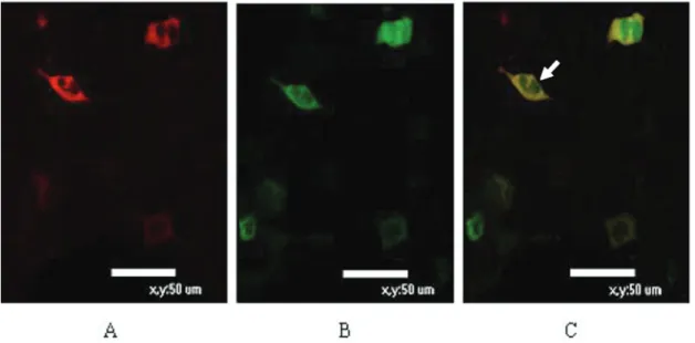
In an immunofluorescent co-localization test, pDsRed/
ATP1B1 was found in the cell membrane, and the pEGFP/ UL136 fusion protein was localized in both the cell mem-brane and the cytoplasm. Results of confocal microscopic analysis revealed that the UL136 fusion protein showed the same location as the ATP1B1 fusion protein on the membrane of mammalian cells (Figure 2).
Discussion
Genes in the HCMV UL/b’ region encode several im -portant proteins including the cell-tropic factors (UL131A -128), virion structure (UL132) (2,7), viral latency determi-nant (UL138), and host cell machinery modulation factors (UL141, 142, 144, and 148) (8-10,12,13). This region is one of the most important targets to elucidate the mechanism
of pathogenesis related to clinical HCMV isolates. So far,
research on targets of the UL136 protein has been found in
only one publication concerning HCMV UL138, but there was
no further functional analysis of the UL136 protein (13).
Host factors may have important roles in viral entry,
replication, budding, release, pathogenesis, and restricting cross-species transmission. Many host factors have been
identified to interact with HCMV proteins and are involved
in all stages of the virus life cycle (14). Similar to other viral
infections, HCMV is known to perturb a number of host cell
functions. The majority of these perturbations presumably optimize host cell functions and conditions to support viral
persistence and productive viral replication (15,16). Earlier studies have demonstrated that both cytomegaly and viral replication strongly depend on the presence of extracellular
Figure 1. Result of a pull-down experiment analyzed by Western
blot. The upper part of the figure was reacted with a goat
anti-GST monoclonal antibody, and the lower part was reacted with a mouse anti-c-Myc monoclonal antibody, separately. Lane 1
shows the protein lysates of transfected DH5α expressing
GST-ATP1B1 bait protein. Lane 2 shows interaction between c-Myc-tagged pUL136 protein and GST-ATP1B1. Lane 3 shows c-Myc-tagged pUL136 protein.
Figure 2. Immunofluorescent co-localization test. A, pDsRed/ATP1B1 localized in the cell membrane; B, the pEGFP/
UL136 fusion protein localized in both the cell membrane and cytoplasm; C, confocal microscopic analysis revealed that
1254 Xin Cui et al.
encoded protein shares the same location with ATP1B1 in cells, the UL136 and ATP1B1 expressing vectors were co-transferred in mammalian cells. Confocal microscopic analysis revealed that the UL136-encoded protein was co-localized with the ATP1B1 protein on the membranes of mammalian cells.
ATP1B1 is an auxiliary subunit of Na+/K+-ATPase, which
is an important regulator of cellular ion homeostasis (23). Recent studies have shown that ATP1B1 can interact with
proteins other than the Na+/K+-ATPase α subunit, such as
NKIP and BKCa (24,25). Reduced expression of
endog-enous ATP1B1 can markedly inhibit evoked BKCa currents
(25). Therefore, we hypothesize that interaction of ATP1B1 with the UL136 protein possibly facilitates the transport and
correct assembly of the Na+/K+-ATPase α subunit in the
cell membrane and affects ion channel activity.
Based on the results of this study, we propose that the UL136-encoded protein may be important for maintaining cell osmotic pressure and intracellular ion homeostasis. Details of the mechanisms involved in this process require further investigation.
Acknowledgments
Research supported by the National Natural Science Foundation of China (#81171581, #81171580, #30801254,
and #30901625) and the Outstanding Scientific Fund of Shengjing Hospital.
References
1. Pellett PE, Roizman B. The family herpesviridae: a brief
introduction, in Fields Virology. In: Knipe DM, Howley PM (Editors), Philadelphia: Lippicott/The Wiliams & Wilkins;
2007. p 2480-2499.
2. Hahn G, Revello MG, Patrone M, Percivalle E, Campanini G, Sarasini A, et al. Human cytomegalovirus UL131-128 genes
are indispensable for virus growth in endothelial cells and
vi-rus transfer to leukocytes. J Virol 2004; 78: 10023-10033.
3. Penfold ME, Dairaghi DJ, Duke GM, Saederup N, Mocarski
ES, Kemble GW, et al. Cytomegalovirus encodes a potent
alpha chemokine. Proc Natl Acad Sci U S A 1999; 96: 9839-9844.
4. Dunn W, Chou C, Li H, Hai R, Patterson D, Stolc V, et al. Functional profiling of a human cytomegalovirus genome.
Proc Natl Acad Sci U S A 2003; 100: 14223-14228.
5. Cha TA, Tom E, Kemble GW, Duke GM, Mocarski ES, Spaete RR. Human cytomegalovirus clinical isolates carry at
least 19 genes not found in laboratory strains. J Virol 1996; 70: 78-83.
6. Wang W, Taylor SL, Leisenfelder SA, Morton R, Moffat JF,
Smirnov S, et al. Human cytomegalovirus genes in the 15-ki -lobase region are required for viral replication in implanted human tissues in SCID mice. J Virol 2005; 79: 2115-2123. 7. Spaderna S, Kropff B, Kodel Y, Shen S, Coley S, Lu S, et
al. Deletion of gpUL132, a structural component of human cytomegalovirus, results in impaired virus replication in
fibroblasts. J Virol 2005; 79: 11837-11847.
8. Tomasec P, Wang EC, Davison AJ, Vojtesek B, Armstrong M, Griffin C, et al. Downregulation of natural killer cell-activating
ligand CD155 by human cytomegalovirus UL141. Nat Im-munol 2005; 6: 181-188.
9. Wills MR, Ashiru O, Reeves MB, Okecha G, Trowsdale J, Tomasec P, et al. Human cytomegalovirus encodes an MHC class I-like molecule (UL142) that functions to inhibit NK cell
lysis. J Immunol 2005; 175: 7457-7465.
10. Poole E, King CA, Sinclair JH, Alcami A. The UL144 gene product of human cytomegalovirus activates NFkappaB via
a TRAF6-dependent mechanism. EMBO J 2006; 25: 4390-4399.
11. Heo J, Petheram S, Demmler G, Murph JR, Adler SP, Bale
J, et al. Polymorphisms within human cytomegalovirus
chemokine (UL146/UL147) and cytokine receptor genes
(UL144) are not predictive of sequelae in congenitally in-fected children. Virology 2008; 378: 86-96.
12. Ji YH, Ruan Q, Lu Y, Qi Y, He R, Liu Q, et al. [Polymorphism
analysis of human cytomegalovirus UL148 gene in low pas-sage clinical isolates]. Zhonghua Shi Yan He Lin Chuang Bing Du Xue Za Zhi 2004; 18: 154-157.
Na+ (17). Therefore, it is not surprising that HCMV infection
is characterized by significant effects on a variety of mem
-brane ion transporters that mediate Na+ transmembrane
movements. Examples of such transporters include the
sodium pump and the Na+/H + exchanger (NHE), as well as
the Cl-/HCO3- exchanger, which works in concert with NHE
to mediate net uptake of inorganic osmolytes promoting
cell volume increase. Previously, Fons et al. (18) reported
that treatment of HCMV-infected cells with an inhibitor of NHE (amiloride) inhibited HCMV replication by almost 2
orders of magnitude.
ATP1B1 is a member of the Na, K-ATPase family of proteins, which are ubiquitous transmembrane proteins that establish and maintain an electrochemical gradient across the plasma membrane in epithelial cells (19). Na,
K-ATPase consists of α and β subunits. The glycosylated β subunit has been proposed to facilitate the assembly and transport of the α subunit from the endoplasmic reticulum
to the plasma membrane (20,21), and the β subunit is the
key factor in polarizing distribution of Na, K-ATPase (22). The results of the present study showed that the HCMV
UL136 protein has the ability to interact with ATP1B1. Bio-technology Information indicated that the UL136 protein
could interact with the C-terminal of ATP1B1 in vitro. It is
predicted that the N-terminal of ATP1B1 located inside the transmembrane region and the UL136-encoded protein
also has a potential transmembrane domain. These findings
suggest that the UL136 protein may interact with ATP1B1
UL136-13. Grainger L, Cicchini L, Rak M, Petrucelli A, Fitzgerald KD,
Semler BL, et al. Stress-inducible alternative translation initiation of human cytomegalovirus latency protein pUL138. J Virol 2010; 84: 9472-9486.
14. Scholz B, Rechter S, Drach JC, Townsend LB, Bogner E.
Identification of the ATP-binding site in the terminase subunit
pUL56 of human cytomegalovirus. Nucleic Acids Res 2003; 31: 1426-1433.
15. Kalejta RF, Shenk T. Manipulation of the cell cycle by human
cytomegalovirus. Front Biosci 2002; 7: d295-d306. 16. Landolfo S, Gariglio M, Gribaudo G, Lembo D. The human
cytomegalovirus. Pharmacol Ther 2003; 98: 269-297. 17. Maglova LM, Crowe WE, Russell JM. Perinuclear
localiza-tion of Na-K-Cl-cotransporter protein after human cyto-megalovirus infection. Am J Physiol Cell Physiol 2004; 286: C1324-C1334.
18. Fons M, Nokta M, Cerruti-Sola S, Albrecht T. Amiloride inhi -bition of human cytomegalovirus replication. Proc Soc Exp Biol Med 1991; 196: 89-96.
19. Lingrel JB, Orlowski J, Shull MM, Price EM. Molecular genet -ics of Na,K-ATPase. Prog Nucleic Acid Res Mol Biol 1990; 38: 37-89.
20. Beggah A, Mathews P, Beguin P, Geering K. Degradation and endoplasmic reticulum retention of unassembled alpha- and beta-subunits of Na,K-ATPase correlate with interaction of BiP. J Biol Chem 1996; 271: 20895-20902.
21. Beggah AT, Beguin P, Bamberg K, Sachs G, Geering K.
beta-subunit assembly is essential for the correct packing and the stable membrane insertion of the H,K-ATPase
alpha-subunit. J Biol Chem 1999; 274: 8217-8223. 22. Morth JP, Pedersen BP, Toustrup-Jensen MS, Sorensen
TL, Petersen J, Andersen JP, et al. Crystal structure of the sodium-potassium pump. Nature 2007; 450: 1043-1049.
23. Xie Z, Wang Y, Liu G, Zolotarjova N, Periyasamy SM, Askari
A. Similarities and differences between the properties of native and recombinant Na+/K+-ATPases. Arch Biochem Biophys 1996; 330: 153-162.
24. Pratscher B, Friedrich C, Goger W, Allen M, Fink D, Thal -linger C, et al. Characterization of NKIP: a novel, Na+/K+ -ATPase interacting protein mediates neural differentiation and apoptosis. Exp Cell Res 2008; 314: 463-477.