Intramolecular Mechanism Implicated in GIT1 Activation
Antonio Totaro1, Veronica Astro1, Diletta Tonoli1, Ivan de Curtis1,2*
1Cell Adhesion Unit, Division of Neuroscience, San Raffaele Scientific Institute, Milano, Italy,2Universita` Vita-Salute San Raffaele, Milano, Italy
Abstract
GIT1 is an ArfGAP and scaffolding protein regulating cell adhesion and migration. The multidomain structure of GIT1 allows the interaction with several partners. Binding of GIT1 to some of its partners requires activation of the GIT1 polypeptide. Our previous studies indicated that binding of paxillin to GIT1 is enhanced by release of an intramolecular interaction between the amino-terminal and carboxy-terminal portions that keeps the protein in a binding-incompetent state. Here we have addressed the mechanism mediating this intramolecular inhibitory mechanism by testing the effects of the mutation of several formerly identified GIT1 phosphorylation sites on the binding to paxillin. We have identified two tyrosines at positions 246 and 293 of the human GIT1 polypeptide that are needed to keep the protein in the inactive conformation. Interestingly, mutation of these residues to phenylalanine did not affect binding to paxillin, while mutation to either alanine or glutamic acid enhanced binding to paxillin, without affecting the constitutive binding to the Rac/Cdc42 exchange factor bPIX. The involvement of the two tyrosine residues in the intramolecular interaction was supported by reconstitution experiments showing that these residues are important for the binding between the amino-terminal fragment and carboxy-terminal portions of GIT1. Either GIT1 or GIT1-N tyrosine phosphorylation by Src and pervanadate treatment to inhibit protein tyrosine phosphatases did not affect the intramolecular binding between the amino- and carboxy-terminal fragments, nor the binding of GIT1 to paxillin. Mutations increasing the binding of GIT1 to paxillin positively affected cell motility, measured both by transwell migration and wound healing assays. Altogether these results show that tyrosines 246 and 293 of GIT1 are required for the intramolecular inhibitory mechanism that prevents the binding of GIT1 to paxillin. The data also suggest that tyrosine phosphorylation may not be sufficient to release the intramolecular interaction that keeps GIT1 in the inactive conformation.
Citation:Totaro A, Astro V, Tonoli D, de Curtis I (2014) Identification of Two Tyrosine Residues Required for the Intramolecular Mechanism Implicated in GIT1 Activation. PLoS ONE 9(4): e93199. doi:10.1371/journal.pone.0093199
Editor:Vania M.M. Braga, Imperial College London, United Kingdom ReceivedJune 8, 2013;AcceptedMarch 3, 2014;PublishedApril 3, 2014
Copyright:ß2014 Totaro et al. This is an open-access article distributed under the terms of the Creative Commons Attribution License, which permits unrestricted use, distribution, and reproduction in any medium, provided the original author and source are credited.
Funding:The following sources of funding to Ivan de Curtis have supported this work: Telethon-Italy, grant GGP12126 (http://www.telethon.it/). AIRC, grant 10321 (http://www.airc.it/). Moreover, a fellowship to Veronica Astro was supported by FIRC (http://www.fondazionefirc.it/). The funders had no role in study design, data collection and analysis, decision to publish, or preparation of the manuscript.
Competing Interests:Coauthor Ivan de Curtis is a PLOS ONE Editorial Board member. This does not alter our adherence to all the PLOS ONE policies on sharing data and materials.
* E-mail: [email protected]
Introduction
During cell migration the intracellular pathways activated in response to extracellular motogenic stimuli need to be spatially and temporally regulated to ensure the synchronization of complex activities such as adhesion, cytoskeleton remodeling, and membrane traffic. Several studies have indicated a role of GIT proteins in cytoskeletal rearrangement and focal adhesion dynamics during motility in distinct cellular contexts, from migrating cells to neurons [1–3]. The members of the GIT family, GIT1 and GIT2, combine the ArfGAP enzymatic activity toward Arf GTPases with the scaffolding function in a variety of signalling complexes [1,3]. GIT proteins include an N-terminal ArfGAP domain to inactivate Arf GTPasesin vitro[4], and Arf6 in
cells [1], a well known regulator of membrane traffic during cell migration [5]. Adjacent to the ArfGAP domain are three ankyrin repeats and a Spa2-homology domain (SHD), a tandem repeat of a sequence homologous to the yeast Spa2. Spa2 is a scaffold-like protein of a polarizing complex involved in actin cytoskeleton reorganization [6]. The SHD domain is important for binding to the PIX proteins [7–10]. The C-terminus of GIT1 contains a
leucine zipper domain involved in homodimerization [11–13], and a paxillin binding sequence (PBS) [14]. GIT1 and paxillin are positive regulators of protrusion at the leading edge of migrating cells, and the recruitment of paxillin to focal adhesions is necessary for their turnover [15]. Paxillin participates in the recruitment of GIT/PIX complexes to focal adhesions by the interaction of the LD4 domain of paxillin with the PBS of GIT [16–20].
Deletion of either the N-terminal ArfGAP or ankyrin domains, as well as the deletion of the C-terminal SHD domain is sufficient to abolish the intramolecular interaction, to increase binding to paxillin, and to affect cell spreading and morphology [23]. Here, we have addressed the mechanisms mediating the intramolecular interaction, and identified two tyrosine residues important to keep the GIT1 protein in the inactive state. Mutation of these residues activates GIT1 binding to paxillin and positively affects cell motility.
Materials and Methods
Plasmids
Plasmids encoding GFP-tagged human GIT1 (770 amino acid residues, NP 001078923.1) [19] and the mutants GIT1-Y392E, GIT1-Y554E, GIT1-S422D/S423D, S457D/S458D, S507D/T508D, GFP-GIT1-S545D/T546D were kindly provided by Alan Rick Horwitz (University of Virginia, Charlottesville, VA). Plasmids GPF-GIT1-Y246E, GIT1-Y293E, GIT1-Y246E/Y293E, GFP-GIT1-Y246A/Y293A, and GFP-GIT1-Y246F/Y293F were ob-tained by site directed mutagenesis starting from GFP-GIT1. Plasmids GIT1-N, GIT1-N-Y246F/Y293F and GFP-GIT1-N-Y246E/Y293E were obtained by PCR amplification of the corresponding GFP-GIT1 full length constructs with the primers 59-cgGAATTCcATGTCCCGAAAGGGGCCGC-39 and 59 -cgcGGATCCTTAGTCGATGATCAAGGTGG-CAAAC-39. The PCR products were digested with EcoR I and BamH I restriction enzymes and cloned in the pEGFP-C1 vector digested with the same enzymes. The constructs FLAG-GIT1 wildtype, FLAG-GIT1-FF and FLAG-GIT1-EE were obtained by digesting the corresponding pEGFP human GIT1 constructs with EcoRI and XmaI enzymes, and subcloning the resulting fragments in pFLAG CMV2 vector digested with the same enzymes. Plasmids FLAG-GIT1-N (residues 1–346), FLAG-GIT1-N2 (res-idues 1–230), C (res(res-idues 346–740), FLAG-GIT1-C2 (residues 229–740) and FLAG-GIT1-FLAG-GIT1-C2-LZ were as described [8,12,23]. The plasmid HA-GIT1-Spa-I (residues 1–321) was obtained by removing with the restriction enzyme ScaI the fragment coding for the carboxy-terminal portion of GIT1 from a pBK-HA chicken GIT1 plasmid. The plasmid encoding the GFP-GIT1-Y246E/Y293E-DPBS mutant lacking the region of inter-action with paxillin (residues 624–770) was obtained by digesting the GFP-GIT1-Y246E/Y293E plasmid with XbaI and by intra-molecular religation. The orange-paxillin plasmid was provided by Alan Rick Horwitz (University of Virginia, Charlottesville, VA).
Antibodies
Monoclonal antibodies (mAbs) against FLAG (clone M2), and polyclonal antibodies (pAbs) against FLAG or mouse IgG were from Sigma-Aldrich (St. Louis, MO). The anti-haemagglutinin mAb (HA, clone 12CA5) was from PRIMM (Milan, Italy). The anti-paxillin mAb (clone 349) was from BD Biosciences (San Jose, CA). The anti-GIT1 pAb raised in goat, was from Santa Cruz Biotechnology (Santa Cruz, CA). The anti-phosphotyrosine mAb (Clone 4G10) was from Millipore (Billerica, MA). The anti-Src mAb (Clone 327) was generously provided by Sara Courtneidge (Sanford Burnham Medical Research Institute, La Jolla, CA). The pAb against GFP, and Alexa-488 anti-rabbit secondary antibodies were from Molecular Probes, Invitrogen. Tha anti-bPIX antibody was described previously (12).
Cell Culture and Transfection
COS7 cells were cultured in DMEM containing 10% FetalClone III (Hyclone PERBIO, Erembodegem, Belgium). Cells seeded in 60 mm diameter dishes were transfected with Lipofec-tamine-2000 (Invitrogen). Cells were processed after 18–24 h.
Biochemical Analysis
Cells washed twice with ice-cold TBS (20 mM Tris-HCl, pH 7.5, 150 mM NaCl) were lysed in lysis buffer (0.5% Triton X-100, 150 mM NaCl, 20 mM Tris-Cl, 2 mM MgCl2, 1 mM sodium orthovanadate, 10 mM sodium fluoride; pH 7.5) in the presence of anti-proteases (16 Complete, Roche, Manheim, Germany). The insoluble material was removed by centrifugation for 15 min at 12000 g at 4uC. Protein determination was done using the Bradford protein assay reagent from BIO-RAD (Hercules, CA, USA).
When indicated, cells were incubated for 20 min at 37uC with 1 mM pervanadate before lysis, to inhibit protein tyrosine phosphatases and increase the levels of protein phosphorylation. Pervanadate was prepared immediately before use by mixing 1 M Na3VO4and 30% H2O2to obtain a 100 mM Na3VO4, 100 mM
H2O2 solution. After 30 min at 37uC, 0.2 mg/ml catalase was
added and incubated for 30 min at 37uto remove H2O2.
For immunoprecipitation, primary Abs were pre-adsorbed to protein A Sepharose beads (Amersham Biosciences). The anti-paxillin mAb was adsorbed to the protein A Sepharose beads preadsorbed with rabbit anti-mouse IgG. Beads were incubated with aliquots of lysate for 2 h at 4uC. After washing with lysis buffer with 0.1% Triton X-100, immunoprecipitates, lysates and unbound fractions were analyzed by immunoblotting.
Haptotactic Migration Assay
COS7 cells transfected with the indicated GFP-GIT1 constructs were starved overnight in serum-free medium (DMEM with 0.1% bovine serum albumin). Transfected cells were trypsinized, resuspended in serum-free medium and seeded in transwell (40000 cells/transwell; 8mm pore PET membrane from Millipore, Billerica, MA). The lower side of the membrane was coated with 10mg/ml of fibronectin. After 6 h at 37uC non-migrating cells were removed from the upper chamber, and cells on the lower side were fixed with 3% paraformaldehyde and detected by fluores-cence. For quantification, GFP-positive cells were counted from different representative fields per well. Control fields with less than 30 cells were not considered. Data were collected from 2–3 independent experiments, each in duplicate. Values of migrated cells were normalized with respect to the percentage of transfected cells (40–60% transfection efficiency). Data are presented as normalized mean values 6SEM. Significance (,0.05%) was established by the Student’s t test.
Wound Healing Assay
Results and Discussion
Specific Mutations in the Amino-terminal Half of GIT1 Enhance Binding to Paxillin
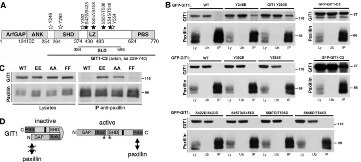
We have previously shown that either endogenous or overex-pressed GIT1 binds very poorly to endogenous paxillin, while deletion of different segments of the amino-terminal portions of the polypeptide induces strong binding of paxillin to the carboxy-terminal portion of the truncated proteins [23]. To limit the possible effects of these deletions on the overall structure of the GIT1 protein, we have tested here the hypothesis that activation may be obtained by site-directed mutagenesis of specific residues of GIT1. A number of studies indicate that GIT1 is phosphor-ylated in response to different stimuli [7,24–26], and that phosphorylation may modulate GIT1 functions in different cells [14,27–28]. For this reason, we have considered several residues that have been shown to be sites of phosphorylation on the GIT1 proteins (Figure 1A, Figure S1) [29], and tested whether one or more of these sites were able to regulate GIT1 function measured as its ability to bind paxillin.
Specific point mutations were introduced into the GFP-GIT1 wildtype construct. In particular, tyrosine, or serine/threonine residues were mutated, in glutamic acid or aspartic acid respectively, to mimic the phosphorylated states of these residues. To limit this analysis, we excluded tyrosine residues located in the PBS region, since their phosphorylation does not appear to be involved in the regulation of GIT1-paxillin interaction [14]. Conversely, residues Y246 and Y293 were selected for being the only two phosphorylated tyrosines in the amino-terminal portion of GIT1 [29]. The phosphorylation sites of GIT1 located in the
central part of the protein (SLD, synaptic localization domain [29– 30]) were also considered for the analysis, including the two tyrosines Y392 and Y554, and the serine/serine or serine/ threonine couples S422/S423, S457/S458, S507/T508, and S54/ T546. The mutations introduced in the GFP-tagged full length protein were tested by immunoprecipitation to look at the effects on the binding of GIT1 to endogenous paxillin. We confirmed the very poor binding of endogenous paxillin to the full length GIT1 protein: immunoblotting on immunoprecipitates with anti-paxillin antibodies performed on lysates of COS7 cells transfected with GFP-tagged GIT1 constructs showed virtually no binding of wildtype GIT1 to endogenous paxillin, while paxillin interacted with the truncated GIT1-C2 protein (Figure 1B). Similarly, several mutants of either tyrosine or serine/threonine residues with glutamic acid or aspartic acid residues respectively (tyrosine mutants GIT1-Y392E and GIT1-Y554E; serine/threonine mu-tants GIT1-S422D/S423D, GIT1-S457D/S458D, GIT1-S507D/ T508D, and S545D/T546D) showed virtually no binding to paxillin. In contrast, mutation of either Y246 or Y293 with glutamic acid (GIT1-Y246E and GIT1-Y293E respectively) resulted in a clear interaction with endogenous paxillin (Figure 1B).
The exchange of either serine or threonine residues with aspartic acid or glutamate residues are considered phosphomi-metic substitutions. The exchange of a tyrosine with a glutamate has also been considered phosphomimetic in several studies. On the other hand, apart from the negative charge, phosphotyrosine is very different from glutamate, since it is shorter than phosphotyr-osine and lacks an aromatic ring. Therefore we have further challenged the hypothesis that the effects of the substitution of
Figure 1. Mutations Y246E and Y293E of GIT1 enhance binding to paxillin.(A) Schematic representation of human GIT1 (NP 001078923.1). GFP-tagged wild type GIT1 was used to introduce phosphomimetic mutations. Tyrosine (Y) or serine (S) and threonine (T) residues were mutated into glutamic acid or aspartic acid, respectively. White and black stars indicate the locations of the mutated tyrosine and serine/threonine residues, respectively. ArfGAP, Arf GTPase-activating protein; ANK, ankyrin repeats; SHD, Spa2-homology domain; LZ, leucine zipper; PBS, paxillin binding site; SLD, synaptic localization domain. (B) Aliquots of lysates (400mg) from cells transfected with the indicated constructs were used for immunoprecipitation of endogenous paxillin. Filters with immunoprecipitates (IP), and equal amounts (80mg) of the respective lysates (Ly) or unbound fractions after immunoprecipitation (Ub) were blotted with anti-GFP (for GIT1) or anti-paxillin antibodies. Molecular weight markers are indicated to the right of each blot. (C) The double substitution of residues Y246 and Y293 with either two glutamic acid (EE) or two alanine residues (AA) enhanced GIT1 binding to paxillin. Aliquots of lysates (200mg protein) from cells transfected with the indicated constructs were immunoprecipitated with anti-paxillin antibody. Filters with immunoprecipitates (IP), and lysates (40mg) were blotted as indicated, using anti-GFP (for GIT1) or anti-paxillin antibodies. (D) Model for GIT1 activation: see details in the text.
Y246 and Y293 by glutamate residues on the binding of GIT1 to paxillin could be due to the phosphomimetic effects of these mutations. The double mutation of Y246 and Y293 with two glutamates enhanced the binding of GIT1-Y246E/Y293E to endogenous paxillin (Figure 1C). Surprisingly, also the substitu-tion of the two tyrosines with alanine residues induced a clear binding of the GIT1-Y246A/Y293A mutant to paxillin, indicating that the change of the biochemical properties observed for these GIT1 mutants was not due to the phosphomimetic effect of the glutamate substitution. Intriguingly, the GIT1-Y246F/Y293F mutant where the two tyrosines were substituted by two phenylalanines behaved as the wildtype protein, since only background binding to paxillin could be detected to this mutant. Altogether these results show that specific point mutations in the region of GIT1 between the ankyrin repeats and the SHD are sufficient to affect the ability of GIT1 to bind paxillin.
Our previous data support the existence of two conformation of the GIT1 polypeptide [23]: a close conformation where the amino-terminal portion of GIT1 interacts with its carboxy-terminal part, and an open conformation that allows the binding of paxillin to the carboxy-terminal part of the polypeptide. In this model the change in the efficiency of association of GIT1 with paxillin is considered as a measure of the release of the intramolecular interaction. Here, the results obtained with the GIT1 mutants fit with the proposed model of GIT1 activation (Figure 1D). The data presented suggest that specific mutations act by allowing the carboxy-terminal PBS to become available for binding to paxillin. Interestingly, the two residues Y246 and Y293, which if mutated alter the biochemical properties of the GIT1 protein, are located in the ankyrin and SHD regions that are necessary to keep the protein in a closed binding-incompetent conformation [23]. On the other hand, substitution of the two tyrosines with phenylalanines that preserve the aromatic ring found also in the tyrosine, maintain the GIT1 protein in the inactive conformation, indicating that the hydrophobic and/or bulky properties of these residues are important for keeping GIT1 in the inactive state.
The Mutations of Tyrosines 246 and 293 do not Affect Binding of GIT1 tobPIX
We have tested if the interaction of GIT1 with other binding partners was affected by mutation of tyrosines 246 and 293. Our previous results have indicated that GIT1 forms stable complexes with the Rac/Cdc42-specific exchanging factorbPIX [23]. The bPIX protein interacts with GIT1 by binding to the SHD domain. We have tested whether the mutations of tyrosines 246 and 293 had an effect on the binding of bPIX to GIT1. We found that binding to endogenous bPIX was similar for wildtype, GIT1-Y246E/Y293E, GIT1-Y246A/Y293A, and GIT1-Y246F/Y293F mutant proteins (Figure 2A). These results show that the modification in the structure of GIT1 introduced by the mutation of the two tyrosines specifically affects the exposure of the paxillin binding region, without affecting the stable association between GIT1 and PIX. Moreover, binding of endogenous bPIX to the active GIT1-EE mutant was not affected by the interaction of GIT1-Y246E/Y293E with the GIT1-N fragment (Figure 2B). These data indicate that GIT1-N does not compete withbPIX for binding to GIT1-Y246E/Y293E. Since tyrosine 293 is in the SHD domain required for binding tobPIX, the results also support the hypothesis that this tyrosine residue is required for the intramo-lecular interaction, but not for the interaction of GIT1 withbPIX (Figure 2C).
Tyrosines 246 and 293 are Required to Keep GIT1 in a Closed Conformation
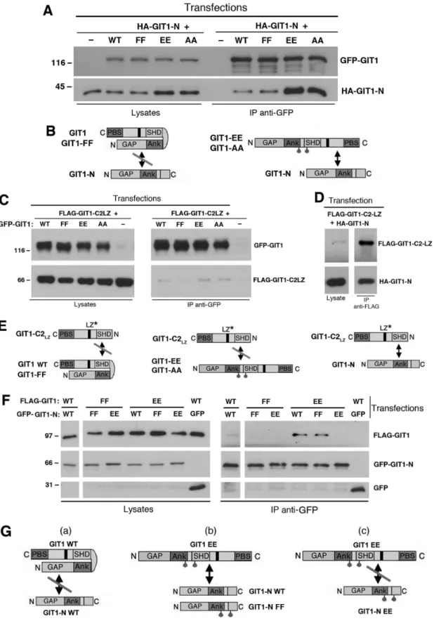
The model proposed for the activation of the GIT1 protein (Figure 1D) is based on a set of experiments with fragments of the GIT1 protein [23]. We confirmed our previous findings that the GIT1-N fragment interacts efficiently with the carboxy-terminal GIT1-C2 fragment, but not with GIT1-C, a carboxy-terminal fragments lacking an intact SHD domain (Figure 3, Figure S1). The GIT1-N2 fragment including the three ankyrin repeats, but lacking the SHD domain interacted only weakly with GIT1-C2. On the other hand, the GIT1-SpaI fragment including the first Spa2 repeat of the two forming the SHD domain can interact efficiently with GIT1-C2, but not with GIT1-C. These data suggest that inclusion of the two tyrosine residues 246 and 293 in the amino-terminal portion of GIT1 is important for the efficient interaction with the carboxy-terminal portion of the GIT1 polypeptide.
To further explore this hypothesis, we have verified if the introduction of specific point mutations in the full length GIT1 polypeptide was sufficient to induce the opening of the full length protein, thus making it available for the interaction with an exogenous amino-terminal portion (GIT1-N). As expected, the amino-terminal GIT1-N fragment interacted poorly with the wildtype GIT1 protein (Figure 4A). On the other hand, GIT1-N interacted more strongly with the full length double mutants GIT1-Y246E/Y293E and GIT1-Y246A/Y293A, but not with GIT1-Y246F/Y293F, indicating that the mutation of the two tyrosines to either alanine or glutamate was sufficient to unlock the closed conformation of the GIT1 polypeptide (Figure 4B), thus making its carboxy-terminal portion available to the binding with the supplied amino-terminal GIT1-N fragment.
Next we tested the hypothesis that the two amino-terminal tyrosines 246 and 293 are required for the binding of the amino-terminal portion of the full length GIT1 to an exogenous carboxy-terminal GIT1 fragment. For this, we compared the interaction of the monomeric carboxy-terminal construct GIT1-C2-LZ (ob-tained by mutating two leucine residues in the carboxy-terminal leucine zipper to prevent homodimerization, [12]) with either wildtype GIT1 or with the full length tyrosine mutants. Interestingly, neither GIT1, nor any of the three mutants GIT1-Y246E/Y293E, GIT1-Y246A/Y293A, and GIT1-Y246F/Y293F were able to bind the carboxy-terminal GIT1-C2-LZ fragment (Figure 4C). As a positive control, GIT1-C2-LZ could bind efficiently the amino-terminal fragment GIT1-N including the tyrosines 246 and 293 (Figure 4D). These results indicate that the two tyrosines in the amino-terminal part of the GIT1 polypeptide are needed for the interaction with the carboxy-terminal portion of the protein to keep the polypeptide in the closed conformation (Figure 4E): the mutant proteins with an open conformation (GIT1-Y246E/Y293E, GIT1-Y246A/Y293A) can not interact with the carboxy-terminal fragment due to the lack of the proper amino-terminal residues; on the other hand in the proteins with a closed conformation (GIT1-Y246F/Y293F and wildtype GIT1) the proper residues are not available because already occupied in intramolecular interactions (Figure 4E).
proteins with GFP-GIT1-N. As expected, the full length wildtype GIT1 protein did not interact with the wildtype GIT1-N fragment, while the full length ‘‘activated’’ GIT1-Y246E/Y293E double mutant interacted with either wildtype GIT1-N, or the mutated Y246F/Y293F, but not with the GIT1-N-Y246E/Y293E fragment (Figure 4F). On the other hand, the full length GIT1-Y246F/Y293F mutant did not interact with either GIT1-Y246E/Y293E or GIT1-N-Y246F/Y293F frag-ments.
These data demonstrate that opening of the full length protein by dysrupting the intramolecular interaction mediated by Y246 and Y293 allows binding of the exposed carboxy-terminal part of the full length mutants to the exogenous wild type GIT1-N fragment (Figure 4B). On the other hand, the substitution of Y246 and Y293 in the GIT1-N fragment with two glutamates prevented the interaction with the exposed carboxy-terminus of the mutant full length protein (Figure 4G, scheme c). Conversely the interaction could still occur if the two tyrosines
in GIT1-N were substituted by two phenylalanines (Figure 4G, scheme b). These findings support the hypothesis that the aromatic rings of tyrosines 246 and 293 are important for the intramolecular interaction between the amino- and carboxy-terminal portions of the GIT1 polypeptide that keeps the protein in the inactive state.
Tyrosine Phosphorylation does not Prevent the Intramolecular Interaction Mediated by Y246 and Y293
We have shown that the substitution of the two amino-terminal tyrosines with two glutamic acid or alanine residues induced the interaction of the full length mutant proteins with the amino-terminal GIT1-N fragment (Figure 4A,B). This result was obtained by immunoprecipitating the full length GFP-tagged proteins with anti-GFP antibodies from cotransfected cells. We then used a pulldown assay to reconstitutein vitro the binding of
the mutated full length protein to the GIT1-N fragment. For this, we transfected the cells with FLAG-GIT1-N, and immunoprecip-Figure 2. Mutation of tyrosines 246 and 293 does not affect the binding of GIT1 tobPIX.(A) Lysates from mock-transfected cells or from cells transfected with the indicated GIT1 constructs were immunoprecipitated for GIT1 with anti-GFP antibody. Filters with immunoprecipitates (IP, from 200mg of protein lysate), and lysates (25mg) were blotted with anti-GFP (for GIT1), anti-paxillin, and anti-bPIX antibodies. (B) Binding of GIT1-N to GIT1-Y246E/Y293E does not affect the interaction of GIT1-Y246E/Y293E tobPIX. Lysates from COS7 cells co-transfected to express the HA-GIT1-N fragment and the full length GFP-GIT1-WT or GIT1-Y246E/Y293E proteins were immunoprecipitated (IP, from 175mg of protein lysate) with anti-GFP. Lysates (25mg, oen the lef) and IP (right) were blotted to reveal the full length proteins (anti-GFP), the HA-GIT1-N fragment (anti-HA mAb 12CA5), and endogenousbPIX. (C) Our data support the hypothesis that the tyrosine 293 of the SHD domain of GIT1 is required for the intramolecular interaction, but not for the interaction of the SHD domain of GIT1 withbPIX.
itated the polypeptide with M2 anti-FLAG antibodies conjugated to beads. After removing the unbound material, the beads with the immunoprecipitates were incubated for 2 h at 4uC with lysates of cells transfected with either wildtype or mutated full length GFP-GIT1 proteins. Under these experimental conditions, we observed the reconstitutionin vitroof the interaction between GIT1-N and
the GIT1-Y246E/Y293E mutant, while no interaction of GIT1-N with either GIT1-Y246F/Y293F or wildtype GIT1 was detected (Figure 5A). This result shows that it is possible to reconstitute
in vitrothe interaction of an exogenous GIT1-N fragment with the
carboxy-terminal part of the full length GIT1 made available by mutation of Y246 and Y293. Together with the previous findings, these results are consistent with an activation model in which Y246 and Y293 are required to keep GIT1 in an inactive state, by means of intramolecular interactions that hold together the amino-and carboxy-terminal portions of the protein.
It has been shown that tyrosines 246 and 293 of GIT1 are phosphorylated by overexpression of Src and pervanadate treatment [29]. Phosphorylation of GIT1-N was achieved by co-expression of c-Src obtained by transfecting the pSGT-c-Src plasmid [31] in combination with pervanadate treatment to inhibit protein tyrosine phosphatases, as previously described [29]. This procedure allowed us to confirm that either one or both tyrosines 246 and 293 are sites of phosphorylation. Indeed, phosphorylation of either GIT1-N-Y246E/Y293E or GIT1-N-Y246F/Y293F fragments was strongly reduced with respect to the wildtype
GIT1-N including both tyrosines (Figure 5B). We tested the possibility that phosphorylation of the two tyrosines by c-Src- and pervanadate-enhanced phosphorylation of GIT1-N blocked its binding to the full length GIT1-Y246E/Y293E. For this, we performedin vitropulldown experiments with immunoprecipitates
of the phosphorylated FLAG-GIT1-N protein (Figure 5C). This treatment resulted in a strong increase of the tyrosine phosphor-ylation of the GIT1-N fragment (Figure 5C). After immunopre-cipitation of the phosphorylated GIT1-N, we incubated the beads carrying the immunoprecipitates with lysates from cells transfected with either wild type or mutated full length GIT1 proteins. Phosphorylation did not prevent GIT1-N to bind the full length GIT1-Y246E/Y293E mutant more efficiently with respect to either wild type or GIT1-Y246F/Y293F full length proteins (Figure 5C). Similar results were obtained when the pulldown was reversed: lysates containing the phosphorylated GIT1-N fragment were incubated with immunoprecipitated full length wild type or mutant GIT1 proteins (Figure 5D). Both control and hyperphosphorylated N bound more strongly to GIT1-Y246E/Y293E than to either the wild type or the GIT1-Y246F/ Y293F full length proteins. These results suggest that phosphor-ylation of the aminoterminal tyrosines 246 and 293 may not be sufficient to prevent the binding to the carboxy-terminal region of GIT1.
Figure 3. Efficient binding of the amino-terminal portion of GIT1 to the carboxy-terminal portion requires the inclusion of the first Spa2 region of the SHD.(A) Scheme of the constructs of avian GIT1 used in the following experiment. To be noted that tyrosine 284 of avian GIT1 corresponds to tyrosine 293 of human GIT1. (B) COS7 cells were cotransfected with the indicated combination of HA-tagged amino-terminal and FLAG-tagged carboxy-terminal fragments of GIT1. Lysates were immunoprecipitated with the anti-FLAG M2-conjugated beads. Filters with immunoprecipitates (IP, from 250mg of protein lysate) and lysates (50mg) were incubated with anti-HA or anti-FLAG antibodies. Amino-terminal fragments in the lysates are indicated by asterisks.
Figure 4. Tyrosines 246 and 293 are required to hold GIT1 in a closed conformation.(A,C,D,F) COS7 cells were cotransfected with the indicated constructs, lysed, and immunoprecipitated with anti-GFP (A,C,F) or anti-FLAG antibodies (D). Filters with immunoprecipitates (IP) and lysates were blotted to reveal the full length GFP-tagged constructs (with anti-GFP), HA-GIT1-N (with mouse 12CA5 mAb), or GIT1-C2-LZ fragment (with anti-FLAG). (B,E,G) Schemes of the interactions proposed from the experiments presented in (A), (C,D), and (F), respectively. The asterisks in (E) indicate the monomeric mutated carboxy-terminal fragment that can not dimerize. See the Results for details.
The Binding of Full Length GIT1 to Paxillin can be Reconstitutedin vitro, and is not Affected by Phosphorylation
We tested if the binding of active GIT1 forms to paxillin could be reconstituted in vitro (Figure 6). Cells were transfected with
either GFP or with each of the wildtype or mutant full length GFP-GIT1 proteins. Lysates from the transfected cells were then used for pulldown experiments, by mixing the beads with the immunoprecipitated GFP-tagged proteins with aliquots of a lysate from cells transfected with orange-paxillin. The results show increased binding of endogenous paxillin and exogenous orange-paxillin to the full length Y246E/Y293E and GIT1-Y246A/Y293A mutants compared to either wildtype GIT1 or GIT1-Y246F/Y293F (Figure 6A). These results indicate that the specific interaction between paxillin and the activated forms of the GIT1 can be reconstitutedin vitro.
We next tested if tyrosine phosphorylation of the full length GIT1 polypeptides would affect the binding to paxillin in vitro.
Cells were cotransfected with each of the wildtype or mutant full length GIT1 proteins together with the tyrosine kinase c-Src, and were incubated before lysis for 20 min with pervanadate, to inhibit protein phosphatases and enhance the levels of GIT1 phosphor-ylation. The combination of c-Src expression and pervanadate
treatment strongly increased the tyrosine phosphorylation of the GIT1 proteins (Figure 6B). Pulldown assays were then performed as described in the previous paragraph. Binding of the activated forms of GIT1 to paxillin was not enhanced by c-Src- and pervanadate-enhanced tyrosine phosphorylation. In contrast, the data suggest that binding to the active forms of GIT1 (GIT1-Y246E/Y293E and GIT1-Y246A/Y293A) was moderately de-creased compared to that observed after pulldown with the same constructs from cells where GIT1 was not hyperphosphorylated. This may be due to the phosphorylation of the carboxy-terminal portion of the GIT1 polypeptide that partially interferes with the binding to paxillin [14].
GIT1-Y246E/Y293E Specifically Enhances Cell Motility To test if the activation of the binding of GIT1 to paxillin reflects a functional outcome, we tested the effects of the overexpression of the different GIT1 full length constructs on cell migration. We used haptotactic cell migration with the lower side of the transwell membrane coated with fibronectin, to induce integrin-dependent migration of the cells plated on the upper chamber of the transwells. The results showed a significant increase (.25%) of the number of cells that migrated to the lower side of the filters only when the cells were transfected with the Figure 5. Hyperphosphorylation of GIT1-N by Src and pervanadate does not affect its bindingin vitroto full length GIT1 proteins. (A–C) COS7 cells were transfected with full length GFP-GIT1 or GIT1-N constructs (WT, FF, or EE), or with wildtype or mutant FLAG-GIT1-N fragments, alone or together with c-Src. The cells cotransfected with c-Src were incubated 20 min at 37uC with 1 mM pervanadate before lysis (pV). Aliquots of the lysates (200mg of protein) were immunoprecipitated (IP) with anti-FLAG (A,C) or anti-GFP (B) antibodies. For pulldowns shown in (A) and (C): FLAG-immunoprecipitates were washed and incubated for 2 h at 4uC with lysates (250mg of protein) from cells transfected with the indicated full length GFP-GIT1 constructs. Equal amounts of lysates (25mg of protein), and the pulldowns performed with GIT1-N without (A) or with c-Src and pervanadate treatment (C) were blotted for the detection of the indicated antigens. In (B) the immunoprecipitations with anti-GFP antibody (right) were immunoblotted to detect the levels of GFP-GIT1-N protein (upper filter) and of its tyrosine phosphorylation (lower filter). (D) COS7 cells were transfected to express the indicated GFP-GIT1 mutants, or transfected with the HA-GIT1-N fragment alone or together with c-Src. The cells co-transfected with c-Src were treated as in (A,C). Aliquots of the lysates from cells expressing GFP-GIT1 mutants (250mg of protein) were immunoprecipitated with anti-GFP. Pulldowns: GFP-immunoprecipitates were washed and incubated for 2 h at 4uC with lysates (400mg of protein) from cells transfected with HA-GIT1-N alone, or together with c-Src. Equal amounts of lysates (25mg of protein), and the pulldowns were blotted for the detection of the indicated antigens.
GIT1-Y246E/Y293E mutant (Figure 7A,B). This increase was significant with respect to either cells overexpressing GFP, or GFP-GIT1-WT. No effects were observed following overexpression of the GIT1-Y246F/Y293F mutant.
Deletion of the carboxy-terminal PBS of GIT1 prevents binding to paxillin [32]. Transfection with GIT1-Y246E/Y293E-DPBS, a GIT1-Y246E/Y293E mutant lacking the PBS (Supplementary Figure S2), induced a decrease in haptotactic migration compared to the full length GIT1-Y246E/Y293E mutant (Figure 7C), suggesting that binding to paxillin is required by the activated GIT1-Y246E/Y293E protein to promote motility.
The specific effects of the GIT1-Y246E/Y293E mutant on cell motility were confirmed by a distinct assay. Wound healing was performed on COS7 cells transfected with GFP alone or in combination with GFP-tagged GIT1 constructs (Figure 7D). Single cell tracking showed an increase in the distance covered by the cells expressing GFP-GIT1-Y246E/Y293E compared to either GFP-GIT1 or GFP transfected cells (Figure 7E). Quantification confirmed the specific enhancement of the euclidean distance covered by GFP-GIT1-Y246E/Y293E-positive cells compared to cells expressing either GFP or full length GIT1 (Figure 7F). Altogether the results show that activation of the binding of GIT1 to paxillin correlates with positive effects on cell motility.
Conclusions
GIT1 is able to assemble a variety of molecular complexes devoted to distinct cellular functions. Given its ability to form large molecular assemblies, the GIT1 protein must be tightly regulated. In this direction, the expression of truncated GIT1 constructs including the PBS enhances cell spreading and protrusion more evidently than full length GIT1 [10,19,23]. On the other hand, the findings that overexpression of specific GIT1 carboxy-terminal fragments cause large intracellular membrane-bound dysfunction-al aggregates [16,33], and that GIT1 carboxy-termindysfunction-al fragments are included in huntingtin aggregates from patients [34] under-score the importance of GIT1 regulation.
Previous studies suggested that GIT1 is regulated by confor-mational changes implicating an intramolecular mechanism [10,23]. According to the model, the N-terminal portion of GIT1 interacts with the C-terminal segment, keeping the molecule less accessible for binding to its partners. Here, we presented new evidence supporting this model, by identifying two tyrosine residues in the amino-terminal part of the molecule that are needed for the interaction with the carboxy-terminal part of GIT1. Different studies point to the relevance of phosphorylation in the regulation of GIT1 function: PAK-mediated phosphorylation on serine 709 enhances GIT1 binding to paxillin and positively Figure 6. Effects of tyrosine phosphorylation on the binding of GIT1 to paxillin.COS7 cells were transfected with GIT1 constructs (A) or cotransfected with the GIT1 constructs together with c-Src (B). The cells cotransfected with c-Src were also treated for 20 min at 37uC with 1 mM pervanadate before lysis (pV). In parallel, a lysate was prepared from cells transfected with orange-paxillin (PXN). After immunoprecipitation of each GFP-GIT1 construct with anti-GFP antibodies (250mg of protein lysate/immunoprecipitation), the beads were incubated for 2 h at 4uC with aliquots of the lysate containing orange-paxillin (180mg). Lysates (25mg) and immunoprecipitates (IP) after pulldown were used for immunoblotting for the indicated antigens.
regulates protrusion [28], while in neurons GIT1 phosphorylation on Y392 by Src family kinases downstream of EphB receptor mediates its translocation to synapses via Grb4 [28]. Tyrosines 246 and 293 are not major sites of phosphorylation following either v-Src expression of treatment with pervanadate [35]. Still, we found here that mutation of these sites strongly reduced the levels of tyrosine phosphorylation of the GIT1-N fragment in COS7 cells
overexpressing c-Src and treated with pervanadate. Hyperpho-sphorylation though did not appear to affect the interaction of the amino-terminal GIT1-N fragment including the two tyrosines, with the carboxy-terminal part of GIT1, nor did it increase the binding of wildtype GIT1 to paxillin, suggesting that GIT1-N tyrosine phosphorylation may not be sufficient to regulate these events. On the other hand further analysis of the stoichiometry of Figure 7. Expression of the ‘‘active’’ GIT1-Y246E/Y293E mutant increases the efficiency of migration.(A) COS7 cells transfected with GFP or with GFP-tagged GIT1 constructs were used for haptotactic cell migration towards fibronectin. Images show fields with cells migrated to the lower side of the filters coated with fibronectin. (B,C) Quantification of the number of cells migrated to the lower side of the wells. Bars are normalized mean values6SEM from 12–19 fields from 2–3 independent experiments. **P,0.02. (D–F) Wound-healing assay. COS7 cells were transfected as in (A). Confluent monolayers were wounded and followed by time lapse imaging. (D) Fluorescent images (left column) showing GFP-positive transfected COS7 cells at time 0. The other columns show the phase contrast images of the same fields, at the beginning (t = 0) and the end (t = 6 h) of the assay. Bar, 100mm. (E) Tracks of GFP-positive cells transfected with either GFP-GIT1 or GFP-GIT1-Y246E/Y293E. Tracks were taken during the wound healing assays and refer to an interval of 8 h. (F) Quantification of the euclidean distance covered by cells during the wound healing assay. Bars are mean values6SEM from 90 cells from 3 different experiments. *P,0.05; ***P,0,002.
phosphorylation of the tyrosines 246 and 293 is required to better define the role of their post-translational modification in the activation of GIT1.
How the intramolecular interactions mediated by Y246 and Y293 can be released to activate GIT1 binding to paxillin remains an open question. One possibility is that the activation of GIT1 may rely on the binding of regulatory proteins that affect its conformation. In this direction, PAK is a suitable candidate that interacts indirectly with GIT1 via PIX [36]. PAK is required for the kinase-independent recruitment of the GIT/PIX complex via paxillin at sites of adhesion to the extracellular matrix [16,19,37]. A PAK mutant unable to bind PIX blocks the localization of the complex to the adhesion sites [18], while a small PAK1 PIX-binding fragment [33] is sufficient to enhance GIT1 PIX-binding to paxillin [23]. Future analysis is needed to define the mechanism by which PAK may unleash the intramolecular mechanism that keeps GIT1 inactive.
Supporting Information
Figure S1 Alignment of GIT1 protein sequences. The two human (NP 001078923.1, NP 054749.2), the chicken (NP 989627.1) and the mouse (NP 001004144.1) GIT1 reference sequences from NCBI databases were aligned using ClustalW. The two tyrosines corresponding to the
residues 246 and 293 of the human GIT1 protein (NP 001078923.1) are indicated.
(TIF)
Figure S2 COS7 cells were transfected with either full length GFP-GIT1-EE or GFP-GIT1-EE-DPBS lacking the carboxy-terminal PBS region required for the interac-tion with paxillin.Lysates were immunoprecipitated with anti-GFP. Filters with immunoprecipitates (150mg of protein lysate, IP) and lysates (20mg) were incubated with anti-GFP and anti-paxillin antibodies, respectively.
(TIF)
Acknowledgments
We thank Alan Rick Horwitz (Department of Cell Biology, University of Virginia, Charlottesville, Virginia, USA) for generously providing the plasmids for orange-paxillin and for several of the GFP-GIT1 constructs used in this study.
Author Contributions
Conceived and designed the experiments: AT IdC. Performed the experiments: AT VA DT. Analyzed the data: AT VA DT IdC. Wrote the paper: IdC.
References
1. Frank SR, Hansen SH (2008) The PIX-GIT complex: a G protein signaling cassette in control of cell shape. Semin Cell Dev Biol 19: 234–44.
2. Hoefen RJ, Berk BC (2006) The multifunctional GIT family of proteins. J Cell Sci 119: 1469–1475.
3. Sabe H, Onodera Y, Mazaki Y, Hashimoto S (2006) ArfGAP family proteins in cell adhesion, migration and tumor invasion. Curr Opin Cell Biol 18: 558–564. 4. Vitale N, Patton WA, Moss J, Vaughan M, Lefkowitz RJ, Premont RT (2000) GIT proteins, A novel family of phosphatidylinositol 3,4, 5-trisphosphatestimu-lated GTPase-activating proteins for ARF6. J Biol Chem 275: 13901–13906. 5. D’Souza-Schorey C, Chavrier P (2006) ARF proteins: roles in membrane traffic
and beyond. Nat Rev Mol Cell Biol 7: 347–358.
6. Shih JL, Reck-Peterson SL, Newitt R, Mooseker MS, Aebersold R, Herskowitz I (2005) Cell polarity protein Spa2P associates with proteins involved in actin function in Saccharomyces cerevisiae. Mol Biol Cell 16: 4595–4608. 7. Bagrodia S, Bailey D, Lenard Z, Hart M, Guan JL, Premont RT, Taylor SJ,
Cerione RA (1999) A tyrosine-phosphorylated protein that binds to an important regulatory region on the cool family of p21-activated kinase-binding proteins. J Biol Chem 274: 22393–22400.
8. Di Cesare A, Paris S, Albertinazzi C, Dariozzi S, Andersen J, Mann M, Longhi R, de Curtis I (2000) p95-APP1 links membrane transport to Rac-mediated reorganization of actin. Nat Cell Biol 2: 521–530.
9. Premont RT, Claing A, Vitale N, Perry SJ, Lefkowitz RJ (2000) The GIT family of ADP-ribosylation factor GTPase-activating proteins. Functional diversity of GIT2 through alternative splicing. J Biol Chem 275: 22373–22380. 10. Zhao ZS, Manser E, Loo TH., Lim L (2000) Coupling of PAK-interacting
exchange factor PIX to GIT1 promotes focal complex disassembly. Mol Cell Biol 20: 6354–6363.
11. Kim S, Lee SH, Park D (2001) Leucine zipper-mediated homodimerization of the p21-activated kinase-interacting factor, beta Pix. Implication for a role in cytoskeletal reorganization. J Biol Chem 276: 10581–10584.
12. Paris S, Longhi R, Santambrogio P, de Curtis I (2003) Leucine-zipper-mediated homo- and hetero-dimerization of GIT family p95-ARF GTPase-activating protein, PIX-, paxillin-interacting proteins 1 and 2. Biochem J 372: 391–398. 13. Premont RT, Perry SJ, Schmalzigaug R, Roseman JT, Xing Y, Claing A (2004)
The GIT/PIX complex: an oligomeric assembly of GIT family ARF GTPaseactivating proteins and PIX family Rac1/Cdc42 guanine nucleotide exchange factors. Cell Signal 16: 1001–1011.
14. Schmalzigaug R, Garron ML, Roseman JT, Xing Y, Davidson CE, Arold ST, Premont RT (2007) GIT1 utilizes a focal adhesion targeting-homology domain to bind paxillin. Cell Signal 19: 1733–1744.
15. Webb DJ, Donais K, Whitmore LA, Thomas SM, Turner CE, Parsons JT, Horwitz AF (2004) FAK-Src signalling through paxillin, ERK and MLCK regulates adhesion disassembly. Nat Cell Biol 6: 154–161.
16. Matafora V, Paris S, Dariozzi S, de Curtis I (2001) Molecular mechanisms regulating the subcellular localization of p95-APP1 between the endosomal recycling compartment and sites of actin organization at the cell surface. J Cell Sci 114: 4509–4520.
17. West KA, Zhang H, Brown MC, Nikolopoulos SN, Riedy MC, Horwitz AF, Turner CE (2001) The LD4 motif of paxillin regulates cell spreading and motility through an interaction with paxillin kinase linker (PKL). J Cell Biol 154: 161–176.
18. Brown MC, West KA, Turner CE (2002) Paxillin-dependent paxillin kinase linker and p21-activated kinase localization to focal adhesions involves a multistep activation pathway. Mol Biol Cell 13: 1550–1565.
19. Manabe R, Kovalenko M, Webb DJ, Horwitz AR (2002) GIT1 functions in a motile, multi-molecular signaling complex that regulates protrusive activity and cell migration. J Cell Sci 115: 1497–1510.
20. Lamorte L, Rodrigues S, Sangwan V, Turner CE, Park M (2003) Crk associates with a multimolecular Paxillin/GIT2/beta-PIX complex and promotes Rac-dependent relocalization of Paxillin to focal contacts. Mol Biol Cell 14: 2818– 2831.
21. Botrugno OA, Paris S, Za L, Gualdoni S, Cattaneo A, Bachi A, de Curtis I (2006) Characterization of the endogenous GIT1-betaPIX complex, and identification of its association to membranes. Eur J Cell Biol 85: 35–46. 22. Turner CE, Brown MC, Perrotta JA, Riedy MC, Nikolopoulos SN, McDonald
AR, Bagrodia S, Thomas S, Leventhal PS (1999) Paxillin LD4 motif binds PAK and PIX through a novel 95-kD ankyrin repeat, ARF-GAP protein: A role in cytoskeletal remodeling. J Cell Biol 145: 851–863.
23. Totaro A, Paris S, Asperti C, de Curtis I (2007) Identification of an intramolecular interaction important for the regulation of GIT1 functions. Mol Biol Cell 18: 5124–5138.
24. Haendeler J, Yin G, Hojo Y, Saito Y, Melaragno M, Yan C, Sharma VK, Heller M, Aebersold R, Berk BC (2003) GIT1 mediates Src-dependent activation of phospholipase C-gamma by angiotensin II and epidermal growth factor. J Biol Chem 278: 49936–49944.
25. van Nieuw Amerongen GP, Natarajan K, Yin G, Hoefen RJ, Osawa M, Haendeler J, Ridley AJ, Fujiwara K, van Hinsbergh VW, Berk BC (2004) GIT1 mediates thrombin signaling in endothelial cells: role in turnover of RhoA-type focal adhesions. Circ Res 94: 1041–1049.
26. Yin G, Haendeler J, Yan C, Berk BC (2004) GIT1 functions as a scaffold for MEK1-extracellular signal-regulated kinase 1 and 2 activation by angiotensin II and epidermal growth factor. Mol Cell Biol 24: 875–885.
27. Segura I, Essmann CL, Weinges S, Acker-Palmer A (2007) Grb4 and GIT1 transduce ephrinB reverse signals modulating spine morphogenesis and synapse formation. Nat Neurosci 10: 301–310.
28. Webb DJ, Kovalenko M, Whitmore L, Horwitz AF (2006) Phosphorylation of serine 709 in GIT1 regulates protrusive activity in cells. Biochem Biophys Res Commun 346: 1284–1288.
29. Webb DJ, Mayhew MW, Kovalenko M, Schroeder MJ, Jeffery ED, Whitmore L, Shabanowitz J, Hunt DF, Horwitz AF (2006) Identification of phosphory-lation sites in GIT1. J Cell Sci 119: 2847–2850.
31. Erpel T., Superti-Furga G., and Courtneidge, S A. (1995). Mutational analysis of the Src SH3 domain: the same residues of the ligand binding surface are important for intra- and inter-molecular interactions. EMBO J 14: 963–975. 32. Schmalzigaug R., Garron, M L., Roseman, J T., Xing Y., Davidson, C E.,
Arold, S T., and Premont, R T. (2007). GIT1 utilizes a focal adhesion targeting-homology domain to bind paxillin. Cell Signal 19: 1733–1744.
33. Za L, Albertinazzi C, Paris S, Gagliani M, Tacchetti C, de Curtis I. (2006) betaPIX controls cell motility and neurite extension by regulating the distribution of GIT1. J Cell Sci. 119: 2654–2666.
34. Goehler H, Lalowski M, Stelzl U, Waelter S, Stroedicke M, Worm U, Droege A, Lindenberg KS, et al. (2004) A protein interaction network links GIT1, an
enhancer of huntingtin aggregation, to Huntington’s disease. Mol Cell 15: 853– 865.
35. Fujikawa A, Fukada M, Makioka Y, Suzuki R, Chow JP, Matsumoto M, Noda M (2011) Consensus substrate sequence for protein-tyrosine phosphatase receptor type Z. J Biol Chem 286: 37137–37146.
36. Hofmann C, Shepelev M, Chernoff J (2004) The genetics of Pak. J Cell Sci 117: 4343–4354.