Influe nce s o f
a
-to co phe ro l o n
cho le ste ro l me tabo lism and fatty
stre ak de ve lo pme nt in apo lipo pro te in
E-de ficie nt mice fe d an athe ro ge nic die t
1Departamento de Bioquímica e Imunologia, and
2Departamento de Patologia, Instituto de Ciências Biomédicas,
Universidade Federal de Minas Gerais, Belo Horizonte, MG, Brasil 3Departamento de Nutrição, Escola de Veterinária,
Universidade Federal de Minas Gerais, Belo Horizonte, MG, Brasil M.C.G. Peluzio1,
A.P.P. Homem1, G.C. Cesar1 , G.S. Azevedo1, R. Amorim1, D.C. Cara2, H. Saliba3, E.C. Vieira1, R.E. Arantes2 and J. Alvarez-Leite1
Abstract
Although the role of oxidized lipoproteins is well known in atherogen-esis, the role of vitamin E supplementation is still controversial. There is also little information about cholesterol metabolism (hepatic con-centration and fecal excretion) in the new models of atherosclerosis. In the present study, we evaluated the effect of moderate vitamin E supplementation on cholesterol metabolism and atherogenesis in apo-lipoprotein E (apo E)-deficient mice. Apo E-deficient mice were fed an atherogenic diet containing 40 or 400 mg/kg of a-tocopherol acetate for 6 weeks. Total cholesterol in serum and liver and 3-OH-a -sterols in feces, and fecal excretion of bile acids were determined and histological analyses of aortic lesion were performed. A vitamin E-rich diet did not affect body weight, food intake or serum cholesterol. Serum and hepatic concentrations of cholesterol as well as sterol concentration in feces were similar in both groups. However, when compared to controls, the a-tocopherol-treated mice showed a reduc-tion of about 60% in the atherosclerotic lesions when both the sum of lesion areas and the average of the largest lesion area were considered. These results demonstrate that supplementation of moderate doses of
a-tocopherol was able to slow atherogenesis in apo E-deficient mice and to reduce atherogenic lipoproteins without modifying the hepatic pool or fecal excretion of cholesterol and bile acids.
Co rre spo nde nce J. Alvarez-Leite
Departamento de Bioquímica e Imunologia, ICB, UFMG Caixa Postal 486
30161-970 Belo Horizonte, MG Brasil
Fax: + 55-31-441-5963 E-mail: alvarez@ mono.icb.ufmg.br
Research supported by CNPq and
CAPES. E.C. Vieira, R.E. Arantes and J.I. Alvares-Leite are recipients of research fellowships from CNPq. A.P.P. Homem and M.C.G. Peluzio are recipients of graduate fellowships from CAPES. Scientific training fellowships were
granted to G.C. Cesar by CNPq and to R. Amorim and G.S. Azevedo by FAPEMIG.
Received June 25, 2001 Accepted September 12, 2001
Ke y words
·Antioxidants
·Atherosclerosis
·Apo E
·Mice
·a-Tocopherol
·Cholesterol
Intro ductio n
Atherosclerosis is one of the major causes of morbidity, disability and premature mor-tality in industrialized countries. The oxida-tive hypothesis postulates that the presence and accumulation of oxidized low-density
ß-caro-tene, flavonoids, isoflavonoids and
organo-sulfur compounds protect LDL against in
vitro oxidation (5).
Vitamin E is a non-enzymatic antioxidant present in plasma and cells that neutralizes
free radicals. In ex-vivo experiments,
supple-mentation with a-tocopherol significantly
re-duced the copper-mediated oxidation of hu-man LDL (6). However, the reduction of ath-erosclerotic lesions by vitamin E in experi-mental models is still controversial (7-10). There is little knowledge of the mechanisms involved in this protection as well as of the levels of vitamin E necessary. The doses of vitamin E administered (800-2000 IU/kg of diet or 40 to 100 times higher than the recom-mendation for mice) in most of the experimen-tal studies are higher than those used for hu-man supplementation (400-800 IU/day). Few studies have tested the effects of vitamin E supplementation at lower concentrations. The amount supplemented in our study was about 20 times higher than the recommendation for mice and equivalent to a human intake of 200 IU/day. We believe that the results obtained with this level of supplementation will be closer to those obtained with the usual human supplementation.
The use of apolipoprotein E (apo E)-deficient mice in this study is justified be-cause they develop lesions similar to those seen in humans and this animal model has been well accepted as an important tool to observe the effects of vitamin E not only on cholesterolemia but also on the evolution of plaques in different stages of the lesion.
The aim of the present study was to evalu-ate the revalu-ate of development of atheroscle-rotic lesions in apo E-deficient mice supple-mented with moderate doses of vitamin E for short periods of time.
Mate rial and Me thods
Animals and e xpe rime ntal de sign
The homozygous apo E-deficient (apoE-/-)
mice (of C57BL/6 background) (11,12) were purchased from Jackson Laboratory (Bar Harbor, ME, USA) and kept in the animal facility of Instituto de Ciências Biológicas, UFMG, Belo Horizonte, MG, Brazil. Forty
7-week-old apoE-/- mice were used in this
experiment. ApoE-/- mice were fed a control
diet (N = 20) or an a-tocopherol-rich diet (N
= 20) for 6 weeks. The animals were distrib-uted based on their initial weight (16.5 ± 1 g) and initial serum cholesterol (470 ± 60 mg/ dl). The mice were housed in groups of 4-6 in plastic cages and kept in a room with a controlled 12:12-h light/dark cycle. Free ac-cess to food and water was provided. The basic composition (g/100 g) of the athero-genic diet was: sucrose (50.2), casein (20), lard (15), salt mixture (5), cellulose (5), vita-min mixture (1), soybean oil (1), choline (1), cholesterol (1), cholic acid (0.5), and
methi-onine (0.3). a-Tocopherol acetate (20 and
400 mg/kg) was added to the control and tocopherol-rich diets, respectively. The di-ets were prepared every two weeks and stored in the dark at -4ºC. Individual body weights were recorded weekly. At the end of the experiment, the animals were fasted for 8 h and killed under ether anesthesia. Blood was collected from the orbital plexus and serum was stored in a nitrogen atmosphere. Serum was frozen at -70ºC for further tocopherol determination. The aorta was removed from the root in the heart to the iliac bifurcation and fixed for histopathological analysis. The liver was removed, weighed and frozen at -20ºC. Feces collected during the last seven days of the experimental period were pooled, dried, weighed and powdered.
Analytical me thods
Blood samples were centrifuged at 12,000
g (3,500 rpm) for 10 min and the sera were
used for cholesterol determination on the same day. For tocopherol determination, sera
were kept in a N2 atmosphere and stored at
tocotrienols in the diets and a-tocopherol in the serum were determined by high-perfor-mance liquid chromatography (13).
Total cholesterol in serum (14) and liver
and 3-OH-a-sterols in feces were determined
enzymatically using commercial kits (Katal, Belo Horizonte, MG, Brazil). Sera from
apoE-/- mice were diluted 1:10 in 0.85% NaCl
before cholesterol determination to keep the absorbance within the proper range of the test. Fecal bile acids were extracted by the method of van der Meer et al. (15) and the supernatants were enzymatically assayed as previously described (16). Hepatic and fecal total lipids were extracted by the method of Folch et al. (17), gravimetrically quantified, resuspended in isopropanol and assayed for total cholesterol.
The heart and proximal section of the aorta were removed from the animals and cleaned of adventitial tissue. The top half of the hearts was obtained under stereoscopic observation and fixed by immersion in 4% paraformaldehyde in 0.1 M phosphate-buff-ered saline at room temperature. The speci-mens were routinely processed for paraffin embedding and histological sections were examined under a light microscope by one individual who did not have access to the codes. The entire specimen was analyzed (250 sections per mouse). The aortic root area was recognized by the proximal pres-ence of aortic valve leaflets. Every consecu-tive section (5 µm thick) throughout the aortic root area (300 µm - 60 sections per mouse) was taken for analysis and stained with hematoxylin and eosin. Of every five sections, one was kept for morphometric analysis using an image analyzer (KS 300 program) attached to a microcamera and Zeiss microscope.
The lesion area of each animal represents the sum of the lesion area obtained from the ten sections analyzed.
In a second experiment, fourteen 4-µm aortic sections (from aortic root) separated by 200-µm intervals were obtained and
stained with hematoxylin-eosin. Morphologi-cal and morphometric analysis was per-formed using the average of the three largest lesion areas from each animal.
Statistical analysis
The Student t-test was used to compare
independent values. P<0.05 was accepted as statistically significant.
Re sults
There were no differences in final body weight or total food intake between groups
(data not shown). The concentration of a
-tocopherol was nearly ten times higher in the experimental diet than that in the control
diet (Table 1). As expected, a-tocopherol
was the major component of total vitamin E
in both diets. a-Tocopherol levels were
two-fold higher in serum of animals receiving the
a-tocopherol-enriched diet than in controls
(Table 1).
At the end of the experiment, apoE
-/-mice fed the control and a-tocopherol diets
had similar serum cholesterol levels (Table 2). No significant differences were found in total lipid, triacylglycerol (data not shown) or cholesterol concentrations in liver (Table 2). Histopathological analysis showed simi-lar fat accumulation in livers of both groups (data not shown).
Hepatic cholesterol and fecal bile acid
and 3-OH-a-sterols were determined to
in-vestigate if the reduction of plasma
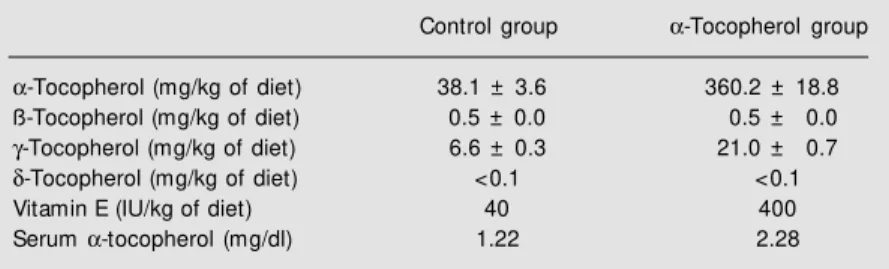
choles-Table 1. Dietary tocopherols and serum a-tocopherol of apoE-/- mice fed an athero-genic diet containing 40 IU/kg (control) or 400 IU/kg a-tocopherol acetate for 6 w eeks.
Control group a-Tocopherol group
a-Tocopherol (mg/kg of diet) 38.1 ± 3.6 360.2 ± 18.8
ß-Tocopherol (mg/kg of diet) 0.5 ± 0.0 0.5 ± 0.0
g-Tocopherol (mg/kg of diet) 6.6 ± 0.3 21.0 ± 0.7
d-Tocopherol (mg/kg of diet) <0.1 <0.1
Vitamin E (IU/kg of diet) 40 400
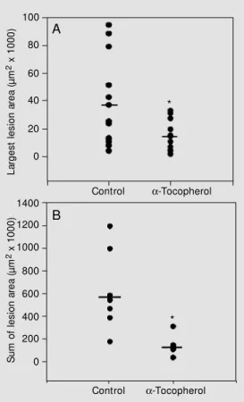
a five-fold increase in the average sum of
lesion areas when compared with the a
toco-pherol group (546,115 ± 338,545 and 110,203
± 49,293 µm2, respectively) (Figure 1).
When the average area of the largest lesion of each animal was considered, the lesion area was four-fold higher in the
con-trol group compared with the a-tocopherol
group (Figure 2).
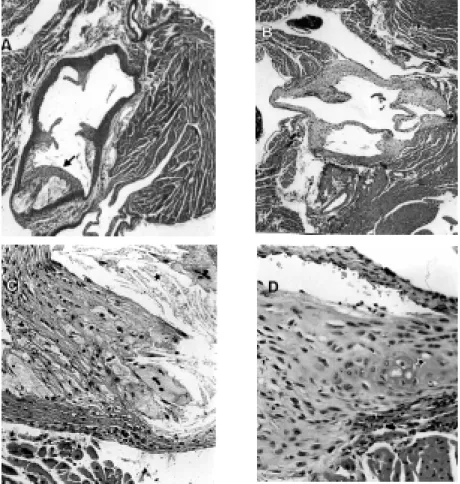
Control animals had intermediate to ad-vanced lesions with the presence of several components such as necrotic core, choles-terol clefts and fibrous cap (Figure 2A and
C). a-Tocopherol supplementation was able
to reduce the lesion area as well as calcium and cholesterol deposits and necrotic core (Figure 2B and D). The predominant lesions observed in this group were fatty streaks and intermediate lesions with foam cells
depos-ited in intima and media. Therefore, a
-toco-pherol supplementation resulted in a reduc-tion in both size and development of lesions: the necrotic core was reduced, and the cal-cium and cholesterol cleft depositions were impaired compared to control.
D iscussio n
The higher levels of a-tocopherol in
plasma of animals from the a-tocopherol
group (Table 1) confirm the efficiency of
dietary supplementation with a-tocopherol
for raising the plasma levels of vitamin E. However, the increase was not proportional to the dietary content of the vitamin. Yoshida et al. (18) isolated and characterized a toco-pherol-binding protein from cytosol of rat
liver. This protein may incorporate a
-toco-pherol in very-low-density lipoprotein (VLDL) particles (19,20). The other forms of vitamin E are similarly absorbed and reach the liver in chylomicron remnants, being
excreted into the bile with excess of a
-toco-pherol (20,21). There is a quantitative limi-tation in tocopherol-binding protein
incor-poration of a-tocopherol in VLDL that could
be explained by the inability to raise plasma Table 2. Serum and hepatic cholesterol, fecal 3-OH-a-sterols and bile acids of apoE
-/-mice fed atherogenic diets containing 40 IU/kg (control) or 400 IU/kg a-tocopherol acetate for 6 w eeks.
Control group a-Tocopherol group
Serum cholesterol (mg/100 ml)* 2593 ± 172 2274 ± 249
Hepatic cholesterol (mg/g liver)* 24.12 ± 2.65 18.74 ± 1.58
Fecal bile acids (µmol/g feces)+ 23.15 ± 7.90 11.50 ± 1.25
Fecal 3-OH-a-sterols (mg/g feces)+ 40.35 ± 3.80 47.50 ± 1.75
* M ean ± SEM of individual values (N = 18). +M ean ± SEM of 3 pooled samples.
terol in the a-tocopherol group was due to an
alteration in cholesterol catabolism. Since sitosterols are not important components of our atherogenic diet, the concentrations of
3-OH-a-sterols reflect concentrations of
cho-lesterol and its catabolic products in feces. No differences were observed in hepatic or fecal parameters (Table 2).
Atherosclerotic lesions were found in both groups. However, the control group had
L
a
rg
e
s
t
le
s
io
n
a
re
a
(
µ
m
2 x
1
0
0
0
)
100
80
60
40
20
1400
1200
1000
800
600
400
200
0
S
u
m
o
f
le
s
io
n
a
re
a
(
µ
m
2 x
1
0
0
0
)
0
Control a-Tocopherol
Control a-Tocopherol A
B Figure 1. Average of the largest
lesion area (A) or of the sum of the atherosclerotic lesion (B) in the proximal aorta of apoE -/-mice fed diets containing 40 IU/ kg (control) or 400 IU/kg a -toco-pherol acet at e f or 6 w eeks. Circles represent individual measurements and bars repre-sent the average of each group. For det ails see M at erial and M ethods. * P<0.05 compared to control group (Student t-test).
*
levels of a-tocopherol beyond two to four times even after excessive ingestion (21). The levels found in the control group (1.22 mg/dl or 28.37 µM) are similar to those reported by Praticò et al. (22) for a group that
was not supplemented with a-tocopherol
(1.29 mg/dl or 30 µM), a fact that can be explained by the similar levels of vitamin E in the diet (40 IU/kg).
After 6 weeks of supplementation with
a-tocopherol, the serum levels of
choles-terol were similar to the control animals. Although reduction of cholesterol induced by vitamin E has been described in rabbits (9,23) and C57BL/6 mice (24), many au-thors have reported no cholesterol-lowering effect of dietary vitamin E (7,10,23,25,26). These reports differ from one another in many respects such as animal models, levels of dietary vitamin E, extent of supplementa-tion, type of diets and simultaneous adminis-tration of other antioxidants - selenium, ß-carotene, and vitamin C (27-29).
Conflicting data have been reported con-cerning the effect of vitamin E on cholester-olemia in humans. Hermann et al. (30)
supple-mented human subjects with 600 IU of a
-tocopherol for 30 days; an increase in HDL-cholesterol and no change in triglycerides or total cholesterol occurred. These results were not confirmed by other investigators who used the same experimental protocol (31,32). Data of cholesterol metabolism in apo E knockout mice are scarce. Since differences in the effect of vitamin E on serum terol can be attributed to changes in terol metabolism, we investigated choles-terol excretion and storage in liver. No dif-ferences were seen in hepatic cholesterol, its fecal catabolic products or fecal bile acids between groups.
In apo E knockout mice atherosclerosis is associated with an increase of atherogenic fractions of lipoproteins and lipid peroxida-tion in plasma as well as their susceptibility to peroxidation in conditions of oxidative stress (22,33-36). Knockout mice fed a
com-mercial diet supplemented with 2000 IU of vitamin E/kg for 16 weeks had significantly lower levels of isoprostanes in urine and plasma when compared with non-supple-mented animals (22). The supplementation also significantly reduced the area of lesion in the aorta of knockout mice. Thus, it seems
that oxidative stress may be reduced by per
os administration of vitamin E to these mice.
These results demonstrate that supple-mentation with vitamin E delays the devel-opment and formation of fatty streaks in apo E-deficient animals and reinforce the
hypo-thesis that supplementation with a
Re fe re nce s
1. Keaney Jr JF & Frei B (1994). Antioxidant protection of low density lipoprotein and its role in the prevention of atheroscle-rotic vascular disease. In: Frei B (Editor),
Natural Antioxidants in Human Health and Disease. Academic Press, San Diego, CA, USA, 303-351.
2. Keaney Jr JF & Vita J (1995). Atheroscle-rosis, oxidative stress and antioxidant pro-tection in endothelium-derived relaxing factor action. Progress in Cardiovascular Diseases, 2: 129-154.
3. Batlouni M (1997). Hipótese oxidativa da aterosclerose e emprego dos antioxidan-tes na doença arterial coronária. Arquivos Brasileiros de Cardiologia, 68: 55-62. 4. Duell PB (1996). Prevention of
atheroscle-rosis w ith dietary antioxidants: fact or fic-tion? Journal of Nutrition, 126: 1067S-1071S.
5. Wiseman H (1996). Dietary influences on membrane function: importance in pro-tection against oxidative damage and dis-ease. Journal of Nutritional Biochemistry, 7: 2-15.
6. Dieber-Rotheneder M , Puhl H, Waeg G, Striegl G & Hermann E (1991). Effect of oral supplementation w ith D-a-tocopherol on the vitamin E content of human low density lipoproteins and resistance to oxi-dation. Journal of Lipid Research, 32: 1325-1332.
7. Craw ford RS, Kirk EA, Rosenfeld M E, LeBoeuf RC & Chait A (1998). Dietary antioxidants inhibit development of fatty streak lesions in the LDL receptor-defi-cient mouse. Arteriosclerosis, Thrombo-sis, and Vascular Biology, 18: 1506-1513. 8. Sun J, Giraud DW, M oxley RA & Driskell JA (1997). ß-Carotene and a-tocopherol inhibit the development of atherosclerotic lesions in hypercholesterolemic rabbits.
International Journal for Vitamin and Nu-trition Research, 67: 155-163.
9. Williams RJ, M otteram JM , Sharp CH & Gallagher PJ (1992). Dietary vitamin E and the attenuation of early lesion develop-ment in modified Watanabe rabbits.
Ath-erosclerosis, 94: 153-159.
10. Fruebis J, Carew TE & Palinski W (1995). Effect of vitamin E on atherogenesis in LDL receptor-deficient rabbits. Athero-sclerosis, 117: 217-224.
11. Plump AS, Smith JD, Hayek T, Aalto-Setälä K, Walsh A, Verstuyft JG, Rubin EM & Breslow JL (1992). Severe hyper-cholesterolemia and atherosclerosis in apolipoprotein E-deficient mice created by homologous recombination in ES cells.
Cell, 71: 343-353.
12. Piedrahita JA, Zhang SH, Hagaman JR, Oliver PM & M aeda N (1992). Generation of mice carrying a mutant apolipoprotein gene inactivated by gene targeting in em-bryonic stem cells. Proceedings of the National Academy of Sciences, USA, 89: 4471-4475.
13. Ueda T & Igarashi O (1990). Determina-tion of vitamin E in biological specimens and foods by HPLC-pretreatment of sam-ples and extraction of tocopherols. Jour-nal of M icronutrient AJour-nalysis, 7: 79-96. 14. Allain CC, Poon LS, Chan CS, Richmond
W & Fu PC (1974). Enzymatic determina-tion of total serum cholesterol. Clinical Chemistry, 20: 470-475.
15. Van der M eer R, De Vries H & Glatz JFC (1985). t-Butanol extraction of feces: a rapid procedure for enzymic determina-tion of fecal bile acids. In: Beynen AC, Geelen M JH, Katan M B & Schouten JA (Edit ors), Cholest erol M et abolism in Health and Disease: Studies in the Neth-erlands. Ponsen & Looijen, Wageningen, Netherlands, 113-137.
16. M ashige F, Tanaka N, M aki A, Kamei S & Yamanaka N (1981). Direct spectropho-tometry of total bile acids in serum. Clini-cal Chemistry, 27: 1352-1356.
17. Folch J, Lees M & Stanley S (1957). A simple method for the isolation and purifi-cation of total lipids from animal tissues.
Journal of Biological Chemistry, 226: 497-509.
18. Yoshida H, Yusin M , Ren I, Kuhlenkamp J, Hirano T, Stolz A & Kaplow itz N (1992).
Identification, purification, and immuno-chemical characterization of a tocopherol-binding protein in rat liver cytosol. Journal of Lipid Research, 33:343-350. 19. Traber M G & Packer L (1995). Vitamin E:
beyond antioxidant function. American Journal of Clinical Nutrition, 62: 1501S-1509S.
20. Dutta-Roy AK, Gordon M J, Campbell FM , Duthie GG & James WPT (1994). Vitamin E requirements, transport and metabo-lism: role of a-tocopherol-binding pro-teins. Journal of Nutritional Biochemistry, 5: 562-570.
21. Kayden HJ & Traber M G (1993). Absorp-tion, lipoprotein transport and regulation of plasma concentrations of vitamin E in humans. Journal of Lipid Research, 34: 343-358.
22. Praticò D, Tangirala RK, Rader DJ, Rokach J & FitzGerald GA (1998). Vitamin E sup-presses isoprostane generation in vivo
and reduces atherosclerosis in Apo E-de-ficient mice. Nature M edicine, 4: 1189-1192.
23. Phonpanichrasamee C, Komaratat P & Wilairat P (1990). Hypocholesterolemic effect of vitamin E on cholesterol-fed rab-bit. International Journal for Vitamin and Nutrition Research, 60: 240-244. 24. M unday JS, Thompson KG, James KAC &
M anktelow BW (1998). Dietary antioxi-dants do not reduce fatty streak forma-tion in the C57BL/6 mouse atherosclero-sis model. Arteriosclerosis, Thrombosis, and Vascular Biology, 18: 114-119. 25. Shaish A, George J, Gilburd B, Keren P,
Levkovitz H & Harats D (1999). Dietary ß-carotene and a-tocopherol combination does not inhibit atherogenesis in an Apo E-deficient mouse model. Arteriosclero-sis, ThromboArteriosclero-sis, and Vascular Biology, 19: 1470-1475.
26. W ojcicki J, Rozew icka L, Barcew -Wiszniew ska B, Samochow iec L, Juzw iak S, Kadlubow ska D, Tustanow ski S & Juzyszyn Z (1991). Effect of selenium and vitamin E on the development of experi-pherol exerts beneficial effects in terms of
prevention of atherosclerosis.
Ackno wle dgm e nts
We are grateful to Dr. Eder Quintão and
mental atherosclerosis in rabbits. Athero-sclerosis, 87: 9-16.
27. Bow ry VW & Ingold KU (1999). The unex-pected role of vitamin E (a-tocopherol) in the peroxidation of human low density lipoprotein. Archives of Chemical Re-search, 32: 27-34.
28. Neuzil J, Christison JK, Iheanacho E, Fragonas JC, Zam m it V, Hunt NH & Stocker R (1998). Radical-induced lipopro-tein and plasma lipid oxidation in normal and apolipoprot ein E gene knockout (apoE-/-) mice: apoE-/- mouse as a model for testing the role of tocopherol-medi-ated peroxidation in atherogenesis. Jour-nal of Lipid Research, 39: 354-368. 29. Thomas SR, Neuzil J, M ohr D & Stocker R
(1995). Coantioxidants make a-tocopherol
an efficient antioxidant for low density lipoprotein. American Journal of Clinical Nutrition, 62: 1357S-1364S.
30. Hermann WJ, Ward K & Faucett J (1979). The effect of tocopherol on high-density lipoprotein cholesterol. American Journal of Clinical Pathology, 72: 848-852. 31. Stampfer M J, Willet W, Castelli WP,
Tay-lor JO, Fine J & Hennekens CH (1983). Effect of vitamin E on lipids. American Journal of Clinical Pathology, 79: 714-716. 32. How ard DR, Rundell CA & Batsakis JG (1982). Vitamin E does not modify HDL-cholesterol. American Journal of Clinical Pathology, 77: 86-89.
33. Zhang SH, Reddick RL, Piedrahita JA & M aeda N (1992). Spontaneous hypercho-lesterolemia and arterial lesions in mice
lacking apolipoprotein E. Science, 258: 468-471.
34. Zhang SH, Reddick RL, Burkey B & M aeda N (1994). Diet-induced atherosclerosis in mice heterozygous and homozygous for apolipoprotein E gene disruption. Journal of Clinical Investigation, 94: 937-945. 35. Hofker M H, van Vlijmen BJM & Havekes
LM (1998). Transgenic mouse models to study the role of APOE in hyperlipidemia and atherosclerosis. Atherosclerosis, 137: 1-11.