Effect of
Lactobacillus delbrueckii
on
cholesterol metabolism in germ-free
mice and on atherogenesis in
apolipoprotein E knock-out mice
1Departamento de Bioquímica e Imunologia,
2Departamento de Patologia Geral, 3Departamento de Microbiogia,
Instituto de Ciências Biológicas, Universidade Federal de Minas Gerais, Belo Horizonte, MG, Brasil
L.R. Portugal1,
J.L. Gonçalves1,
L.R. Fernandes1,
H.P.S. Silva1,
R.M.E. Arantes2,
J.R. Nicoli3, L.Q. Vieira1
and J.I. Alvarez-Leite1
Abstract
Elevated blood cholesterol is an important risk factor associated with atherosclerosis and coronary heart disease. Several studies have re-ported a decrease in serum cholesterol during the consumption of large doses of fermented dairy products or lactobacillus strains. The proposed mechanism for this effect is the removal or assimilation of intestinal cholesterol by the bacteria, reducing cholesterol absorption. Although this effect was demonstrated in vitro, its relevance in vivo is still controversial. Furthermore, few studies have investigated the role of lactobacilli in atherogenesis. The aim of the present study was to determine the effect of Lactobacillus delbrueckii on cholesterol me-tabolism in germ-free mice and the possible hypocholesterolemic and antiatherogenic action of these bacteria using atherosclerosis-prone apolipoprotein E (apo E) knock-out (KO) mice. For this purpose, Swiss/NIH germ-free mice were monoassociated with L. delbrueckii
and fed a hypercholesterolemic diet for four weeks. In addition, apo E KO mice were fed a normalchow diet and treated with L. delbrueckii
for6 weeks. There was a reduction in cholesterol excretion in germ-free mice, which was not associated with changes in blood or liver cholesterol concentration. In apo E KO mice, no effect of L. delbrueckii
was detected in blood, liver or fecal cholesterol. The atherosclerotic lesion in the aorta was also similar in mice receiving or not these bacteria. In conclusion, these results suggest that, although L. delbrueckii treatment was able to reduce cholesterol excretion in germ-free mice, no hypocholesterolemic or antiatherogenic effect was observed in apo E KO mice.
Correspondence J.I. Alvarez-Leite Laboratório de Bioquímica Nutricional
Departamento de Bioquímica e Imunologia, ICB, UFMG Caixa Postal 486
30161-970 Belo Horizonte, MG Brasil
Fax: +55-31-3499-2614 E-mail: alvarez@ufmg.br
Research supported by FAPEMIG (Orc CBB2818/97). L.Q. Vieira, J.R. Nicoli, J.L. Gonçalves, L.R. Fernandes, and J.I. Alvarez-Leite are recipients of CNPq fellowships. H.P.S. Silva is the recipient of a PROBIC/FAPEMIG fellowship. L.R. Portugal is the recipient of a CAPES fellowship.
Received May 19, 2005 Accepted November 18, 2005
Key words
Introduction
Elevated serum cholesterol is an impor-tant risk factor associated with atherosclero-sis and coronary heart disease. Ingestion of probiotics such as lactic acid bacteria is a potential natural method to treat and prevent hypercholesterolemia.
Several studies (1-8) have reported a de-crease in serum cholesterol during the con-sumption of lactic bacteria such as Lactoba-cillus acidophilus and L. reuteri and their fermented dairy products. However, these results cannot be extrapolated to the condi-tions of human consumption.
The cholesterol-lowering potential of L. acidophilus has been extensively studied in humans. Massey (9) showed that, ini-tially, yogurt consumption significantly re-duced cholesterol by 10 to 12% in human adult males, but 2 weeks later concentra-tions returned to the control values even with continued yogurt consumption. Lin et al. (10) conducted a large double-blind, placebo-controlled trial with a crossover de-sign on 334 volunteers. The results did not show a significant effect of lactobacilli on serum cholesterol. de Roos et al. (11) found no effect of the consumption of yogurt en-riched with L. acidophilus L-1 at a dose of 1010 colony-forming units/day (CFU/day)
in a placebo-controlled study of 78 healthy men and women. Thus, the effects of lacto-bacilli on blood cholesterol are not consist-ent.
The present study investigated the effect of L. delbrueckii on total blood cho-lesterol and total lipids and on chocho-lesterol in liver and feces in germ-free mice fed a hypercholesterolemic diet. In addition, we investigated the effect of L. delbrueckii on cholesterol metabolism, on the lipopro-tein fractions and atherosclerotic lesions of the aorta in apolipoprotein E (apo E) knock-out (KO) mice that spontaneously develop hypercholesterolemia and athero-sclerosis.
Material and Methods
Animals and diet
Thirty-one 8-week-old germ-free mice fed a hypercholesterolemic diet (1% choles-terol and 0.5% cholic acid) were divided into three groups: control group (N = 10) kept in the axenic condition, EL group (N = 13) simultaneously receiving Escherichia coli EMO and L. delbrueckii in a mixture of De Man, Rogosa and Sharpe (MRS) and brain heart infusion (BHI) culture broth contain-ing 1012 CFU/mL of both bacteria, and Ec
group (N = 8) receiving 1012 CFU/mL E. coli
EMO (as a control of colonization with a bacterial strain with no hypocholesterolemic effect). The animals were kept in Trexler type isolators (Standard Safety Company, McHenry, IL, USA) in rooms on a 12-h light-dark cycle and constant temperature (22ºC). The animals had free access to ster-ile diet and water. After confirmation of cell viability, culture broth was introduced asep-tically in the isolators and given orally to the mice separately or mixed with water and food. The experimental diet was introduced only after confirmation of intestinal coloni-zation by feces culture. All animals were sacrificed under anesthesia after 4 weeks of the experiment.
To investigate atherosclerosis develop-ment, 4-week-old apo E KO mice with simi-lar body weight (about 17 g) and serum cholesterol concentrations (about 300 mg/ dL) were used. Since these animals sponta-neously develop hypercholesterolemia and atherosclerosis, they received commercial diet (Nuvital, Curitiba, PR, Brazil) ad libi-tum during the 6 weeks of the experiment. Body weight and food intake were measured weekly. The animals were divided into a control group (N = 7) receiving water and a Lac group (N = 7) receiving saline solution containing L. delbrueckii (1012 CFU/mL).
Laboratory Animals of the National Research Council (12).
Bacteria
L. delbrueckii UFV H2B20 and E. coli EMO were isolated and identified at the Federal University of Viçosa (MG, Brazil) and in the Laboratory of Microbial Ecology (INRA, Jouy-en-Josas, France), respective-ly. The strains were inoculated respectively into MRS and BHI culture media and incu-bated at 37ºC for 18 h before inoculation.
Blood, liver and feces samples. Serum lipoproteins were separated by fast protein liquid chromatography (FPLC) as previously described (13). The liver was removed, washed in saline solution, dried on filter paper, weighed, and frozen at -20ºC. Feces from the last experimental day were col-lected, homogenized and frozen at -20ºC. The hepatic and fecal lipids were extracted as described (14). Total blood cholesterol and cholesterol in FPLC fractions and in hepatic and fecal extracts were determined using commercial kits (KATAL, Belo Hori-zonte, MG, Brazil)
Histological analysis. The heart and proximal section of the aorta of apo E KO mice were removed and cleaned of adventi-tial tissue. The top half of the hearts was obtained under stereoscopic observation and fixed by immersion in 4% paraformalde-hyde in 0.1 M phosphate-buffered saline at room temperature. The specimens were rou-tinely processed for paraffin embedding and analyzed by the method of Paigen et al. (15) with modifications, as briefly described be-low. Ten-micrometer thick cross-sections were cut with a microtome (Spencer, #820, Buffalo, NY, USA) all along the aortic root area (300 µm), stained with hematoxylin-eosin, coded and examined by a single pa-thologist, who was unaware of the experi-mental conditions for each group. The slides were decoded only after the report had been written. The aortic root area was recognized
by the proximal presence of aortic valve leaflets. One of every three sections (for a total of ten sections per mouse) was kept for morphometric analysis and the images ob-tained from a microscope coupled to a digi-tal camera were processed with an image analyzer (Kontron Electronic 300 software, Fremont, CA, USA). The total lesion area of each animal was the sum of lesion areas obtained from the ten selected sections. Data are reported as mean total lesion area per group of 7 control and 7 Lactobacillus-treated animals.
Statistical analysis
Student t-test was used to compare con-trol and experimental groups, with the level of significance set at P < 0.05.
Results
Germ-free mice
To examine the effect of L. delbrueckii on cholesterol absorption and metabolism, germ-free mice or germ-free mice colonized with E. coli EMO (without a hypocholester-olemic effect) or E. coli plus L. delbrueckii were fed a hypercholesterolemic diet for 4 weeks. Feces culture confirmed the axenic status of control animals, the monoassocia-tion in the Ec group and the diassociamonoassocia-tion in the EL group until the end of experiment.
Body weight was similar in both groups, suggesting no differences in food intake or nutritional alterations caused by bacterial colonization (data not shown).
(Table 1). However, although the liver cho-lesterol level was lower in both colonized groups than in the control group, there was no difference between the Ec and EL groups, suggesting an effect of colonization but not specifically of L. delbrueckii on liver cho-lesterol. These results suggest that, although it had no effect on blood cholesterol, L.
delbrueckii may be able to reduce the
ab-sorption or increase the release of lipids from the systemic circulation.
A hypercholesterolemic diet can contami-nate the feces and consequently cause an overestimation of the fecal lipid and choles-terol concentration. For this reason, we ana-lyzed lipids and cholesterol in cecum con-tents. No differences were observed in the concentration of cecal lipids among groups (Table 1). However, the EL group showed a reduction of cecal cholesterol concentration compared to the Ec group (P = 0.047).
Apolipoprotein E knock-out mice
As germ-free Swiss/NIH mice do not de-velop spontaneous atherosclerosis, we studied the effect of L. delbrueckii in a model of atherosclerosis. Apo E KO animals fed on a regularchow diet develop spontaneous hyper-cholesterolemia and aortic atherosclerotic le-sions after the age of 8 weeks. In this experi-ment, the animals received L. delbrueckii daily to maintain the intestinal colonization.
There was no difference between groups when food and water intake and body weight were analyzed (data not shown).
Liver lipids and cholesterol were un-changed after lactobacillus supplementation (Table 2). Similarly, fecal lipids and 3-α -hydroxy sterols (as indicators of cholesterol excretion) were also similar when the Lac group was compared to control animals, sug-gesting that L. delbrueckii has no effect on cholesterol absorption and metabolism un-der these experimental conditions.
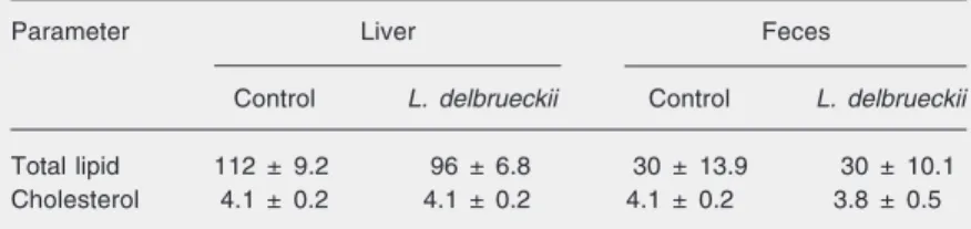
As expected, blood cholesterol was higher than in wild-type animals (about 80 mg/dL), confirming the spontaneous hypercholester-olemia occurring in these animals. However, no statistically significant differences were seen between groups at the beginning (300 mg/dL for both groups) or after 6 weeks of the experiment (356 ± 26 and 399 ± 35 mg/dL for control and L. delbrueckii-supplemented groups, respectively). These results demon-strate, as observed in germ-free mice, no hy-pocholesterolemic effect of L. delbrueckii. Likewise, lipoprotein analysis showed a simi-lar profile of atherogenic (VLDL + IDL + LDL) fractions and also of the protective (HDL) fraction for the various groups (Figure 1).
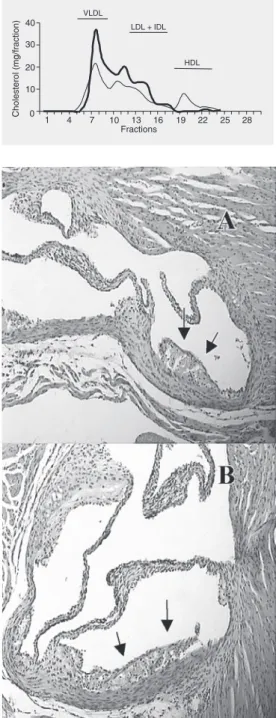
We then analyzed atherosclerotic lesions in the aorta of mice from both groups. When the aortic valve was analyzed, no reduction in lesion area was observed in mice from the Lac group (278 ± 76 x 1000 µm2) compared
to control (199 ± 95 x 1000 µm2). The
ath-erosclerotic lesions in mice of both groups showed an initial lesion (fatty streak) com-Table 1. Total lipids and cholesterol in liver and cecum of NIH mice colonized with
Escherichia coli or E. coli and Lactobacillus delbrueckii for 4 weeks or maintained axenic (control).
Control E. coli E. coli + L. delbrueckii
(N = 10) (N = 8) (N = 13)
Liver lipids 322.0 ± 4.16a 194.2 ± 21.81b 105.3 ± 5.01c Liver cholesterol 24.4 ± 2.5a 9.6 ± 0.8b 8.1 ± 1.91b
Cecum lipids ND 20.0 ± 4.08 16.6 ± 0.55
Cecum cholesterol ND 2.4 ± 0.22a 1.9 ± 0.14b
Data are reported as mean ± SEM in mg/g. ND = not determined. Different letters on the same line indicate statistically significant differences (P < 0.05, Student t-test).
Table 2. Hepatic and fecal total lipid and cholesterol of apolipoprotein E knock-out mice receiving Lactobacillus delbrueckii for 6 weeks (N = 7 mice/group).
Parameter Liver Feces
Control L. delbrueckii Control L. delbrueckii
Total lipid 112 ± 9.2 96 ± 6.8 30 ± 13.9 30 ± 10.1
Cholesterol 4.1 ± 0.2 4.1 ± 0.2 4.1 ± 0.2 3.8 ± 0.5
posed of well-defined foam cells and sparse inflammatory cells (Figure 2). Since there was no difference in blood cholesterol or in atherosclerotic lesions between groups, it was concluded that L. delbrueckii does not have a positive effect in preventing athero-sclerotic lesions under these experimental conditions.
Discussion
We investigated the effect of L.
delbru-eckii on cholesterol metabolism in two
dif-ferent models. In germ-free mice fed a hy-percholesterolemic diet, we studied the spe-cific effects of this bacterium on cholesterol absorption and liver and blood cholesterol concentrations without the interference of indigenous microbiota. However, wild-type (conventional or germ-free) mice are not the best models to study atherogenesis, since they do not develop atherosclerosis because of their high levels of the protective lipopro-tein fraction (HDL). On the other hand, apo E KO mice are good models to study athero-sclerosis, since their profile of blood choles-terol and lipoprotein fractions as well as atherosclerotic lesions are closely similar to the human (16). For this reason, we used apo E KO mice to study the influence of L.
delbrueckii specifically on atherogenesis.
Our results showed that colonization by L. delbrueckii did not reduce blood cholesterol in germ-free mice, although a reduction of cholesterol excretion was seen in monoasso-ciated animals. The reduction of fecal excre-tion may indicate a higher intestinal absorp-tion or assimilaabsorp-tion of cholesterol by bacte-ria. As the liver and blood cholesterol con-centration was not increased in our experi-ment, it is probable that L. delbrueckii as-similated cholesterol in the intestines.
It has been reported that cholesterol can be removed from culture media by precipitation with free bile acids because of the activity of the bacterial enzyme bile salt hydrolase (17). Enhanced bile salt hydrolase activity in vivo
may increase cholesterol excretion. However, this hypothesis was not confirmed by our in vivo experiment since there was a reduction of cecal cholesterol excretion. Several in vitro studies have shown that some bacteria such as lactobacilli assimilate cholesterol from cul-ture media (18-20). This indicates that certain strains could act directly on cholesterol in the gastrointestinal tract, assimilating it from the
0 10 20 30 40
1 4 7
VLDL
LDL + IDL
HDL
10 13 16 19 22 25 28
Cholesterol (mg/fraction)
Fractions
Figure 1. Lipoprotein profile of control (pool of 7 mice, fine line) or Lactobacillus delbrueckii apo-lipoprotein E knock-out mice (pool of 7 mice, bold line) after 6 weeks of the experiment (VLDL, fractions 6 to 9; LDL + IDL, frac-tions 10 to 19; HDL, fracfrac-tions 20 to 26).
intestines and this could be associated with a reduction in blood cholesterol levels. This mechanism is consistent with our results show-ing a reduction of cholesterol excretion in EL animals. However, in our experiment this ef-fect was not associated with lower blood cho-lesterol levels.
It is well known that hepatic cholesterol concentration depends on liver uptake of lipoproteins (via LDL and LRP receptors), cholesterol synthesis (via HMG CoA reduc-tase activity), and bile acid synthesis (via cholesterol 7-α-hydroxylase activity). We believe that after 4 weeks the reduction of exogenous cholesterol following L.
delbru-eckii cholesterol assimilation was
compen-sated mostly by a higher hepatic cholestero-genesis that kept liver cholesterol at levels similar to those of the control group, pre-venting a reduction of blood cholesterol.
Zacconi et al. (21) investigated the effect of L. acidophilus N5 in axenic mice and in conventionalized flora. Lactobacillus in-duced a discrete decrease of blood choles-terol in axenic mice, but had no effect in conventionalized mice. The discrepancy be-tween this result and ours may be due to the different species of lactobacillus used in each experiment (L. acidophilus and L. delbru-eckii, respectively).
Despite the large number of studies asso-ciating lactobacilli and blood cholesterol, there are no studies addressing the effect of these bacteria on atherogenesis. For this rea-son, we investigated the effect of L. delbru-eckii in apo E KO mice that spontaneously develop atherosclerosis. To the best of our knowledge, this is the first study directly correlating Lactobacillus inoculation and the development of atherosclerosis.
Contrary to the results obtained for germ-free mice, we did not detect differences in fecal cholesterol excretion in apo E KO mice. This may be due to the differences between mouse strains and diets: germ-free mice fed a hypercholesterolemic diet have a higher concentration of cholesterol in the intestines
compared to apo E KO mice fed a regular chow diet. Moreover, germ-free mice were diassociated (E. coli and L. delbrueckii) while “conventional” apo E KO mice had an indig-enous microbiota. Consequently, in germ-free mice, the effect of cholesterol assimila-tion could be more evident due to the higher cholesterol concentration in the diet and could be detected as a reduction of fecal excretion. In apo E mice, the lower intestinal concen-tration of cholesterol and the presence of other cholesterol-metabolizing bacterial strains possibly masked the effect of L. delbrueckii observed with germ-free mice.
Our main finding was that L. delbrueckii did not affect the lipoprotein profile or the development of aortic lesion. We directly showed that the atherosclerotic lesions of L. delbrueckii-colonized animals were qualita-tively and quantitaqualita-tively similar to those of control mice. It is possible that other lactoba-cillus strains have a more potent effect on cholesterol metabolism (or on other compo-nents of plaque development) and would be able to impair atherosclerosis formation. For this reason, more studies on bacterial inocula-tion and atherosclerotic lesions should be per-formed.
In conclusion, we believe that L. del-brueckii can assimilate intestinal cholesterol, but this effect did not impair atherogenesis in apo E KO mice. Since experimental mod-els of atherosclerosis are available, the study of the hypocholesterolemic effect of probiotic bacteria should be performed in these mod-els to determine both the prevention of risk factors and the reduction of atherosclerosis.
Acknowledgments
References
1. Mann GV (1974). Studies of a surfactant and cholesteremia in the Maasai. American Journal of Clinical Nutrition, 27: 464-469. 2. McNamara DJ, Lowell AM & Sabb JE (1989). Effect of yogurt intake
on plasma lipid and lipoprotein levels in normolipidemic males. Atherosclerosis, 79: 167-171.
3. Rao DR, Chawan CB & Pulusani SR (1981). Influence of milk and thermophilus milk on plasma cholesterol levels and hepatic cholesterogenesis in rats. Journal of Food Science,46: 1339-1341. 4. De Rodas BZ, Gilliland SE & Maxwell CV (1996). Hypocholesterol-emic action of Lactobacillus acidophilus ATCC 43121 and calcium in swine with hypercholesterolemia induced by diet. Journal of Dairy Science, 79: 2121-2128.
5. Akalin AS, Gonc S & Duzel S (1997). Influence of yogurt and acidophilus yogurt on serum cholesterol levels in mice. Journal of Dairy Science,80: 2721-2725.
6. Fukushima M & Nakano M (1996). Effects of a mixture of organisms, Lactobacillus acidophilus or Streptococcus faecalis on cholesterol metabolism in rats fed on a fat- and cholesterol-enriched diet. British Journal of Nutrition,76: 857-867.
7. Taranto MP, Medici M, Perdigon G et al. (2000). Effect of Lactobacil-lus reuteri on the prevention of hypercholesterolemia in mice. Jour-nal of Dairy Science, 83: 401-403.
8. Taranto MP, Medici M, Perdigon G et al. (1998). Evidence for hypocholesterolemic effect of Lactobacillus reuteri in hypercholes-terolemic mice. Journal of Dairy Science, 81: 2336-2340.
9. Massey L (1984). Effect of changing milk and yogurt consumption on human nutrient intake and serum lipoprotein. Journal of Dairy Science, 67: 255-262.
10. Lin SY, Ayres JW, Winkler Jr W et al. (1989). Lactobacillus effects on cholesterol: in vitro and in vivo results. Journal of Dairy Science, 72: 2885-2899.
11. de Roos NM, Schouten G & Katan MB (1999). Yogurt enriched with Lactobacillus acidophilus does not lower blood lipids in healthy men and women with normal to borderline high serum cholesterol levels.
European Journal of Clinical Nutrition, 53: 277-280.
12. National Research Council (1996). Guide for the Care and Use of Laboratory Animals. National Research Council, Washington, DC, USA.
13. Peluzio MC, Miguel Jr E, Drumond TC et al. (2003). Monocyte chemoattractant protein-1 involvement in the alpha-tocopherol-in-duced reduction of atherosclerosis lesions in apo E knock out mice. British Journal of Nutrition, 90: 3-11.
14. Oliveira DR, Portugal LR, Cara DC et al. (2001). Gelatin intake increases the atheroma formation in apo E knock out mice. Athero-sclerosis, 154: 71-77.
15. Paigen B, Morrow A, Holmes PA et al. (1987). Quantitative assess-ment of atherosclerosis lesions in mice. Atherosclerosis, 68: 231-240.
16. Nakashima Y, Plump S, Raines W et al. (1994). Apo E-deficient mice develop lesions of all phases of atherosclerosis throughout the arterial tree. Arteriosclerosis and Thrombosis,14: 133-140. 17. Klaver FA & van der Meer R (1993). The assumed assimilation of
cholesterol by Lactobacilli and Bifidobacterium bifidum is due to their bile salt-deconjugating activity. Applied and Environmental Microbiology,59: 1120-1124.
18. Gilliland SE (1990). Health and nutritional benefits from lactic acid bacteria. FEMS Microbiology Reviews, 7: 175-188.
19. Gilliland SE, Nelson CR & Maxwell C (1985). Assimilation of choles-terol by Lactobacillus acidophilus. Applied and Environmental Mi-crobiology, 49: 377-381.
20. Gilliland SE & Walker DK (1990). Factors to consider when selecting a culture of Lactobacillus acidophilus as a dietary adjunct to produce a hypocholesterolemic effect in humans. Journal of Dairy Science, 73: 905-911.