Tacrolimus (FK506) reduces
ischemia-induced hippocampal
damage in rats: a 7- and 30-day study
Departamento de Farmácia e Farmacologia, Centro de Ciências da Saúde, Universidade Estadual de Maringá, Maringá, PR, Brasil
F. Giordani, A. Benetolli, L.A. Favero-Filho, K.C.M. Lima, L. Cestari Junior and H. Milani
Abstract
The neuroprotective effect of the immunosuppressant agent FK506 was evaluated in rats after brain ischemia induced for 15 min in the 4-vessel occlusion model. In the first experimental series, single doses of 1.0, 3.0 or 6.0 mg FK506/kg were given intravenously (iv) immedi-ately after ischemia. In the second series, FK506 (1.0 mg/kg) was given iv at the beginning of reperfusion, followed by doses applied intraperitoneally (ip) 6, 24, 48, and 72 h post-ischemia. The same protocol was used in the third series except that all 5 doses were given iv. Damage to the hippocampal field CA1 was assessed 7 or 30 days post-ischemia on three different stereotaxic planes along the septotemporal axis of the hippocampus. Ischemia caused marked neurodegeneration on all planes (P<0.001). FK506 failed to provide neuroprotection to CA1 both when applied iv as a single dose of 1.0, 3.0 or 6.0 mg/kg (experiment 1), and after five iv injections of 1.0 mg/ kg (experiment 3). In contrast, the repeated administration of FK506 combining iv plus ip administration reduced CA1 cell death on all stereotaxic planes both 7 and 30 days post-ischemia (experiment 2; P£0.01). Compared to vehicle alone, FK506 reduced rectal tempera-ture in a dose-dependent manner (P£0.05); however, this effect did not alter normothermia (37ºC). FK506 reduced ischemic brain damage, an effect sustained over time and apparently dependent on repeated doses and on delivery route. The present data extend previous findings on the rat 4-vessel occlusion model, further supporting the possible use of FK506 in the treatment of ischemic brain damage.
Correspondence H. Milani
Departamento de Farmácia e Farmacologia, CCS, UEM Av. Colombo, 5790 87020-900 Maringá, PR Brasil
Fax: +55-44-263-6231 E-mail: [email protected]
Research supported by CNPq and UEM.
Received January 3, 2002 Accepted November 5, 2002
Key words
·Cerebral ischemia ·Hippocampal cell death ·FK506
·Sustained neuroprotection
Introduction
Humans often suffer transient, global ce-rebral ischemia resulting from cardiocircula-tory arrest or cardiopulmonary bypass sur-gery. In humans (1,2) and in experimental animals (3,4), brief interruptions of cerebral blood flow cause irreversible neuronal dam-age to vulnerable brain regions such as the
hippocampus, striatum and cortex.
of focal cerebral ischemia employing intra-cerebrally applied endothelin-1 (6). These findings have been extended to models of transient global forebrain ischemia (7-10). FK506 is a fungal-derived macrolide exhib-iting a potent immunosuppressant action, and has been recently introduced in clinical practice to prevent allograft rejection. The mechanism by which FK506 prevents is-chemic brain damage is not understood, and may depend on de novo protein synthesis (11) rather than on the inhibition of calci-neurin (11,12). Since FK506 is already per-mitted for human use, and considering that it crosses the blood-brain barrier (13), it could be a promising candidate to enter clinical trials as a neuroprotective agent.
However, data obtained from animal models of global cerebral ischemia in vivo are still limited. Three studies employing FK506 have been performed using the gerbil model of global ischemia (7-9) and one study used the rat 2-vessel occlusion + hypoten-sion model (10). The possible influence of drug-induced hypothermia in these studies is controversial. Only a single study has inves-tigated whether the neuroprotective effects of FK506 are sustained for post-ischemic periods longer than the commonly used 4-7-day survival time (7). This is an important aspect of neuroprotection since some treat-ments may merely delay, rather than prevent ischemia-induced cell loss (14).
There is a need for further investigation of the neuroprotective effects of FK506 in the rat, using the 4-vessel occlusion (4-VO) model (15). In an initial experiment, we attempted to reproduce a previous finding of robust neuroprotection in the gerbil after a single intravenous (iv) dose of FK506 (7). Given the unexpected negative results, sub-sequently we assessed the effects of FK506 applied in repeated doses, employing a com-bination of iv and intraperitoneal (ip) admin-istration. The effects of FK506 were as-sessed at two different post-ischemic sur-vival times, and evaluated in different
rostro-caudal portions of the CA1 subfield. In a third experiment, we investigated improve-ment in FK506 neuroprotection by restrict-ing the administration of repeated doses to the iv route.
Material and Methods
Animals
Adult male Wistar rats weighing 270-300 g were housed in groups of three to four in plastic cages (39 x 33 x 16 cm) at con-trolled temperature (22 ± 1ºC) on a 12-h light/dark cycle (lights on at 7:00 h) with constant air exchange. Food and water were offered ad libitum. These housing condi-tions were maintained until the end of the experiments.
Ischemia
Transient global forebrain ischemia was induced by the 4-VO method (15) with modi-fications (16). Briefly, the animals were anes-thetized with ether plus local application of 2% xylocaine, and were fixed in a stereo-taxic-like frame; the vertebral arteries were then bilaterally electrocoagulated. Subse-quently, the animals were placed in the su-pine position and the carotid arteries were carefully isolated from the adjacent tissues and loosely snared with a silk thread.
monitored but not controlled during the is-chemic period, and up to 3.5 h of reperfu-sion, using a rectal probe inserted to a depth of approximately 6 cm.
Sham-operated animals were submitted to the same manipulations, except for verte-bral and carotid occlusion. Experimental pro-cedures followed the ethical principles es-tablished by the Brazilian College of Animal Experimentation (www.meusite.com.br/ COBEA/index.htm).
Drug treatment
In the first experiment, FK506 was ad-ministered iv via the penile vein in single doses (1.0, 3.0, or 6.0 mg/kg, in a volume of 0.1 ml/100 g body weight) immediately after the beginning of reperfusion. In a second experiment, 1.0 mg FK506/kg was given iv at reperfusion (time = 0 h) followed by ip injections applied 6, 24, 48 and 72 h post-ischemia. In the third study, repeated doses of FK506 (1.0 mg/kg) were administered only by the iv route, but at the same intervals as in the second experiment. Control ani-mals received vehicle alone (0.1 ml/100 g) as single or repeated injections where appro-priate. Both FK506 (solution, 10 mg/ml am-poule) and vehicle (polyoxyethylene hydro-genated castor oil 60 and anhydrous ethanol) were kindly supplied by the Fujisawa Phar-maceutical Co., Osaka, Japan.
Histological analysis
Seven or thirty days after ischemia, the brain was removed for histological assess-ment of ischemic lesions in the hippocampus. The animals were deeply anesthetized with ether and perfused transcardiacally with 0.9% saline followed by Bouins fixative (20 ml/ min for 7-10 min). Following decapitation, the head was immersed in crushed ice (1-2ºC) for 1 h. The brain was then carefully removed and kept in Bouins fluid for 3 days.
In the first experiment (dose-response
curve), ischemic damage was assessed at a level corresponding to approximately 4.52 mm posterior to bregma (18). Eight to twelve paraffin-embedded coronal sections (5-µm thickness) were taken from each brain and stained with celestine blue/acid fuchsin. Three sections from each stereotaxic level were chosen for bilateral counts of normal-appearing neurons in the CA1 pyramidal stratum.Thus, six fields/level were counted for each rat, the number of cells in each level being expressed as the mean of the six fields. Fields were chosen by centering the 400X microscopic field laterally and close to the apex of the CA1 sectors in each respective level. The number of intact pyramidal cells with a distinct nucleus and nucleolus was counted along a transect of 450-µm length (approximately 0.160 mm2). In subsequent
experiments, the histological analysis was expanded to include two additional stereo-taxic coronal planes, which corresponded approximately to 3.60 and 5.30 mm poste-rior to bregma. The identity of the treatment groups was not revealed during histological assessment.
Statistical analysis
The Kruskal-Wallis test was used to evalu-ate the effects of FK506 on ischemia-in-duced hippocampal CA1 neurodegeneration. In the second and third experiments, a sepa-rate test was used for data obtained at each stereotaxic level. When a significant main effect appeared, Dunns multiple range test was performed to determine differences among the treatment groups. The effect of FK506 on rectal temperature was analyzed by MANOVA for repeated measures. In the case of a significant group effect, the New-man-Keuls multiple range test was used to distinguish the groups.
Results
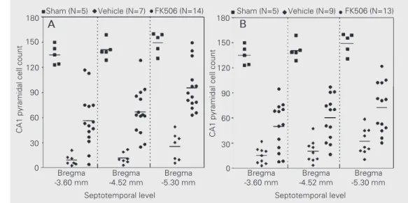
relation-utes of 4-VO caused pronounced CA1 cell loss at all coronal levels analyzed (77.4-93.2%, P<0.001, sham vs vehicle). When given repeatedly (1 iv injection + 4 ip injec-tions), FK506 reduced the degree of cell death at all levels, independently of whether the animals were sacrificed 7 or 30 days post-ischemia (P<0.01-0.001, vehicle vs FK506, Dunns multiple comparison test). In animals analyzed 30 days post-ischemia, neuroprotection was seen at the medial and temporal levels of the hippocampus (Figure 2B: P<0.05, vehicle vs FK506), but not at the most rostral level (P>0.05). The degree of neuroprotection provided by FK506 varied as a function of the septotemporal level; this differential effect was clearly evident in the 7-day post-ischemic group (Figure 2A; F1,40
= 12.36, P = 0.0011), but was reduced in the 30-day group (Figure 2B, F1,37 = 3.64, P =
0.064). Regression analysis for the vehicle-treated groups also revealed that the severity of ischemia decreased from the septal to the temporal pole of the hippocampus in both the 7-day and 30-day post-ischemic groups (Figure 2A: F1,19 = 7.75, P = 0.012; Figure
2B: F1,25 = 7.61; P<0.011). In the
sham-CA1 pyramidal cell count
180
150
120
90
60
30
0
Sham (N=5) Vehicle (N=7) FK506 (N=14) Sham (N=5) Vehicle (N=9) FK506 (N=13)
CA1 pyramidal cell count
180
150
120
90
60
30
0 Bregma
-3.60 mm
Bregma -4.52 mm
Bregma -5.30 mm
Bregma -3.60 mm
Bregma -4.52 mm
Bregma -5.30 mm
Septotemporal level Septotemporal level
A B
Figure 2. Effect of repeated iv plus
ip doses of FK506 (1.0 mg/kg) on hippocampal pyramidal CA1 cell loss caused by ischemia. The first injection (iv) was given immediately after the beginning of reperfusion. Additional ip injections were given 6, 24, 48 and 72 h post-ischemia. Histological analysis was per-formed 7 (panel A) or 30 (panel B) days post-ischemia. Fifteen min-utes of 4-vessel occlusion caused pronounced CA1 cell loss at all coro-nal levels acoro-nalyzed (P<0.001, sham
vs vehicle). FK506 reduced the de-gree of cell death in all stereotaxic planes studied, independently of whether the animals were sacri-ficed 7 or 30 days post-ischemia (P<0.01-0.001, vehicle vs FK506, Dunn’s test). The severity of
ische-mia decreased (panel A: P<0.05; panel B: P<0.011), while FK506 neuroprotection increased (panel A: P<0.005; panel B: P = 0.064) along the septotemporal axis of the hippocampus. The sham-operated rats (N = 5) are the same in panels A and B.
Figure 1. Dose-response effect of FK506 on hippocampal pyramidal CA1 cell loss caused by ischemia. FK506 (1.0, 3.0 or 6.0 mg/kg) was given iv immediately after the be-ginning of reperfusion. Hippocam-pal damage was assessed 7 days after ischemia and is expressed as a reduction in the number (mean ± SEM) of intact-appearing pyrami-dal cells. Compared to the vehicle-treated group (92.6% cell loss), FK506 did not reduce CA1 dam-age, independently of the dose used (P>0.05, Dunn’s test). Each point represents one individual; the horizontal lines indicate the mean values of the respective groups. The numbers in parentheses indi-cate sample size.
ship of FK506 on ischemia-induced CA1 cell death. Fifteen minutes of 4-VO caused marked CA1 cell loss (92.6%) (P<0.001, sham vs vehicle, Dunns multiple compari-son test). This neurodegenerative effect of ischemia was not mitigated by a single iv dose of FK506, whatever the concentration used (P>0.05, vehicle vs FK506). In con-trast, repeated doses of FK506 were effec-tive in reducing the neurodegeneraeffec-tive effect of ischemia (Figure 2A and B). Fifteen
min-CA1 pyramidal cell count
150
120
90
60
30
0 (16)
(17) (10)
(12) (12)
Sham Vehicle 1 mg/kg 3 mg/kg 6 mg/kg
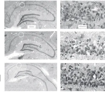
Figure 3. Photomicrographs of coronal sections of the hippo-campus at stereotaxic levels corresponding to -3.60 mm (I), -4.52 mm (II) and -5.30 mm (III) in rats subjected to sham operation (A), ischemia plus vehicle (B) or ischemia plus FK506 (C), at magnifications of 20X (left) and 400X (right). Hippocampal damage was assessed 7 days after ischemia. White circles (left panels) indicate the approximate location of the cell counts in the CA1 field. Intact-appearing pyramidal neurons are indicated by arrows. Bars = 100 µm (20X) and 20 µm (400X).
A B
C
operated group, the number of CA1 pyrami-dal neurons was unaltered among the vari-ous stereotaxic planes (linear regression: P>0.05). Representative photomicrographs of the hippocampus of rats treated with ve-hicle or FK506 are illustrated in Figure 3.
Figure 4 shows the results of repeated iv administration of FK506 (1.0 mg/kg), given 0, 6, 24, 48 and 72 h post-ischemia. As seen previously, 15 min of ischemia led to pro-nounced CA1 neurodegeneration at all septotemporal levels (P<0.01, sham vs ve-hicle, Dunns multiple comparison test), with the severity of ischemia decreasing from the
septal to the temporal pole of the hippocam-pus (F1,25 = 13.4, P = 0.0012). Repeated iv
injections of FK506 failed to reduce ische-mia-induced CA1 cell death (P>0.05) at all septotemporal levels.
protection, whatever the concentration used (1.0, 3.0, or 6.0 mg/kg). This result does not confirm a previous finding that a single (3.0 or 10.0 mg/kg) dose injected iv immediately after ischemia resulted in robust neuropro-tection of CA1 pyramidal cells (7). This discrepancy may be partially explained by the differences observed in the effect of FK506 on core temperature in each study. In the study by Ide et al. (7), FK506 caused a substantial and dose-dependent reduction in core temperature, i.e., 34º and 32ºC, respec-tively. In the present experiment, FK506 did not induce hypothermia.
Significant and sustained neuroprotec-tion was provided, however, when FK506 was given repeatedly, as a combination of iv (1x) plus ip (4x) injections. Again, although rectal temperature was reduced by up to 1ºC compared to vehicle-treated animals, this decrease was far from that at which hypo-thermia is considered to be neuroprotective, i.e., less than 33oC . Thus, the
neuroprotec-tive effect does not appear to be due to the influence of FK506 on core temperature. This finding agrees with other studies. In the gerbil, a single injection of FK506 (1.0 mg/ kg) ip did not prevent ischemia-induced CA1 cell death. In contrast, the same dose given daily for four days provided neuroprotec-tion, without hypothermia during the 24 h following FK506 administration (8,9). In rats, FK506 prevented ischemia-induced brain damage without reducing core temperature when given daily for 4 (10) or 14 days (19). In the present study, rectal temperature was recorded for up to 3.5 h post-ischemia (Fig-ure 5); we cannot rule out the possibility that hypothermia may have occurred subsequent to FK506 given 6, 24, 48 or 72 h post-ischemia.
In the present study, the neuroprotective efficacy of FK506 increased from the septal to the temporal pole of the hippocampus. In contrast, the severity of ischemia declined slightly but significantly along the same axis. This may influence the degree of
neuropro-Rectal temperature (
º
C)
38.5
38.0
37.5
37.0
36.5
Vehicle (N=42) 3 mg/kg (N=12)
1 mg/kg (N=47)
6 mg/kg (N=12)
Ischemia Reperfusion
-30 0 30 60 90 120 150 180 210
Time (min)
Figure 5. Change in rectal temperature (mean ± SEM) of rats subjected to cerebral ischemia and treated with vehicle or FK506. The groups receiving vehicle or FK506 (1 mg/kg) in the three experiments were pooled, since rectal temperature was measured under the same treatment conditions. FK506 significantly reduced rectal temperature at all doses compared to the group receiving vehicle alone (P<0.05-0.001, Newman-Keuls test). However, this effect did not alter the degree of normothermia (37-38ºC).
Figure 4. Effect of repeated doses of FK506 (1.0 mg/kg) iv on hippocampal pyramidal CA1 cell loss caused by ischemia. FK506 was given 0, 6, 24, 48 or 72 h post-ischemia. Histological anal-ysis was performed 7 days post-ischemia. Fifteen minutes of 4-vessel occlusion caused pro-nounced CA1 cell loss at all coro-nal levels acoro-nalyzed (P<0.01, sham vs vehicle). Repeated iv
injections of FK506 failed to pre-vent CA1 cell death at all coronal levels studied (P>0.05, vehicle
vs FK506, Dunn’s test).
CA1 pyramidal cell count
180
150
120
90
60
Bregma -3.60 mm
Septotemporal level 30
0
Bregma -4.52 mm
Bregma -5.30 mm Sham (N=7) Vehicle (N=9) FK506 (N=10)
Discussion
The present study demonstrates that FK506 can attenuate hippocampal damage in models of transient global forebrain ische-mia. Our data extend previous findings to the rat 4-VO model and suggest that such an effect may depend on a repeated dose proto-col.
neuro-tection by FK506 in an inverse fashion, i.e., FK506 prevented CA1 cell death most effec-tively in the region of the hippocampus least sensitive to ischemia. Similar results have been observed following hypothermia in the gerbil model (20). Differential sensitivity of CA1 pyramidal cells to ischemia, decreasing from the septal to the temporal pole of the hippocampus, has been demonstrated by oth-ers (21,22). Since sensitivity to ischemia may vary within the same brain region, the neuroprotective efficacy of drugs will not necessarily be the same in different parts of the brain, or even within the same brain structure. In this respect, our data are inter-esting given that the hippocampus is func-tionally differentiated into several parts, and that behavioral impairments following hip-pocampal lesion also vary following a septotemporal lesion gradient (23). Our ob-servations thus emphasize the importance of a multilevel histological analysis associated with behavioral measurements when assess-ing the neuroprotective efficacy of drugs in future studies.
A major question in neuroprotection re-fers to the maintenance of the neuroprotec-tive effect over time. In the present study, the neuroprotective effect of FK506 was sus-tained at least up to 30 days after the begin-ning of treatment; however, neuroprotection was less than, but not statistically different from, that measured 7 days after ischemia. Sustained neuroprotection for approximately 30 days of recovery has also been obtained with benzodiazepine-related compounds (24) and felbamate (25) in the gerbil, and after mild hypothermia in the rat (26). In contrast, the neuroprotective effect of the GABA reuptake inhibitor tiagabine is evident at 4 days, but not after 21 days of recovery (14). These findings emphasize the importance of considering different post-ischemic survival times when the neuroprotective efficacy of drugs is evaluated under conditions of cere-bral ischemia.
Despite the neuroprotective effect of
FK506 following repeated and combined iv plus ip injections (Figure 2), it must be em-phasized that some animals showed no ben-efit from FK506. Such a bimodal distribu-tion of ischemic CA1 damage has also been reported after treatment with different com-pounds, such as the AMPA receptor antnist, NBQX (27,28), the benzodiazepine ago-nist, diazepam (24,29,30), and the partial benzodiazepine agonist, imidazenil (24). Such variability in response is thought to result from a combination of at least two factors, i.e., normal variation of damage in the model, and differences in the real con-centration of the drug which reaches the brain in each individual (27). We considered this last factor in the present study, and the third experiment (Figure 4) was designed to test whether FK506 efficacy could be in-creased when all five doses were adminis-tered by the iv route alone. The rationale was that by increasing FK506 bioavailability, greater neuroprotective efficacy might be provided. Unexpectedly, however, no neu-roprotection was obtained using this sched-ule. At present, we have no explanation for this finding.
References
1. Zola-Morgan S, Squire LR & Amaral DG (1986). Human amnesia and the medial temporal region: enduring memory impairment following a bilateral lesion limited to field CA1 of the hippocampus. Journal of Neuroscience,6: 2950-2967.
2. Petito CK, Feldmann E, Pulsinelli WA & Plum F (1987). Delayed hippocampal damage in humans following cardiorespiratory arrest.
Neurology, 37: 1281-1286.
3. Pulsinelli WA, Brierley JB & Plum F (1982). Temporal profile of neuronal damage in a model of transient forebrain ischemia. Annals of Neurology, 11: 491-498.
4. Kirino T, Tamura A & Sano K (1984). Delayed neuronal death in the rat hippocampus following transient forebrain ischemia. Acta Neuro-pathologica, 64: 139-147.
5. Dawson TM, Steiner JP, Dawson VL, Dinerman JL, Uhl GR & Snyder SH (1993). Immunosuppressant FK506 enhances phosphorylation of nitric oxide synthase and protects against glutamate neurotoxic-ity. Proceedings of the National Academy of Sciences, USA, 90: 9808-9812.
6. Sharkey J & Butcher SP (1994). Immunophilins mediate the neuro-protective effects of FK506 in focal cerebral ischaemia. Nature, 371: 336-339.
7. Ide T, Morikawa E & Kirino T (1996). An immunosuppressant, FK506, protects hippocampal neurons from forebrain ischemia in the Mon-golian gerbil. Neuroscience Letters, 204: 157-160.
8. Tokime T, Nozaki K & Kikuchi H (1996). Neuroprotective effect of FK506, an immunosuppressant, on transient global ischemia in ger-bil. Neuroscience Letters, 206: 81-84.
9. Yagita Y, Kitagawa K, Matsushita K, Taguchi A, Mabuchi T, Ohtsuki T, Yanagihara T & Matsumoto M (1996). Effect of immunosuppres-sant FK506 on ischemia-induced degeneration of hippocampal neu-rons in gerbils. Life Sciences, 59: 1643-1650.
10. Drake M, Friberg H, Boris-Moller F, Sakata K & Wieloch T (1996). The immunosuppressant FK506 ameliorates ischaemic damage in the rat brain. Acta Physiologica Scandinavica, 158: 155-159. 11. Klettner A, Baumgrass R, Zhang Y, Fischer G, Burger E, Herdegen T
& Mielke K (2001). The neuroprotective actions of FK506 binding protein ligands: neuronal survival is triggered by de novo RNA syn-thesis, but is independent of inhibition of JNK and calcineurin.
Molecular Brain Research, 97: 21-31.
12. Morioka M, Fukunaga K, Kai Y, Todaka T, Yano S, Hamada J-I, Miyamoto E & Ushio Y (2001). Intravenously injected FK506 failed to inhibit hippocampal calcineurin. Biochemical and Biophysical Re-search Communications, 286: 802-806.
13. Yoshimoto T & Siesjo BK (1999). Posttreatment with the immuno-suppressant cyclosporin A in transient focal ischemia. Brain Re-search, 839: 283-291.
14. Inglefield JR, Perry JM & Schwartz RD (1995). Postischemic inhibi-tion of GABA reuptake by tiagabine slows neuronal death in the gerbil hippocampus. Hippocampus, 5: 460-468.
15. Pulsinelli WA & Brierley JB (1979). A new model of bilateral hemi-spheric ischemia in the unanesthetized rat. Stroke, 10: 267-272. 16. Milani H, Lepri ER, Giordani F& Favero-Filho LA (1999). Magnesium
chloride alone or in combination with diazepam fails to prevent hippocampal damage following transient forebrain ischemia. Brazil-ian Journal of Medical and Biological Research, 32: 1285-1293. 17. Seif el Nasr M, Nuglish J & Krieglstein J (1992). Prevention of
ischemia-induced cerebral hypothermia by controlling the environ-mental temperature. Journal of Pharmacological Methods, 27: 23-26.
18. Paxinos G & Watson C (1986). The Rat Brain in Stereotaxic Coordi-nates. 2nd edn. Academic Press, Orlando, FL, USA.
19. Wakita H, Tomimoto H, Akiguchi I & Kimura J (1998). Dose-depend-ent, protective effect of FK506 against white matter changes in the rat brain after chronic cerebral ischemia. Brain Research,792: 105-113.
20. Corbett D, Nurse S & Colbourne F (1997). Hypothermic neuroprotec-tion. A global ischemia study using 18- to 20-month-old gerbils.
Stroke, 28: 2238-2243.
21. Crain BJ, Westerkam WD, Harrison AH & Nadler JV (1988). Selec-tive neuronal death after transient forebrain ischemia in the Mongo-lian gerbil: A silver impregnation study. Neuroscience, 27: 387-402. 22. Auer RN, Jensen ML & Whishaw IQ (1989). Neurobehavioral deficit due to ischemic brain damage limited to half of the CA1 sector of the hippocampus. Journal of Neuroscience, 9: 1641-1647. 23. Moser M-B & Moser EI (1998). Functional differentiation in the
hippocampus. Hippocampus, 8: 608-619.
24. Schwartz-Bloom RD, McDonough KJ, Chase PJ, Chadwick LE, Inglefield JR & Levin ED (1998). Long-term neuroprotection by benzodiazepine full versus partial agonists after transient cerebral ischemia in the gerbil. Journal of Cerebral Blood Flow and Metabo-lism, 18: 548-558.
25. Shuaib A, Waqaar T, Ijaz MS, Kanthan R, Wishart T & Howlett W (1996). Neuroprotection with felbamate: a 7- and 28-day study in transient forebrain ischemia in gerbils. Brain Research,727: 65-70. 26. Colbourne F, Li H & Buchan AM (1999). Indefatigable CA1 sector
neuroprotection with mild hypothermia induced 6 h after severe forebrain ischemia in rats. Journal of Cerebral Blood Flow and Me-tabolism, 19: 742-749.
27. Buchan AM, Li H, Cho S & Pulsinelli WA (1991). Blockade of the AMPA receptor prevents CA1 hippocampal injury following severe but transient forebrain ischemia in adult rats. Neuroscience Letters, 132: 255-258.
28. Li H & Buchan AM (1993). Treatment with an AMPA antagonist 12 h following severe normothermic forebrain ischemia prevents CA1 neuronal injury. Journal of Cerebral Blood Flow and Metabolism, 13: 933-939.
29. Schwartz RD, Yu X, Katzman MR, Hyden-Hixson DM & Perry JM (1995). Diazepam, given postischemia, protects selectively vulner-able neurons in the hippocampus and striatum. Journal of Neurosci-ence,15: 529-539.
30. Dowden J, Reid C, Dooley P & Corbett D (1999). Diazepam-induced neuroprotection: dissociating the effects of hypothermia following global ischemia. Brain Research, 829: 1-6.
Acknowledgments
The authors gratefully acknowledge the