Research Article
Omega-3 Fatty Acids: Possible Neuroprotective Mechanisms in
the Model of Global Ischemia in Rats
Maria Elizabeth Pereira Nobre,
1Alyne Oliveira Correia,
1Francisco Nilson Maciel Mendonça,
1Luiz Ricardo Araújo Uchoa,
1Jessica Tamara Nunes Vasconcelos,
1Carlos Ney Alencar de Araújo,
1Gerly Anne de Castro Brito,
2Rafaelly Maria Pinheiro Siqueira,
2Gilberto dos Santos Cerqueira,
2Kelly Rose Tavares Neves,
2Ricardo Mário Arida,
3and Glauce Socorro de Barros Viana
1,21Faculty of Medicine, Est´acio of Juazeiro do Norte (FMJ), Rua Tenente Raimundo Rocha 515, 63040-360 Juazeiro do Norte, CE, Brazil 2Federal University of Cear´a (UFC), Rua Coronel Nunes de Melo 1127, 60430-270 Fortaleza, CE, Brazil
3Federal University of S˜ao Paulo (UNIFESP), Rua Pedro de Toledo 669, 04039-032 S˜ao Paulo, SP, Brazil
Correspondence should be addressed to Glauce Socorro de Barros Viana; [email protected]
Received 17 December 2015; Revised 1 April 2016; Accepted 4 April 2016
Academic Editor: Duo Li
Copyright © 2016 Maria Elizabeth Pereira Nobre et al. his is an open access article distributed under the Creative Commons Attribution License, which permits unrestricted use, distribution, and reproduction in any medium, provided the original work is properly cited.
Background. Omega-3 (�3) administration was shown to protect against hypoxic-ischemic injury. he objectives were to study
the neuroprotective efects of�3, in a model of global ischemia.Methods. Male Wistar rats were subjected to carotid occlusion
(30 min), followed by reperfusion. he groups were SO, untreated ischemic and ischemic treated rats with�3 (5 and 10 mg/kg, 7
days). he SO and untreated ischemic animals were orally treated with 1% cremophor and, 1 h ater the last administration, they were behaviorally tested and euthanized for neurochemical (DA, DOPAC, and NE determinations), histological (Fluoro jade staining), and immunohistochemical (TNF-alpha, COX-2 and iNOS) evaluations. he data were analyzed by ANOVA and Newman-Keuls as thepost hoctest.Results. Ischemia increased the locomotor activity and rearing behavior that were partly reversed by�3. Ischemia
decreased striatal DA and DOPAC contents and increased NE contents, efects reversed by�3. his drug protected hippocampal
neuron degeneration, as observed by Fluoro-Jade staining, and the increased immunostainings for TNF-alpha, COX-2, and iNOS
were partly or totally blocked by�3.Conclusion. his study showed a neuroprotective efect of�3, in great part due to its
anti-inlammatory properties, stimulating translational studies focusing on its use in clinic for stroke managing.
1. Background
Ischemic stroke is a pathologic condition and a major cause of death and disability worldwide. Because of its huge socioe-conomic burden and considering the global life expectancy increases, one can assume that stroke is already the most challenging disease [1]. Although animal stroke models have shed light on the pathophysiology of ischemic stroke, the translation of these results from bench to bedside has been somewhat disappointing [2, 3].
he most common cause of stroke is the sudden occlusion of a blood vessel by a thrombus or embolism, resulting
in an almost immediate loss of oxygen and glucose to the cerebral tissue. Brain is almost exclusively dependent on the continuous steady low of glucose and oxygen to undergo oxidative phosphorylation for energy production, since it has no energetic stores. Within minutes of vascular occlusion, a complex sequence of pathophysiological events called ischemic cascade occurs. he irst consequence is the depletion of substrates, particularly oxygen and glucose, that causes the accumulation of lactate via anaerobic glycolysis. Acidosis may enhance free-radical formation, interfering with intracellular protein synthesis and worsening ischemic brain injury [4, 5]. Energy failure leads to perturbation of the
Na+/K+-ATPases and Ca2+/H-ATPases pumps and, in addi-tion, the Na+-Ca2+transporter is reversed [6]. Subsequently, an alteration of ion homeostasis causes cytotoxic edema, leading to events triggered by intracellular Ca2+excess [1].
�3 fatty acids are a group of essential fatty acids that serve as energy substrates and integral membrane components and, therefore, play crucial roles in the maintenance of normal neurological functions. Recent studies show that �3 fatty acids display neuroprotective properties and may exert ben-eicial efects on cerebral ischemia and other brain disorders [7]. It is well recognized that cerebral ischemia induces exces-sive release of excitatory amino acids, as glutamate and as-partate, which provoke enzymatic processes leading to irre-versible neuronal injury [8]. On the other hand, several years ago, the behavioral efects of�3 fatty acids deiciency were proposed to be mediated through monoaminergic neurotransmission, including the dopaminergic system, what was evidenced years later [9, 10].
hus, the objectives of the present work were to study the possible neuroprotective efects of�3 fatty acids in a model of global brain ischemia in rats, focusing on behavior and striatal DA, DOPAC, and NE levels. Besides, considering that inlammation plays an important role in the pathogenesis of ischemic stroke [11, 12] and the anti-inlammatory properties of�3 [13–16] also observed by us [17], we decided to analyze the action of�3 fatty acids on proinlammatory cytokines and enzymes, in hippocampi from ischemic animals, through TTC and Fluoro-Jade staining and with immunohistochem-ical assays.
2. Material and Methods
2.1. Drugs and Reagents. Proepa (Ach´e Laborat´orios Far-macˆeuticos SA) was the source of �3 fatty acids, while sodium thiopental was from Laborat´orio Crist´alia, Brazil. Cremophor EL, 2,3,5-triphenyltetrazolium chloride (TTC), standard monoamines, and standard amino acids were from Sigma-Aldrich, USA. All other reagents were of analytical grade.
2.2. Animals. Male Wistar rats (200–250 g) were obtained from the Animal House of the Faculty of Medicine Est´acio of Juazeiro do Norte, Brazil. he animals were housed at 24± 2∘C, under a 12 h light/12 h dark cycle and had free access to a standard pellet diet (Purina chow) and tap water. hey were deprived of food for 8 h, before the experiments, except for drinking water. he animals were treated in accordance with the current law and the NIH Guide for the Care and Use of Laboratory Animals. he project was previously approved by the Animal’s Ethics Committee of the Faculty of Medicine of the Federal University of Cear´a at the city of Barbalha, Brazil.
2.3. Experimental Protocol. he animals were anesthetized with sodium thiopental (50 mg/kg, i.p.) and submitted to the model of transitory global ischemia, by the bilateral occlusion of both carotids, for 30 min. Ater reperfusion, the incision was sutured and the animals placed in individual plastic cages for recovery, with water and food ad libitum. he sham-operated groups (SO, controls) were submitted to the same
procedure, except for the clamping of the carotids. Ater surgery, the animals were orally treated (by gavage) with
�3 fatty acids, at the doses of 5 and 10 mg/kg, daily for 7 days. he SO and ischemic groups received 1% Cremophor (1 mL/kg). he animals were distributed into 4 groups (except for COX-2 immunostaining, where the�3 dose of 2.5 mg/kg was also used) as follows: SO, untreated ischemic, and ischemic groups ater treatments with�3 (5 and 10 mg/kg, p.o.). At the 7th day of treatment and 1 h ater the last drug administration, the animals were submitted to behavioral tests (open ield and water maze tests) and sacriiced for striatadissection and hippocampal slicing. hestriatawere used for homogenate preparations and DA, DOPAC, and NE determinations. Hippocampal slices were processed for immunohistochemistry assays.
2.4. Behavioral Testing
2.4.1. Open Field Test. his test is used to measure locomotor activity in rodents and can also serve to determine motor deicits and anxiety. It was originally described for the study of emotionality in rats and now is one of the most popular models in animal psychology [18]. Locomotor activity is measured by determining the amount of distance traveled and stereotyped behaviors, as rearing and grooming. he test is sensitive to motor dysfunction, as well as hippocampal and basal ganglia damage, and we used an arena (50×50×20cm) divided into four equal quadrants. Each animal was evaluated for 5 min, at the 7th day ater ischemia, and we determined the number of quadrants crossed by the animal with all 4 paws.
2.4.2. Morris Water Maze Test. his is a test of spatial learning for rodents that relies on distal cues to navigate from start locations around the perimeter of an open swimming arena for locating a submerged escape platform. he original pro-cedure was described as Morris water maze [19, 20]. Spatial learning is assessed across repeated trials and reference memory. he test has proven to be a robust and reliable one and is strongly correlated with hippocampal synaptic plasticity and NMDA receptor function. For that, we used a circular black pool (1.7 m of diameter and 1 m height), illed with water (0.59 m deep), at 25∘C temperature. he pool is divided into four quadrants and has a 10 cm diameter platform immersed 0.5 cm below the water surface. he room is provided with four cues located on North, South, East, and West of the walls. he animals were subjected to two trials, for two consecutive days (pretraining), and, 48 h ater the last trial, to the test in the water maze. he animals had a maximum time of 54 s (cut-of time) for inding the platform, where they can stay for 15 s but would have a second chance, in case of failure.
Twenty-microliter samples were then injected into a high-performance liquid chromatograph (HPLC) column. he mobile phase was 0.163 M citric acid, pH 3.0, containing 0.02 mM EDTA with 0.69 mM sodium octanesulfonic acid (SOS), as an ion pairing reagent, 4% v/v acetonitrile, and 1.7% v/v tetrahydrofuran. he monoamines were electrochemi-cally detected, using an amperometric detector (Shimadzu, Japan), by oxidation on a glassy carbon electrode at 0.85 V relative to the Ag-AgCl reference electrode. heir concentra-tions were determined by comparison with standards injected into the HPLC column at the day of experiment and the values expressed as ng/g tissue.
2.4.4. TTC Staining. TTC staining is considered a reliable method for detection of cerebral infarction in rats ater ischemia [21]. he animals were decapitated and their brains removed for TTC (2,3,5-triphenyltetrazolium chloride) stain-ing. hen, 2 mm coronal sections were obtained with the rodent brain matrix apparatus (Harvard, USA). he slices were incubated with TTC for 30 min at 37∘C in the dark and ixed by immersion in 4% bufered (7.4) formaldehyde solution for 24 h. Aterwards, the sections were photographed and ischemic areas were quantiied by the Image J sotware (NIH, USA). In those cells populations where the electron mitochondrial transport is maintained, the TTC shows a red color that is greatly decreased in brain ischemic areas.
2.4.5. Fluoro-Jade Staining. Fluoro-Jade is an anionic luores-cein derivative, used for the histological staining of neurons which become luorescent ater degeneration. Ater parain removal (by immersion in xylol), hippocampus sections (5�m) were mounted on slides surrounded by gelatin and rehydrated by immersion in ethanol for 3 min, followed by immersions in 70 and 50% ethanol solutions and distilled water. he slices were placed into a 0.06% potassium per-manganate solution, for 15 min, washed in distilled water, and transferred to a Fluoro-Jade solution where they stayed for 30 min (with gentle stirring). Ater staining, the slices were washed in distilled water (3 times, 2 min each time). he excess of water was discarded and the dry slices mounted in Fluoromountmedia and examined with a luorescence microscope. he data were quantiied by the Image J sotware (NIH, USA).
2.4.6. Immunohistochemistry Assays for TNF-Alpha, COX-2, and iNOS. Brain hippocampal sections were ixed in 10% bufered formol, for 24 h, followed by a 70% alcohol solution and embedded into parain wax for slices processing on appropriate glass slides. hese were placed into the oven at 58∘C, for 10 min, followed by deparainization in xylol, rehydration in alcohol at decreasing concentrations, and washing in distilled water and PBS (0.1 M sodium phosphate bufer, pH 7.2), for 10 min. he endogenous peroxidase was blocked with a 3% hydrogen peroxide solution, followed by incubation with the appropriate primary anti-antibody for TNF-alpha, iNOS, and COX-2, and diluted according to the manufacturers’ instructions (Santa Cruz or Millipore, USA), for 2 h, at room temperature in a moist chamber. he glass
slides were then washed with PBS (3 times, 5 min each) and incubated with the biotinylated secondary antibody, for 1 h, at room temperature in a moist chamber. hen, they were washed again in PBS and incubated with streptavidin-peroxidase, for 30 min, at room temperature (in a moist chamber) and, ater a inal wash in PBS, incubated in 0.1% DAB solution (in 3% hydrogen peroxide). Finally, the glass slides were washed in distilled water and counterstained with Mayer’s hematoxylin, washed in tap water, dehydrated in alcohol (at increasing concentrations), diaphonized in xylol, and mounted on Entelanfor optic microscopy examination. he data were quantiied by the Image J sotware (NIH, USA).
2.5. Statistical Analyses. he data are presented as means
± SEM and analyzed by one-way ANOVA, followed by Newman-Keuls test as the post hoctest. Whenever needed the data were analyzed by two-tailed unpaired Student’s � -test. he diferences were considered statistically signiicant at� < 0.05.
3. Results
3.1. Behavioral Tests
3.1.1. Open Field Test. We showed a 1.9-fold increase in locomotor activity in the untreated ischemic group, as related to the sham-operated one (SO, controls). he treatment of ischemic groups with�3 (5 and 10 mg/kg, p.o.) signiicantly reversed the locomotor activity to values close to normality (Figure 1(a)). A similar proile was seen with the rearing behavior (Figure 1(b)). hese efects were reversed ater�3 treatments and, in both cases, the values were even lower than those of the sham-operated group.
3.1.2. Morris Water Maze Test. We showed a signiicant 2-fold increase in the time to ind the platform ater ischemia, as related to controls (SO), indicating an impairment of spatial learning and hipocampal dysfunction. On the other hand, the repeated treatment of ischemic groups with �3 (5 and 10 mg/kg), for 7 days, completely reversed the efects, showing an improvement on spatial memory due to�3 treatments (Figure 2).
3.2. Neurochemical Determinations
A, B, C
D
0 5 10 15 20
N
u
m
b
er o
f cr
ossin
gs/
5
min
SO ISC ISC+ �3 (5) ISC+ �3 (10)
(a)
0 5 10 15 20
SO ISC A, B, C
N
u
m
b
er o
f r
ea
rin
g b
eha
vio
rs/
5
min
ISC+ �3 (5) ISC+ �3 (10)
(b)
Figure 1: Evaluations of the number of crossing (a) and rearing (b) behaviors by the open ield test. he columns represent means±SEM
(number of crossings/5 min) from SO and rats subjected to cerebral ischemia untreated or ater oral treatments with�3, at the doses of 5 and
10 mg/kg (number of animals per group: SO = 27; ISC = 35; ISC +�3 (5) = 14; ISC +�3 (10) = 17). (a) (A) versus SO, q =6.591∗∗∗; (B) versus
ISC +�3 (5), q =8.916∗∗∗; (C) versus ISC +�3 (10), q =8.288∗∗∗; (D) versus SO, q =3.435∗. (b) (A) versus SO, q =5.533∗∗∗; (B) versus ISC
+�3 (5), q =6.808∗∗∗; (C) versus ISC +�3 (10), q =7.246∗∗∗(one-way ANOVA and Newman-Keuls test as thepost hoctest).
A, B, C
0 10 20 30
T
ime t
o
find t
h
e p
la
tf
o
rm
(s)
SO ISC ISC+ �3 (5) ISC+ �3 (10)
Figure 2: Evaluation of the spatial memory by the water maze
test. he columns represent means±SEM of the time (s) to ind
the platform. he studied groups were SO and ischemic animals
untreated or ater oral treatments with�3, at the doses of 5 and
10 mg/kg (number of animals per group: SO = 28; ISC = 27; ISC +�3
(5) = 14; ISC +�3 (10) = 14). (A) versus SO, q =5.159∗∗; (B) versus
�3 (5), q =4.016∗; (C) versus�3 (10), q =3.897∗∗(one-way ANOVA
and Newman-Keuls test as thepost hoctest).
lower than those of the SO group, in ischemic animals ater
�3 treatments with the doses of 5 and 10 mg/kg (Figure 3(c)).
3.3. Histological and Immunohistochemistry Assays
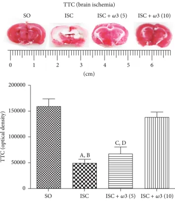
3.3.1. TTC Staining in the Hippocampus. he ischemic group (ISC) showed a 69% decrease in TTC staining as related to the SO group. his decrease was of only 13% in the ischemic group ater�3 treatment with the dose of 10 mg/kg
(ISC +�3 (10)). On the other hand, no signiicant diferences were observed in the ISC group (ISC) without and ater treatment with �3 at the dose of 5 mg/kg (ISC + �3 (5)) (Figure 4).
3.3.2. Fluoro-Jade Staining in the Hippocampus. A greater number of luorescent cells were observed in the CA1 hip-pocampal subield of the untreated ischemic group, indicat-ing neuronal degeneration. his change is quantiied by the Image J sotware as a decrease (50%) in optical density, as related to the SO group whose neurons appear darker. A similar picture was observed in the CA3 area (51% decrease) and in the dentate gyrus (56% decrease). In all cases, these alterations were completely reversed ater �3 treatments (Figure 5).
3.3.3. Immunohistochemistry for TNF-Alpha, COX-2, and iNOS in Hippocampus and Temporal Cortex. he immuno-histochemistry data for TNF-alpha showed a higher number of immunopositive cells in CA1, CA3, and dentate gyrus areas in the ischemic group (ISC) as related to the SO group. he efects were much more intense in the CA1 (313-fold increase), followed by DG (30-(313-fold increase) and CA3 (14-fold increase) areas. In all cases, these changes were signiicantly decreased ater�3 treatments (Figure 6).
A
B C
0 500 1000 1500 2000 2500 3000 3500
D
A
(n
g/g
tissue)
ISC
SO ISC+ �3 (5) ISC+ �3 (10)
(a)
A, B, C
0 500 1000 1500
D
O
PA
C (n
g/g
tissue)
SO ISC ISC+ �3 (5) ISC+ �3 (10)
(b)
A
B
C 0
200 400 600
NE (n
g/g
tissue)
SO ISC ISC+ �3 (5) ISC+ �3 (10)
(c)
Figure 3: Evaluation of�3 treatments at the doses of 5 and 10 mg/kg on the striatal contents of DA (a), DOPAC (b), and NE (c), in rats
subjected to brain ischemia and reperfusion for 7 days. he numbers of animals per group were the following: for DA, SO = 14, ISC = 9, ISC
+�3 (5) = 19, and ISC +�3 (10) = 15; for DOPAC, SO = 14, ISC = 11, ISC +�3 (5) = 15, and ISC +�3 (10) = 14; for NE, SO = 8, ISC = 10, ISC
+�3 (5) = 8, and ISC +�3 (10) = 6. he columns represent means±SEM and the groups are sham-operated (SO); ischemic (ISC) untreated;
and ischemic ater�3 treatments for 7 days. DA: (A) versus SO, q =3.830∗∗; (B) versus�3 (5), q =5.145∗∗; (C) versus�3 (10), q =4.570∗∗.
DOPAC: (A) versus SO, t = 2.484, df = 23; (B) versus�3 (5), q =4.707∗∗; (C) versus�3 (10), t = 2.085, df = 23. NE: (A) versus SO, q =16.52∗∗∗;
(B) versus�3 (2.5), q =18.79∗∗∗; (C) versus�3 (5), q =17.09∗∗∗(one-way ANOVA and Newman-Keuls test as thepost hoctest).
4. Discussion
Global cerebral ischemia in rodents is characterized mor-phologically by a selective neuronal damage, particularly in the hippocampus, but also in the striatum and cortex [22]. he resultant damage to vulnerable cells, notably in the CA1 and hilar hippocampal ields, is frequently associated with memory deicits [23]. he most common model for global cerebral ischemia in rodents uses brain ischemia associated with hypotension [24], which results in higher brain damage
mainly in the hippocampus. However, in the present work we used the common carotids occlusion for 30 min without hypotension, which has been shown to cause alterations in hippocampal CA1 neurons [25].
TTC (brain ischemia) SO ISC
1 2 3 4 5 6
0
(cm)
SO ISC A, B
C, D
T
T
C (o
p
tical den
si
ty)
0 50000 100000 150000 200000
ISC+ �3 (5) ISC+ �3 (10)
ISC+ �3 (5) ISC+ �3 (10)
Figure 4: Efects of�3 treatments in ischemic groups, visualized by the 2,3,5-triphenyltetrazolium chloride (TTC) staining as related to the
SO group. (A) versus SO, q =9.546∗∗∗; (B) versus ISC +�3 (10), q =8.194∗∗∗; (C) versus SO, q =7.894∗∗∗; (D) versus ISC +�3 (10), q =
6.452∗∗∗(one-way ANOVA and Newman-Keuls test as thepost hoctest).
A, B
A, B
C
0 100000 200000 300000
FJ
, CA3 (o
p
tical den
si
ty)
0 50000 100000 150000 200000 250000
FJ
, CA1 (o
p
tical den
si
ty)
ISC SO
SO ISC
SO ISC
FJ (CA1,×400)
SO ISC
FJ (CA3,×400)
ISC+ �3 (5) ISC+ �3 (5)
ISC+ �3 ISC+ �3
Figure 5:�3 treatments of ischemic animals (3 animals per group) drastically decreased the neuronal degeneration (visualized by an intense
luorescence to Fluoro-Jade) in hippocampus and cortex. he efects in CA1 and CA3 and DG are represented by photomicrographs and quantitative measurements performed with the Image J sotware, from 3 to 6 ields. he groups are sham-operated (SO); ischemic (ISC)
untreated, and ischemic ater�3 treatments (5 mg/kg, for 7 days). CA1: (A) versus SO, q =10.98∗∗∗; (B) versus ISC +�3, q =10.34∗∗∗. CA3:
(A) versus SO, q =14.57∗∗∗; (B) versus ISC +�3, q =20.55∗∗∗; (C) versus SO, q =5.979∗∗(one-way ANOVA and Newman-Keuls test as the
SO ISC
TNF-alpha (DG,×400) TNF-alpha (CA1,×100)
SO ISC
TNF-alpha (CA3,×100) SO ISC
ISC+ �3 (5) ISC+ �3 (10) ISC+ �3 (5) ISC+ �3 (10)
ISC+ �3 (10)
A, B, C A, B, C
D, E
0 50000 100000 150000 200000 250000
TNF
-al
p
h
a, CA3 (o
p
tical den
si
ty)
0 50000 100000 150000 200000 250000
TNF
-al
p
h
a, CA1 (o
p
tical den
si
ty)
ISC SO
ISC
SO ISC+ �3 (5) ISC+ �3 (10) ISC+ �3 (5) ISC+ �3 (10)
A, B
0 50000 100000 150000 200000
TNF
-al
p
h
a, D
G
(o
p
tical den
si
ty)
ISC
SO ISC+ �3 (10)
Figure 6:�3 treatments of ischemic animals (3 animals per group) reduced the immunostaining for TNF-alpha, mainly in CA1 and CA3
hippocampal subields and in the dentate gyrus (DG). Representative photomicrographs and quantitative measurements performed with the
Image J sotware from 3 to 5 ields. he groups are SO, untreated ischemic (ISC) and ischemic ater�3 treatments (5 and 10 mg/kg, 7 days).
CA1: (A) versus SO, q =41.76∗∗∗; (B) versus ISC +�3 (5), q =40.44∗∗∗; (C) versus ISC +�3 (10), q =40.63∗∗∗. CA3: (A) versus SO, q =
32.86∗∗∗; (B) versus ISC +�3 (5), q =7.494∗∗∗; (C) versus ISC +�3 (10), q =33.31∗∗∗; (D) versus SO, q =7.494∗∗∗; (E) versus ISC +�3 (10),
q =23.91∗∗∗. DG: (A) versus SO, q =30.26∗∗∗; (B) versus ISC +�3 (10), q =27.34∗∗∗(one-way ANOVA and Newman-Keuls test as thepost
DG
Hilus
COX-2(×100)
A, B, C
D
E
A, B, C
D E
0 20000 40000 60000 80000
C
O
X, hil
u
s (o
p
tical den
si
ty)
0 20000 40000 60000
C
O
X-2, D
G
(o
p
tical den
si
ty)
ISC
SO ISC+ �3 (2.5) ISC+ �3 (10) SO ISC ISC+ �3 (2.5) ISC+ �3 (10)
Figure 7:�3 treatments of ischemic animals (3 animals per group) reduced the immunostaining for COX-2, mainly in the dentate gyrus
(DG) and hilus. Representative photomicrographs and quantitative measurements performed with the Image J sotware, from 3 to 5 ields.
he groups are untreated ischemic (ISC) and ischemic ater treatments with�3 (2.5 and 10 mg/kg, 7 days). DG: (A) versus SO, q =14.88∗∗∗;
(B) versus ISC +�3 (2.5), q =7.135∗∗∗; (C) versus ISC +�3 (10), q =4.979∗∗. Hilus: (A) versus SO, q =13.95∗∗∗; (B) versus ISC +�3
(2.5), q =7.418∗∗∗; (C) versus ISC +�3 (10), q =6.943∗∗∗; (D) versus SO, q =6.527∗∗∗; (E) versus SO, q =7.003∗∗∗(one-way ANOVA and
Newman-Keuls test as thepost hoctest).
chain polyunsaturated fatty acid of the�3 series, is accom-panied by anxiety and learning and memory impairments that have been associated with changes in neurotransmission processes [10].
Most of the literature data deal with animals chronically submitted to �3 supplemented diet [29–33] or �3 high dose, as seen in preclinical [34, 35] and clinical studies as well [36]. As far as we know, ours is the irst study dealing with �3 administration for a short period and at a lower dose range. We focused on behavioral changes, determinations of striatal monoamines, and immunohisto-chemistry assays in the hippocampus of animals submitted
to global brain ischemia untreated or treated with�3 fatty acids.
A, B
C
0 50000 100000 150000 200000 250000
iN
OS (o
p
tical den
si
ty)
ISC SO
iNOS (CA3,×100)
SO ISC ISC+ �3 (5)
ISC+ �3 (5)
Figure 8:�3 treatments (3 animals per group) reduced the immunostaining for iNOS mainly in the CA3 hippocampal subield. Representative
photomicrographs and measurements performed with the Image J sotware, from 3 to 5 ields. he groups are sham-operated (SO), untreated
ischemic (ISC), and ischemic ater�3 treatment (5 mg/kg, 7 days). (A) versus SO, q =16.03∗∗∗; (B) versus ISC +�3 (5), q =19.49∗∗∗; (C)
versus SO, q =3.456∗(one-way ANOVA and Newman-Keuls test as thepost hoctest).
doses of�3 was clearly observed by us ater the TTC staining, a reliable method for the detection of brain ischemic areas in the rat [21].
In the open ield test, we showed that ater global brain ischemia the animals presented not only an increased locomotor activity, but also an increased number of rearing behaviors, as related to controls (SO). On the other hand, the treatment of ischemic groups with�3 signiicantly decreased all these behavioral parameters to values close to those of controls. Brain ischemia is known to increase locomotor activity and anxiety [43–47] and the degree of hippocampal damage has been positively correlated with the increase in motor activity.
Our data showed that brain ischemia signiicantly impaired spatial memory, and this efect was completely reversed ater�3 treatments. Evidences indicate that tran-sient global ischemia may lead to severe impairments in learning and memory [48–50]. he pyramidal CA1 neu-rons of the hippocampus are critically involved in spatial learning and memory and are also especially vulnerable to cerebral ischemia. he transient global cerebral ischemia model results in neurodegeneration, known to induce both hippocampal neuronal loss and learning deicits in rats [48, 50, 51]. Reduction of memory deicits by PUFA, ater global ischemia, has been already observed by others [30, 34, 52]. However, in most of these data, the animals were chronically subjected to a rich-in-�3 fatty acids diet, while in the present study the animals were orally given lower doses of�3, for a short period of time.
We also showed a 37% reduction in DA levels in the striata of ischemic rats, as related to the SO group. On the other hand, these efects were completely reverted
ater �3 treatments and a similar picture was noticed in DOPAC levels. Ischemic conditions can induce the release of brain neurotransmitters. Earlier studies showed that the DA metabolism is signiicantly altered during and ater ischemia in the rabbit retina [53]. On the other hand, biochemical consequences of ischemia in the hippocampus, cortex, and striatum have been object of much attention, because these brain structures are particularly vulnerable to ischemia [54]. he extracellular levels of DA have been shown to increase drastically, immediately ater ischemia. his increase is fol-lowed by a great decrease, probably relecting that the release of DA is not compensated by an increased synthesis of this neurotransmitter [55–60].
Evidences indicate that DA release exerts an important role in the striatal damage observed ater ischemia. As a matter of fact, the depletion of DA by alpha-methyltyrosine or 6-OHDA before the ischemic process reduces the degree of neuronal damage [61–63], using an experimental model similar to ours, showing by microdialysis that the extracel-lular concentrations of DA increased abruptly, around 3 min ater the ischemic insult, reaching a maximum value between 20 and 40 min ater the insult and decreasing subsequently. hese authors concluded that, during ischemia, the great increase in DA concentrations occurs probably as a result of energy failure in cellular membranes. hen, the DA release could be the cause of the neuronal damage, associated with brain ischemia.
Others [7] showed that�3 fatty acids present neuroprotective properties, exerting beneits on cognitive functions, neurode-generative diseases, and brain ischemia.
Evidences [64] have indicated the efects of a diet which was deicient in alpha-linolenic acid, precursor of long chains unsaturated fatty acids as �3, on the dopaminergic neurotransmission in the core of the ratnucleus accumbens. hese in vivo microdialysis experiments showed increased basal levels of dopamine and their metabolites (DOPAC and HVA), in rats subjected to a fatty-acid-deicient diet, as com-pared to controls, indicating alterations in the dopaminergic neurotransmission in thenucleus accumbens.
For the assessment of neuronal degeneration, we used the staining with Fluoro-Jade which is a luorescein derivative that speciically binds to degenerating neurons. Fluoro-Jade stains cell bodies, dendrites, axons, and their terminals but does not stain healthy neurons [65]. We showed a high number of degenerating neurons in the CA1, CA3,dentate gyrus, and temporal cortex from untreated ischemic rats, and this proile was completely reversed ater �3 treat-ments, towards that of SO (controls), indicating the drug neuroprotective efects. Interestingly, Fluoro-Jade staining demonstrating neuronal damage was shown in some brain areas of animals submitted to PUFAs dietary restriction [66], corroborating with our indings.
While PUFAs are known to present brain immunomod-ulatory properties, n-3 PUFAs are able to reduce inlamma-tion and n-6 PUFAs are more proinlammatory [67]. hese authors showed that the central n-3 PUFA increase modulates the brain innate immune system, leading to the protection against LPS-induced proinlammatory cytokine production and spatial memory impairment, in transgenic mice. Recently [68],�3 was shown to reduce cytokine-induced expression of proatherogenic and proinlammatory proteins, in human endothelial cells, and these properties may contribute to the antiatherogenic and anti-inlammatory efects of n-3 PUFAs. In the present study, the treatment of ischemic animals with �3 drastically decreased immunoreactivity for TNF-alpha in the hippocampus and temporal cortex. A previous work [69] demonstrated that�3 pretreatments signiicantly attenuated LPS-stimulated TNF-alpha production. Others [70] showed that the inhibition by�3 of proinlammatory cytokines as TNF-alpha, in murine macrophages, is in part mediated by the inactivation of NF-kappaB signaling. Besides, Nielsen et al., 2005 [71], observed that �3 fatty acids were able to inhibit the increase in proinlammatory cytokines, in patients with Crohn’s disease. �3 fatty acids are known to afect immune response, partly by inluencing cytokine secretion [72].
Furthermore, we also showed that the treatments with�3 of ischemic animals signiicantly decreased COX-2 and iNOS immunoreactivity in hippocampal brain areas. Evidences indicate that�3 fatty acids dampen inlammation, through multiple pathways, and directly or indirectly suppress the activity of nuclear transcription factors, as NF-kappaB, and reduce the production of proinlammatory enzymes and cytokines, including COX-2, Talpha, and IL-1B [73]. NF-kappaB is one of the most important transcription factors involved in inlammatory response and upregulation of gene
encoding of inlammatory cytokines, adhesion molecules, and COX-2. Evidences indicated that�3 decreases expression of adhesion molecules and production of inlammatory cytokines and COX-2 metabolites, and a common mecha-nism would be the impact on the NFkB system [74]. his could well be the way�3 is acting in our study.
Besides decreasing COX-2 immunostaining,�3 attenu-ates hippocampal iNOS immunoreactivity. It has been shown that a diet rich in�3 fatty acids decreases eNOS and iNOS in the diabetic kidney [75]. Interestingly, the attenuation of iNOS in LPS-stimulated macrophages by�3 was shown to be independent of COX-2 derived PGE2, contradicting the hypothesis that the decrease in NO production associated with�3 treatments occurs through a COX-2 derived PGE2 dependent mechanism [76]. he expression of iNOS is one direct consequence of inlammatory processes [77]. One important step during inlammation is leukocyte iniltration, mainly controlled by chemokines. he production of these chemokines is positively or negatively regulated by iNOS-derived NO [78]. Furthermore, a clinical study demonstrated that ish oil decreased serum levels of TNF-alpha, IL-1beta, IL-6, and nitric oxide metabolites in multiple sclerosis patients treated with IFN-1beta [79].
5. Conclusion
�3 fatty acids by their anti-inlammatory and antioxidant properties that contribute to the neuroprotective property might be a therapeutic target to be pursued for the prevention or treatment of inlammation-related diseases.�3 deiciency increases the brain’s vulnerability, representing an important risk factor for the development of neuropathologies [80]. Fur-thermore, understanding the precise roles of�3 fatty acids, as a possible disease modiier, will permit the development of new therapies for neurological diseases.
Competing Interests
he authors declare no conlict of interests.
Authors’ Contributions
Maria Elizabeth Pereira Nobre supervised the experimental protocol; Alyne Oliveira Correia, Francisco Nilson Maciel Mendonc¸a, and Luiz Ricardo Ara´ujo Uchoa performed the brain ischemia experiments; Jessica Tamara Nunes Vascon-celos and Carlos Ney Alencar de Ara´ujo carried out the daily animals treatments; Gerly Anne de Castro Brito supervised the immunohistochemical assays; Rafaelly Maria Pinheiro Siqueira, Gilberto dos Santos Cerqueira, and Kelly Rose Tavares Neves performed the immunohistochemical assays; Ricardo M´ario Arida and Glauce Socorro de Barros Viana were responsible for the design, statistical analysis, and drat of the paper. All authors read and approved the inal version of the paper.
Acknowledgments
(FUNCAP) of the Cear´a State, Brazil, for inancial support; Maria Vilani Rodrigues Bastos for her technical assistance; and Professor M. O. L. Viana for the orthographic revision of the paper.
References
[1] A. Durukan and T. Tatlisumak, “Acute ischemic stroke: over-view of major experimental rodent models, pathophysiology,
and therapy of focal cerebral ischemia,”Pharmacology
Biochem-istry and Behavior, vol. 87, no. 1, pp. 179–197, 2007.
[2] S. Braeuninger and C. Kleinschnitz, “Rodent models of focal cerebral ischemia: procedural pitfalls and translational
prob-lems,”Experimental & Translational Stroke Medicine, vol. 1, pp.
1–11, 2009.
[3] M. Bacigaluppi, G. Comi, and D. M. Hermann, “Animal models
of ischemic stroke. part two: modeling cerebral ischemia,”he
Open Neurology Journal, vol. 4, pp. 34–38, 2010.
[4] Y. Huang and J. O. McNamara, “Ischemic stroke: ‘Acidotoxicity’
is a perpetrator,”Cell, vol. 118, no. 6, pp. 665–666, 2004.
[5] P. Mergenthaler, U. Dirnagl, and A. Meisel, “Pathophysiology of
stroke: lessons from animal models,”Metabolic Brain Disease,
vol. 19, no. 3-4, pp. 151–167, 2004.
[6] T. G. Phan, P. M. Wright, R. Markus, D. W. Howells, S. M. Davis, and G. A. Donnan, “Salvaging the ischaemic penumbra: more
than just reperfusion?”Clinical and Experimental
Pharmacol-ogy and PhysiolPharmacol-ogy, vol. 29, no. 1-2, pp. 1–10, 2002.
[7] W. Zhang, P. Li, X. Hu, F. Zhang, J. Chen, and Y. Gao, “Omega-3 polyunsaturated fatty acids in the brain: metabolism and
neuroprotection,”Frontiers in Bioscience, vol. 16, no. 7, pp. 2653–
2670, 2011.
[8] J. W. Phillis and M. H. O’Regan, “Characterization of modes of release of amino acids in the ischemic/reperfused rat cerebral
cortex,”Neurochemistry International, vol. 43, no. 4-5, pp. 461–
467, 2003.
[9] S. Chalon, S. Delion-Vancassel, C. Belzung et al., “Dietary ish oil afects monoaminergic neurotransmission and behavior in
rats,”Journal of Nutrition, vol. 128, no. 12, pp. 2512–2519, 1998.
[10] S. Chalon, “Omega-3 fatty acids and monoamine
neurotrans-mission,”Prostaglandins Leukotrienes and Essential Fatty Acids,
vol. 75, no. 4-5, pp. 259–269, 2006.
[11] C. J. S. Price, E. A. Warburton, and D. K. Menon, “Human cellu-lar inlammation in the pathology of acute cerebral ischaemia,”
Journal of Neurology, Neurosurgery and Psychiatry, vol. 74, no. 11, pp. 1476–1484, 2003.
[12] R. Jin, G. Yang, and G. Li, “Inlammatory mechanisms in
ischemic stroke: role of inlammatory cells,”Journal of
Leuko-cyte Biology, vol. 87, no. 5, pp. 779–789, 2010.
[13] P. C. Calder, “Dietary modiication of inlammation with lipids,”
Proceedings of the Nutrition Society, vol. 61, no. 3, pp. 345–358, 2002.
[14] A. P. Simopoulos, “Omega-3 fatty acids in inlammation and
autoimmune diseases,” Journal of the American College of
Nutrition, vol. 21, no. 6, pp. 495–505, 2002.
[15] P. C. Calder, “Omega-3 fatty acids and inlammatory processes,”
Nutrients, vol. 2, no. 3, pp. 355–374, 2010.
[16] P. C. Calder, “Fatty acids and inlammation: the cutting edge
between food and pharma,”European Journal of Pharmacology,
vol. 668, no. 1, pp. S50–S58, 2011.
[17] M. E. P. Nobre, A. O. Correia, M. B. Borges et al., “Eicosapen-taenoic acid and docosahexaenoic acid exert anti-inlammatory
and antinociceptive efects in rodents at low doses,”Nutrition
Research, vol. 34, pp. 251–263, 2013.
[18] L. Prut and C. Belzung, “he open ield as a paradigm to mea-sure the efects of drugs on anxiety-like behaviors: a review,”
European Journal of Pharmacology, vol. 463, no. 1–3, pp. 3–33, 2003.
[19] R. G. M. Morris, “Spatial localization does not require the
pres-ence of local cues,”Learning and Motivation, vol. 12, no. 2, pp.
239–260, 1981.
[20] C. V. Vorhees and M. T. Williams, “Morris water maze: pro-cedures for assessing spatial and related forms of learning and
memory,”Nature Protocols, vol. 1, no. 2, pp. 848–858, 2006.
[21] J. B. Bederson, L. H. Pitts, S. M. Germano, M. C. Nishimura, R. L. Davis, and H. M. Bartkowski, “Evaluation of 2,3,5-triphenyl-tetrazolium chloride as a stain for detection and quantiication
of experimental cerebral infarction in rats,”Stroke, vol. 17, no. 6,
pp. 1304–1308, 1986.
[22] F. Block, “Global ischemia and behavioural deicits,”Progress in
Neurobiology, vol. 58, no. 3, pp. 279–295, 1999.
[23] H. Hodges, A. Nelson, D. Virley, T. R. Kershaw, and J. D. Sin-den, “Cognitive deicits induced by global cerebral ischaemia:
prospects for transplant therapy,”Pharmacology Biochemistry
and Behavior, vol. 56, no. 4, pp. 763–780, 1997.
[24] W. A. Pulsinelli, J. B. Brierley, and F. Plum, “Temporal proile of neuronal damage in a model of transient forebrain ischemia,”
Annals of Neurology, vol. 11, no. 5, pp. 491–498, 1982.
[25] M. A. Atlasi, H. Naderian, M. Noureddini, E. Fakharian, and A. Azami, “he morphology of rat hippocampus CA1 neurons following modiied two and four vessel global ischemia models,”
Archives of Trauma Research, vol. 2, no. 3, pp. 124–128, 2013. [26] I. Fedorova and N. Salem Jr., “Omega-3 fatty acids and rodent
behavior,”Prostaglandins Leukotrienes and Essential Fatty Acids,
vol. 75, no. 4-5, pp. 271–289, 2006.
[27] T. Takeuchi, M. Iwanaga, and E. Harada, “Possible regulatory
mechanism of DHA-induced anti-stress reaction in rats,”Brain
Research, vol. 964, no. 1, pp. 136–143, 2003.
[28] A. Harauma and T. Moriguchi, “Dietary n-3 fatty acid deiciency in mice enhances anxiety induced by chronic mild stress,”
Lipids, vol. 46, no. 5, pp. 409–416, 2011.
[29] N. Vinot, M. Jouin, A. Lhomme-Duchadeuil et al., “Omega-3 fatty acids from ish oil lower anxiety, improve cognitive functions and reduce spontaneous locomotor activity in a
non-human primate,”PLoS ONE, vol. 6, no. 6, Article ID e20491,
2011.
[30] H. Plamondon and M.-C. Roberge, “Dietary PUFA supple-ments reduce memory deicits but not CA1 ischemic injury in
rats,”Physiology and Behavior, vol. 95, no. 3, pp. 492–500, 2008.
[31] H.-M. Su, “Mechanisms of n-3 fatty acid-mediated development
and maintenance of learning memory performance,”Journal of
Nutritional Biochemistry, vol. 21, no. 5, pp. 364–373, 2010. [32] M. Hashimoto, S. Hossain, Y. Tanabe et al., “he protective efect
of dietary eicosapentaenoic acid against impairment of spatial
cognition learning ability in rats infused with amyloid�(1–40),”
Journal of Nutritional Biochemistry, vol. 20, no. 12, pp. 965–973, 2009.
[33] S. C. Heinrichs, “Dietary�3 fatty acid supplementation for
opti-mizing neuronal structure and function,”Molecular Nutrition
and Food Research, vol. 54, no. 4, pp. 447–456, 2010.
transient, global cerebral ischemia,”Nutrition Research, vol. 28, no. 11, pp. 798–808, 2008.
[35] N. Okabe, T. Nakamura, T. Toyoshima, O. Miyamoto, F. Lu, and T. Itano, “Eicosapentaenoic acid prevents memory impairment ater ischemia by inhibiting inlammatory response and
oxida-tive damage,”Journal of Stroke and Cerebrovascular Diseases,
vol. 20, no. 3, pp. 188–195, 2011.
[36] N. F. A. Sopian, M. Ajat, N. I. Shaie et al., “Does short-term dietary omega-3 fatty acid supplementation inluence brain hippocampus gene expression of zinc transporter-3?”
International Journal of Molecular Sciences, vol. 16, no. 7, pp. 15800–15810, 2015.
[37] L. G. Pusk´as, K. Kitajka, C. Nyakas, G. Barcelo-Coblijn, and T. Farkas, “Short-term administration of omega 3 fatty acids from ish oil results in increased transthyretin transcription in old rat
hippocampus,”Proceedings of the National Academy of Sciences
of the United States of America, vol. 100, no. 4, pp. 1580–1585, 2003.
[38] W. Zhang, X. Hu, W. Yang, Y. Gao, and J. Chen, “Omega-3 polyunsaturated fatty acid supplementation confers long-term neuroprotection against neonatal hypoxic-ischemic brain
injury through anti-inlammatory actions,”Stroke, vol. 41, no.
10, pp. 2341–2347, 2010.
[39] X. Tang, Z.-J. Li, J. Xu et al., “Short term efects of diferent omega-3 fatty acid formulation on lipid metabolism in mice fed
high or low fat diet,”Lipids in Health and Disease, vol. 11, article
70, 2012.
[40] M. Ajami, S. Eghtesadi, R. Habibey et al., “Efect of short- and long-term treatment with Omega-3 fatty acids on
scopolamine-induced amn´esia,”Iranian Journal of Pharmaceutical Research,
vol. 11, no. 2, pp. 533–540, 2012.
[41] S. K. Raatz, J. B. Redmon, N. Wimmergren, J. V. Donadio, and D. M. Bibus, “Enhanced absorption of n-3 fatty acids from
emulsiied compared with encapsulated ish oil,”Journal of the
American Dietetic Association, vol. 109, no. 6, pp. 1076–1081, 2009.
[42] K. Yurko-Mauro, D. McCarthy, D. Rom et al., “Beneicial efects of docosahexaenoic acid on cognition in age-related cognitive
decline,”Alzheimer’s and Dementia, vol. 6, no. 6, pp. 456–464,
2010.
[43] S. C. Gerhardt and C. A. Boast, “Motor activity changes follow-ing cerebral ischemia in gerbils are correlated with the degree
of neuronal degeneration in hippocampus,”Behavioral
Neuro-science, vol. 102, no. 2, pp. 301–303, 1988.
[44] Y. Karasawa, H. Araki, and S. Otomo, “Changes in locomotor activity and passive avoidance task performance induced by
cerebral ischemia in mongolian gerbils,”Stroke, vol. 25, no. 3,
pp. 645–650, 1994.
[45] M. Milot and H. Plamondon, “Ischemia-induced hyperactiv-ity: efects of dim versus bright illumination on open-ield exploration and habituation following global ischemia in rats,”
Behavioural Brain Research, vol. 192, no. 2, pp. 166–172, 2008. [46] M. R. Milot and H. Plamondon, “Time-dependent efects of
global cerebral ischemia on anxiety, locomotion, and
habitua-tion in rats,”Behavioural Brain Research, vol. 200, no. 1, pp. 173–
180, 2009.
[47] T. G. Ohk, K.-Y. Yoo, S. M. Park et al., “Neuronal damage using luoro-jade B histoluorescence and gliosis in the striatum ater various durations of transient cerebral ischemia in gerbils,”
Neurochemical Research, vol. 37, no. 4, pp. 826–834, 2012. [48] R. E. Hartman, J. M. Lee, G. J. Zipfel, and D. F. Wozniak,
“Characterizing learning deicits and hippocampal neuron loss
following transient global cerebral ischemia in rats,” Brain
Research, vol. 1043, no. 1-2, pp. 48–56, 2005.
[49] M. Von Euler, O. Bendel, T. Bueters, J. Sandin, and G. Von Euler, “Profound but transient deicits in learning and memory
ater global ischemia using a novel water maze test,”Behavioural
Brain Research, vol. 166, no. 2, pp. 204–210, 2006.
[50] K. D. Langdon, S. Granter-Button, and D. Corbett, “Persis-tent behavioral impairments and neuroinlammation following
global ischemia in the rat,”European Journal of Neuroscience,
vol. 28, no. 11, pp. 2310–2318, 2008.
[51] T. Bueters, M. von Euler, O. Bendel, and G. von Euler, “Degen-eration of newly formed CA1 neurons following global ischemia
in the rat,”Experimental Neurology, vol. 209, no. 1, pp. 114–124,
2008.
[52] C. Luo, H. Ren, J.-B. Wan et al., “Enriched endogenous omega-3 fatty acids in mice protect against global ischemia injury,”
Journal of Lipid Research, vol. 55, no. 7, pp. 1288–1297, 2014. [53] W. Cao, A. Drumheller, M. Zaharia, G. Lafond, J.-R. Brunette,
and F. B. Jolicoeur, “Efects of experimentally induced ischemia
on dopamine metabolism in rabbit retina,”Investigative
Oph-thalmology & Visual Science, vol. 34, no. 11, pp. 3140–3146, 1993. [54] W. A. Pulsinelli and T. E. Dufy, “Regional energy balance in rat
brain ater transient forebrain ischemia,”Journal of
Neurochem-istry, vol. 40, no. 5, pp. 1500–1503, 1983.
[55] L. A. Phebus and J. A. Clemens, “Efects of transient, global, cerebral ischemia on striatal extracellular dopamine, serotonin
and their metabolites,”Life Sciences, vol. 44, no. 19, pp. 1335–
1342, 1989.
[56] M. Y.-T. Globus, “Role of dopamine in ischemic neuronal
damage,”Stroke, vol. 20, no. 6, pp. 827–828, 1989.
[57] T. Kawano, K. Tsutsumi, H. Miyake, and K. Mori, “Striatal dopamine in acute cerebral ischemia of stroke-resistant rats,”
Stroke, vol. 19, no. 12, pp. 1540–1543, 1988.
[58] A. Slivka, T. S. Brannan, J. Weinberger, P. J. Knott, and G. Cohen, “Increase in extracellular dopamine in the striatum during cerebral ischemia: a study utilizing cerebral microdialysis,”
Journal of Neurochemistry, vol. 50, no. 6, pp. 1714–1718, 1988. [59] A. Kuruvilla, R. Cherian, D. R. heodore, and J. Abraham,
“Temporal proile of tissue levels of dopamine and its metabo-lites, HVA and DOPAC following focal cerebral ischaemia in
anaesthetized primates,”Clinical and Experimental
Pharmacol-ogy and PhysiolPharmacol-ogy, vol. 13, no. 7, pp. 519–524, 1986.
[60] K. Ogura, M. Shibuya, Y. Suzuki, M. Kanamori, and I. Ikegaki, “Changes in striatal dopamine metabolism measured by in vivo
voltammetry during transient brain ischemia in rats,”Stroke,
vol. 20, no. 6, pp. 783–787, 1989.
[61] T. Brannan, J. Weinberger, P. Knott et al., “Direct evidence of acute, massive striatal dopamine release in gerbils with
unilateral strokes,”Stroke, vol. 18, no. 1, pp. 108–110, 1987.
[62] J. Weinberger, J. Nieves-Rosa, and G. Cohen, “Nerve terminal damage in cerebral ischemia: protective efect of
alpha-methyl-para-tyrosine,”Stroke, vol. 16, no. 5, pp. 864–870, 1985.
[63] J. A. Clemens and L. A. Phebus, “Dopamine depletion
pro-tects striatal neurons from ischemia-induced cell death,”Life
Sciences, vol. 42, no. 6, pp. 707–713, 1988.
[64] L. Zimmer, S. Delion-Vancassel, G. Durand et al., “Modiication of dopamine neurotransmission in the nucleus accumbens of
rats deicient in n-3 polyunsaturated fatty acids,”Journal of Lipid
Research, vol. 41, no. 1, pp. 32–40, 2000.
[65] L. C. Schmued and K. J. Hopkins, “Fluoro-Jade B: a high ainity luorescent marker for the localization of neuronal
[66] H. D. Cardoso, P. P. Passos, C. J. Lagranha et al., “Diferential
vulnerability ofSubstantia nigraandCorpus striatumto
oxida-tive insult induced by reduced dietary levels of essential fatty
acids,”Frontiers in Human Neuroscience, vol. 6, article 249, 2012.
[67] J.-C. Delpech, C. Madore, C. Jofre et al., “Transgenic increase in n-3/n-6 fatty acid ratio protects against cognitive deicits induced by an immune challenge through decrease of
neuroin-lammation,”Neuropsychopharmacology, vol. 40, no. 3, pp. 525–
536, 2015.
[68] R. De Caterina, M. I. Cybulsky, S. K. Clinton, M. A. Gimbrone Jr., and P. Libby, “he omega-3 fatty acid docosahexaenoate reduces cytokine-induced expression of proatherogenic and
proinlammatory proteins in human endothelial cells,”
Arte-riosclerosis, hrombosis, and Vascular Biology, vol. 14, no. 11, pp. 1829–1836, 1994.
[69] T. A. Babcock, W. S. Helton, D. Hong, and N. J. Espat, “Omega-3 fatty acid lipid emulsion reduces LPS-stimulated macrophage
TNF-�production,”Surgical Infections, vol. 3, no. 2, pp. 145–149,
2002.
[70] T. E. Novak, T. A. Babcock, D. H. Jho, W. S. Helton, and N.
J. Espat, “NF-�B inhibition by�-3 fatty acids modulates
LPS-stimulated macrophage TNF-�-transcription,”American
Jour-nal of Physiology—Lung Cellular and Molecular Physiology, vol. 284, no. 1, pp. L84–L89, 2003.
[71] A. A. Nielsen, L. G. M. Jørgensen, J. N. Nielsen et al., “Omega-3 fatty acids inhibit an increase of proinlammatory cytokines in patients with active Crohn’s disease compared with omega-6
fatty acids,”Alimentary Pharmacology and herapeutics, vol. 22,
no. 11-12, pp. 1121–1128, 2005.
[72] I. H. Skuladottir, D. H. Petursdottir, and I. Hardardottir, “he
efects of omega-3 polyunsaturated fatty acids on TNF-�and
IL-10 secretion by murine peritoneal cells in vitro,”Lipids, vol.
42, no. 8, pp. 699–706, 2007.
[73] J. X. Kang and K. H. Weylandt, “Modulation of inlammatory
cytokines by omega-3 fatty acids,”Sub-Cellular Biochemistry,
vol. 49, pp. 133–143, 2008.
[74] P. C. Calder, “Omega-3 polyunsaturated fatty acids and
inlam-matory processes: nutrition or pharmacology?”British Journal
of Clinical Pharmacology, vol. 75, no. 3, pp. 645–662, 2013. [75] J. H. Garman, S. Mulroney, M. Manigrasso, E. Flynn, and C.
Maric, “Omega-3 fatty acid rich diet prevents diabetic renal
disease,”American Journal of Physiology—Renal Physiology, vol.
296, no. 2, pp. F306–F316, 2009.
[76] A. Razzak, C. Aldrich, T. A. Babcock, A. Saied, and N. J. Espat, “Attenuation of iNOS in an LPS-stimulated macrophage model by omega-3 fatty acids is independent of COX-2 derived PGE2,”
Journal of Surgical Research, vol. 145, no. 2, pp. 244–250, 2008. [77] C. Suschek, O. Schnorr, and V. Kolb-Bachofen, “he role of
iNOS in chronic inlammatory processes in vivo: Is it
damage-promoting, protective, or active at all?” Current Molecular
Medicine, vol. 4, no. 7, pp. 763–775, 2004.
[78] Y. Kobayashi, “he regulatory role of nitric oxide in proin-lammatory cytokine expression during the induction and
resolution of inlammation,”Journal of Leukocyte Biology, vol.
88, no. 6, pp. 1157–1162, 2010.
[79] V. Ramirez-Ramirez, M. A. Macias-Islas, G. G. Ortiz et al.,
“Ei-cacy of ish oil on serum of TNF�, IL-1�, and IL-6 oxidative
stress markers in multiple sclerosis treated with interferon beta-1b,”Oxidative Medicine and Cellular Longevity, vol. 2013, Article ID 709493, 8 pages, 2013.
[80] N. Blondeau, R. H. Lipsky, M. Bourourou, M. W. Duncan, P. B. Gorelick, and A. M. Marini, “Alpha-linolenic acid: an omega-3
fatty acid with neuroprotective properties—Ready for use in the
stroke clinic?”BioMed Research International, vol. 2015, Article
Submit your manuscripts at
http://www.hindawi.com
Stem Cells
International
Hindawi Publishing Corporationhttp://www.hindawi.com Volume 2014
Hindawi Publishing Corporation
http://www.hindawi.com Volume 2014
INFLAMMATION
Hindawi Publishing Corporation
http://www.hindawi.com Volume 2014
Behavioural
Neurology
Endocrinology
International Journal ofHindawi Publishing Corporation
http://www.hindawi.com Volume 2014 Hindawi Publishing Corporation
http://www.hindawi.com Volume 2014
Disease Markers
Hindawi Publishing Corporation
http://www.hindawi.com Volume 2014
BioMed
Research International
Oncology
Journal of Hindawi Publishing Corporationhttp://www.hindawi.com Volume 2014
Hindawi Publishing Corporation
http://www.hindawi.com Volume 2014
Oxidative Medicine and Cellular Longevity
Hindawi Publishing Corporation
http://www.hindawi.com Volume 2014
PPAR Research
The Scientiic
World Journal
Hindawi Publishing Corporation
http://www.hindawi.com Volume 2014
Immunology Research
Hindawi Publishing Corporation
http://www.hindawi.com Volume 2014
Journal of
Obesity
Journal ofHindawi Publishing Corporation
http://www.hindawi.com Volume 2014
Hindawi Publishing Corporation
http://www.hindawi.com Volume 2014 Computational and Mathematical Methods in Medicine
Ophthalmology
Journal ofHindawi Publishing Corporation
http://www.hindawi.com Volume 2014
Diabetes Research
Journal ofHindawi Publishing Corporation
http://www.hindawi.com Volume 2014
Hindawi Publishing Corporation
http://www.hindawi.com Volume 2014 Research and Treatment
AIDS
Hindawi Publishing Corporation
http://www.hindawi.com Volume 2014
Gastroenterology Research and Practice
Hindawi Publishing Corporation
http://www.hindawi.com Volume 2014
Parkinson’s
Disease
Evidence-Based Complementary and Alternative Medicine
Volume 2014 Hindawi Publishing Corporation