http://dx.doi.org/10.1590/1678-77572016retr002
The Editorial Board of the Journal Applied Oral Science communicates the formal retraction of the manuscript:
Cintra LTA, Benetti F, Ferreira LL, Rahal V, Ervolino E, Jacinto RC, et al . Evaluation of an experimental rat model for comparative studies of bleaching agents. J Appl Oral Sci. 2016;24(2):171-80. http://dx.doi.org/10.1590/1678-775720150393
Since it comprises a duplicated version of a manuscript previously published in the preceding edition of the Journal Applied Oral Science:
Cintra LTA, Benetti F, Ferreira LL, Rahal V, Ervolino E, Jacinto RC, et al . Evaluation of an experimental rat model for comparative studies of bleaching agents. J Appl Oral Sci. 2016;24(1):95-104. http://dx.doi.org/10.1590/1678-775720150393.
ABSTRACT
www.scielo.br/jaos
KWWSG[GRLRUJ
Evaluation of an experimental rat model for
comparative studies of bleaching agents
Luciano Tavares Angelo CINTRA1, Francine BENETTI1, Luciana Louzada FERREIRA1, Vanessa RAHAL2, Edilson
ERVOLINO, Rogério de Castilho JACINTO1, João Eduardo GOMES FILHO1, André Luiz Fraga BRISO2
1- Univ. Estadual Paulista - UNESP, Faculdade de Odontologia de Araçatuba, Departamento de Endodontia, Araçatuba, SP, Brasil.
2- Univ. Estadual Paulista - UNESP, Faculdade de Odontologia de Araçatuba, Departamento de Odontologia Restauradora, Araçatuba, SP, Brasil. 3- Univ. Estadual Paulista - UNESP, Faculdade de Odontologia de Araçatuba, Departamento de Ciências Básicas, Araçatuba, SP, Brasil.
Corresponding address: Luciano Tavares Angelo Cintra - Departamento de Odontologia Restauradora - Faculdade de Odontologia de Araçatuba - UNESP
- Univ. Estadual Paulista - R: José Bonifácio, 1193 - Araçatuba - São Paulo - Brazil - Phone: (0055) 18 36362867 - Fax: (0055) 18 36363253 - e-mail: [email protected]
6XEPLWWHG6HSWHPEHU0RGL¿FDWLRQ-DQXDU\$FFHSWHG-DQXDU\
D
ental materials in general are tested in different animal models prior to the clinical use in humans, except for bleaching agents. Objectives: To evaluate an experimentaldifferent concentrations and application times of H2O2
bleaching of rats’ vital teeth. Material and Methods: The right and left maxillary molars of 50 Wistar rats were bleached with 20% and 35% H2O2 gels, respectively, for 5, 10, 15, 30, or 45 min (n=10 rats/group). Ten animals were untreated (control). The rats were killed
and radicular thirds of the pulp. Fibroblasts were also counted. Scores were attributed to odontoblastic layer and vascular changes. Tertiary dentin area and pulp chamber central area were measured histomorphometrically. Data were compared by analysis of variance
increased in the coronal pulp occlusal third up to the 15-min application groups of each bleaching gel. In the groups exposed to each concentration for 30 and 45 min, the number
reduction on the pulp chamber central area and enlargement of the tertiary dentin area
extracoronal bleaching showed to be adequate for studies of bleaching protocols, as it was possible to observe alterations in the pulp tissues and tooth structure caused by different concentrations and application periods of bleaching agents.
Ke y w or ds: Bleaching agents. Animal models. Hydrogen peroxide.
I N TROD UCTI ON
2O2 gel is considered
to be a conservative and affordable aesthetic treatment18. Its effectiveness is attributable to the
low molecular mass of the main active compound, H2O2, which easily diffuses through enamel and dentin, and releases reactive oxygen species (ROS), thus oxidizing organic structures2.
Importantly, H2O2 and its by-products have varying biological effects on human oral tissues30.
ROS-induced oxidative stress can cause mutation,
enzyme inactivation, protein degradation, and fragmentation in pulp cells, which might manifest as pulpitis and tooth sensitivity3. The severity
bleaching protocol used, and this procedure has been increasingly questioned2,6,8.
An increase in vascular permeability depending on the duration of the bleaching procedures has been observed in rats’ incisors12. A 30-min bleaching
session using 35% H2O2 gel, with or without heat, caused a severe inflammatory reaction in the dental pulp of dogs, including increased deposition
of reparative dentin, thinning of the odontoblastic layer, inflammatory infiltration, and internal root resorption. Some of the changes, such as inflammation and bleeding, reversed after 60 days25
incisors by using the abovementioned protocol caused partial necrosis in the coronal pulp and a
8.
Moreover, 45-min bleaching with 35% H2O2 gel resulted in necrosis near to the pulp horns in rats6.
On the other hand, the application of 38% H2O2 gel on human premolars did not cause pathological changes in the dental pulp17. Therefore, it is evident
that anatomical characteristics of the teeth and the in vivo model analyzed, as well as the bleaching protocols employed, determined different results.
Thus, the lesser thickness of enamel and dentin in teeth of rats might allow greater penetration of H2O2, and consequently more damage to pulp tissues8. Therefore, it is essential to characterize
appropriate protocol to be applied in this model and to allow the conduction of further studies on H2O2 damage to pulp tissues. This model will enable the evaluation of new dosages, formulations and concentrations of bleaching agents that arise in the market, in addition to the evaluation of potential therapeutic agents that may be used to minimize the damage caused by H2O2 to the pulp tissue, in different application protocols6,9.
The choice of rats was due to the ease of standardization and control of these animals, and the possibility of performing other tests7,9. Thus, it is
possible to study different variables in order to, in a second stage, with results already standardized and evaluated in animals, propose the validation of these results in humans, with smaller groups, following ethical principles9. Researches involving both dog
and human teeth to study bleaching protocols are
the required sample as well as ethical principles. Furthermore, Cintra, et al.6 (2013), when analyzing
on pulp tissues, indicated the possibility of using teeth of rats for the study of bleaching protocols. Using the rat model for studying bleaching agents is relatively simple and easy to reproduce.
Therefore, the purpose of this study was to characterize an experimental animal model for comparative studies of bleaching agents, by
and application times of H2O2
bleaching of rats’ vital teeth. It was hypothesized that: (I) the H2O2 in bleaching gel is capable of penetrating pulp tissue and causing greater damages with increasing time of application and H2O2 concentration; (II) pulp tissue is capable of recuperating from the damages caused by H2O2
after long periods of time.
M ATERI AL AN D M ETH OD S
An im a ls
Sixty male Wistar rats (180-200g) were used in this study. The animals were housed in a temperature-controlled environment (22°C±1°C) on a standard light–dark schedule with unrestricted access to food and water. The experimental protocol was approved by the Ethics Committee (CEUA
2013-for the Care and Use of Laboratory Animals of the National Institutes of Health (Bethesda, MD).
Toot h ble a ch in g
The rats were anesthetized with intramuscular injections of ketamine (87 mg/kg; Francotar, Virbac do Brasil Ind e Com Ltda, Roseira, SP, Brazil) and xylazine (13 mg/kg; Rompum, Bayer SA, São Paulo, SP, Brazil). The right and left molars in every animal
Dental Products, Joinville, SC, Brazil) and 35% H2O2
Joinville, SC, Brazil), respectively, for 5, 10, 15, 30, or 45 min (n=10 rats/group). Ten animals (controls) did not receive any treatment.
H ist ology
Animals were killed with an overdose of the anesthetic solution 2 or 30 days after the bleaching sessions. Their bilateral maxillae were separated,
in a 10% ethylenediaminetetraacetic acid (EDTA) solution for three months, and then dehydrated in
hematoxylin and eosin (H&E).
The serial histological sections of each specimen were selected from the point where the mesial root
extension.
The coronal pulp was divided into occlusal, middle, and cervical thirds and the radicular pulp was divided into cervical, middle, and apical thirds6. Inflammation was evaluated through
count in the coronal and radicular thirds of the pulp. Fibroblasts were also counted. The cell count
2
the pulp tissue of each specimen, examined under
Scores were attributed to the odontoblastic layer in each third of the pulp tissue, as follows: 1- intact odontoblastic layer; 2- disorganized odontoblastic layer; or 3-disruption of the odontoblastic layer.
Scores for vascular changes were also assigned as follows: 1- normality; 2- increase in the number of blood vessels; or 3- necrosis.
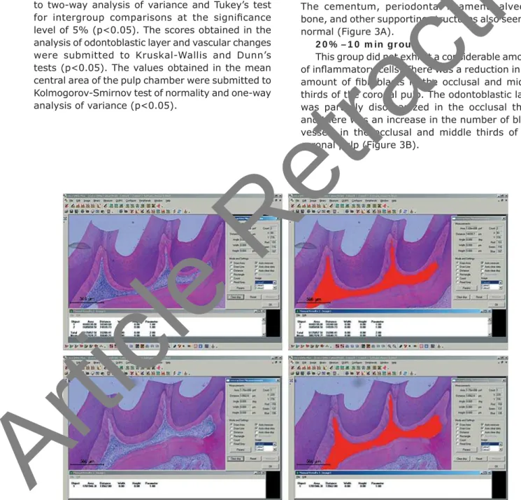
The mean central area of the pulp chamber was measured by image processing software (Leica QWin V3, Leica Microsystems, Wetzlar, Hesse,
was possible to calculate the percentage reduction in the central area of the pulp chamber in the treated groups, considering the central area of the control group.
After the application of the Kolmogorov-Smirnov test of normality, the data obtained in counts of
to two-way analysis of variance and Tukey’s test
level of 5% (p<0.05). The scores obtained in the analysis of odontoblastic layer and vascular changes were submitted to Kruskal-Wallis and Dunn’s tests (p<0.05). The values obtained in the mean central area of the pulp chamber were submitted to Kolmogorov-Smirnov test of normality and one-way analysis of variance (p<0.05).
RESULTS
,QÀDPPDWRU\UHVSRQVH Con t r ol gr ou p
The dental pulp of the control animals exhibited
intact odontoblastic layer and an even distribution of cells, blood vessels, and extracellular matrix structures (Figure 2).
2 0 % – 5 m in gr ou p
The dental pulp appeared similar to that of the control group. The odontoblastic layer was intact and the blood vessels showed normal characteristics. The cementum, periodontal ligament, alveolar bone, and other supporting structures also seemed normal (Figure 3A).
2 0 % – 1 0 m in gr ou p
This group did not exhibit a considerable amount
thirds of the coronal pulp. The odontoblastic layer was partially disorganized in the occlusal third, and there was an increase in the number of blood vessels in the occlusal and middle thirds of the coronal pulp (Figure 3B).
Figure 1- Central area measurement of the pulp chamber in the experimental groups using the Leica QWin V3 Image Processing and Analysis Software (Leica Microsystems, Wetzlar, Hesse, Germany). The values obtained were analyzed E\WKH.ROPRJRURY6PLUQRYQRUPDOLW\WHVWDQGWKHRQHZD\DQDO\VLVRIYDULDQFHS
CINTRA LTA, BENETTI F, FERREIRA LL, RAHAL V, ERVOLINO E, JACINTO RC, GOMES FILHO JE, BRISO ALF
2 0 % – 1 5 m in gr ou p
cells in the occlusal and middle thirds of the coronal
there was an increased amount of blood vessels. The odontoblastic layer was partially disorganized in the occlusal third (Figure 3C).
2 0 % – 3 0 m in gr ou p
found in the middle third of the coronal pulp in this group. There was a large reduction in the amount
disruption of the odontoblastic layer. The amount of blood vessels increased in the occlusal and middle thirds of the coronal pulp (Figure 3D). The radicular pulp seemed normal in all cases.
2 0 % – 4 5 m in gr ou p
This group showed an increased number of
of the coronal pulp. The occlusal third showed necrotic areas. A reduction in the number of
crown. The odontoblastic layer was absent in the occlusal third and partly disorganized in the middle
of the radicular pulp (Figure 3E).
3 5 % – 5 m in gr ou p
reduced in the occlusal third of the coronal pulp, where the odontoblastic layer was partially disorganized. An increase in the number of blood vessels was observed in all areas of the coronal pulp (Figure 3F).
3 5 % – 1 0 m in gr ou p
There was an increase in the number of
of the coronal pulp in this group. The amount
odontoblastic layer was absent in the occlusal third, and partly disorganized in the middle third of the coronal pulp. There was an increase in the number of blood vessels throughout the coronal
3 5 % – 1 5 m in gr ou p
There was an increase in the number of
reduced. The odontoblastic layer was absent in the occlusal third, and partly disorganized in the middle third of the coronary pulp. There was an increase in the number of blood vessels throughout the coronal pulp (Figure 3H).
Figure 2- Representative images of hematoxylin & eosin-stained sections showing the coronal pulp of the controls. Panels DQGDUHPDJQL¿HGLPDJHVîRIWKHUHVSHFWLYHLQVHWVLQWKHXSSHUOHIWSDQHOîPDJQL¿FDWLRQ7KHEODFN arrows indicate the odontoblastic layer and the white arrows show the distribution of cells and blood vessels in the subjacent tissue. Asterisks indicate the predentin layer
3 5 % – 3 0 m in gr ou p
reduced in the occlusal third, where necrotic areas were present. There was an increase in the
cervical thirds of the coronary pulp. The amount of
coronary pulp. The odontoblastic layer was absent in the occlusal and middle thirds of the crown. There was an increase in the number of blood vessels in the middle and cervical thirds of the coronal pulp. The occlusal third was characterized as necrotic. A
the cervical third of the radicular pulp (Figure 3I).
3 5 % – 4 5 m in gr ou p
There was necrosis in the occlusal third of this
fibroblasts. The number of inflammatory cells increased in the cervical and middle thirds of the coronal pulp, and in the cervical third of the
in these thirds. The odontoblastic layer was absent in the occlusal and middle thirds of the crown, and partially disorganized in the cervical third. The number of blood vessels increased in the cervical
third of the coronary and radicular pulp. The remaining thirds seemed normal (Figure 3J).
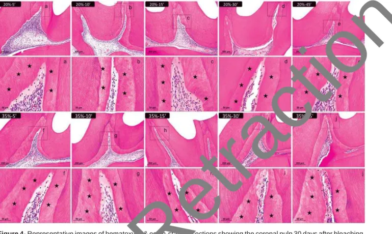
Re pa r a t iv e de n t in a r e a
Thirty days after the bleaching sessions, all the specimens showed normal dental pulp. However, the central area of the pulp chamber reduced, and the tertiary dentin area increased (Figure 4).
I n t e r gr ou p com pa r ison s
of the experimental groups. The most predominant inflammatory cells found were mononuclear cells, such as lymphocytes, macrophages and
infiltrate. The amount of inflammatory cells gradually increased with increasing concentrations and application time of the bleaching gel, up to the 15-min application groups of each bleaching gel, in the occlusal third of the coronal pulp. The groups that received the application of 30 and 45-min of each bleaching agent showed areas of necrosis in the occlusal third with a decrease
differences were observed between the bleached
)LJXUH Representative images of hematoxylin & eosin-stained sections showing the coronal pulp 2 days after bleaching. Panels A, B, C, D, and E represent the groups treated with 20% H2O2 gel and panels F, G, H, I, and J represent those
treated with 35% H2O2JHOIRUDQGPLQUHVSHFWLYHO\îPDJQL¿FDWLRQ3DQHOVD±MDUHPDJQL¿HGLPDJHV
îRIWKHLQVHWVLQSDQHOV$±-UHVSHFWLYHO\$VWHULVNVLQGLFDWHWKHSUHGHQWLQOD\HU7KHQXPEHURILQÀDPPDWRU\FHOOV DQG¿EUREODVWVZDVREWDLQHGLQHDFKWKLUGRIWKHSXOSWLVVXHDWîPDJQL¿FDWLRQDQGVXEMHFWHGWRWKH.ROPRJRURY 6PLUQRYQRUPDOLW\WHVWWZRZD\DQDO\VLVRIYDULDQFHDQG7XNH\¶VWHVWSWKHVFRUHVRIRGRQWREODVWLFOD\HUDQG YDVFXODUFKDQJHVXQGHUZHQW.UXVNDO:DOOLVDQG'XQQ¶VWHVWVS
CINTRA LTA, BENETTI F, FERREIRA LL, RAHAL V, ERVOLINO E, JACINTO RC, GOMES FILHO JE, BRISO ALF
groups and the control group in the occlusal third (p<0.05), except for the 35%–45 min group, which
in the middle third of the coronal pulp were noted between the control group and the 20%–10 to 45
min and 35%–5 to 45 min groups (p<0.05). In the cervical third, the difference from the control group was also present in the 20%–15 to 45 min and 35%–10 to 45 min groups (p<0.05). In the cervical
Figure 4- Representative images of hematoxylin & eosin-stained sections showing the coronal pulp 30 days after bleaching. Panels A, B, C, D, and E represent the groups treated with 20% H2O2 gel and panels F, G, H, I, and J represent those
treated with 35% H2O2JHOIRUDQGPLQUHVSHFWLYHO\îPDJQL¿FDWLRQ3DQHOVD±MDUHPDJQL¿HGLPDJHV
îRIWKHLQVHWVLQSDQHOV$±-UHVSHFWLYHO\6WDUVLQGLFDWHWKHUHSDUDWLYHGHQWLQOD\HUDVWHULVNVLQGLFDWHWKHSUHGHQWLQ layer. The values of the pulp chamber area were obtained as shown in Figure 1 to carry out the statistical analysis
Group Coronal Radicular
Occlusal Middle Cervical Cervical Middle Apical
Control 0.0 ±0.0a 0.0 ±0.0a 0.0 ±0.0a 0.0 ±0.0a 0.0 ±0.0a 0.0 ±0.0a
20% H2O2 gel 5 min 3.6 ±0.5b 2.2 ±0.4ab 1.0 ±0.4ab 0.0 ±0.0a 0.0 ±0.0a 0.0 ±0.0a
10 min 5.6 ±1.1bc 4.6 ±1.1bcd 2.2 ±0.5abc 0.0 ±0.0a 0.0 ±0.0a 0.0 ±0.0a
15 min 7.6 ±1.5c 6.0 ±1.4ce 3.4 ±0.9bd 0.0 ±0.0a 0.0 ±0.0a 0.0 ±0.0a
30 min 5.2 ±0.8bc 8.2 ±1.3e 5.2 ±1.1d 0.0 ±0.0a 0.0 ±0.0a 0.0 ±0.0a
45 min 3.2 ±0.8b 13.0 ±2.9fg 9.0 ±2.3e 5.6 ±0.9b 0.0 ±0.0a 0.0 ±0.0a
35% H2O2 gel 5 min 4.0 ±0.7b 4 ±1.0bc 2.2 ±0.5abc 0.0 ±0.0a 0.0 ±0.0a 0.0 ±0.0a
10 min 10.6 ±2.3d 7.4 ±1.1de 4.4 ±1.1cd 0.0 ±0.0a 0.0 ±0.0a 0.0 ±0.0a
15 min 14.6 ±3.6e 11.8 ±2.3f 5.2 ±1.3d 0.0 ±0.0a 0.0 ±0.0a 0.0 ±0.0a
30 min 6.0 ±1.4bc 15.6 ±1.3g 9.8 ±2.6e 5.8 ±0.8b 0.0 ±0.0a 0.0 ±0.0a
45 min 0.0 ±0.0a 12.0 ±2.9f 11.0 ±2.6e 7.6 ±1.5c 0.0 ±0.0a 0.0 ±0.0a
'LIIHUHQWOHWWHUVLQWKHFROXPQVLQGLFDWHVLJQL¿FDQWGLIIHUHQFHEHWZHHQWKHJURXSV.ROPRJRURY6PLUQRYQRUPDOLW\WHVWDQG WKH7ZR:D\$129$DQG7XNH\WHVW3
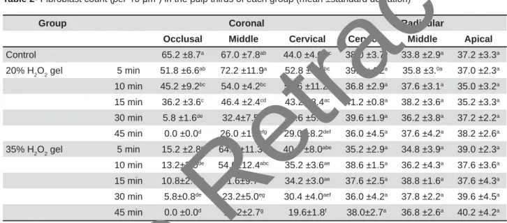
Table 1-,QÀDPPDWRU\FHOOFRXQWperȝP2) in the pulp thirds of each group (mean ±standard deviation)
were noted between the 20%–45 min and 35%–30 min groups from the other groups, and between
differences were not observed in the other radicular thirds (p>0.05).
the experimental groups. The 20%–10 to 45 min
compared with the control group (p<0.05). This decrease was also present in the middle third of the coronal pulp in the 20%–15 to 45 min and 35%–15 to 45 min groups (p<0.05). In the cervical third, only the groups that received the bleaching gels
were not observed in the radicular thirds of any group (p>0.05).
Table 3 shows the scores assigned to the odontoblast layer of each experimental group. In the occlusal third, the 20%–45 min and 35%–30
control and 20%–5 min groups (p<0.05). In the middle third of the coronal pulp, the 35%–45 min
and 20%–5 to 30 min groups (p<0.05). There were
radicular thirds (p>0.05).
Table 4 shows the scores assigned to the vascular changes in each experimental group. The 20%–45
Group Coronal Radicular
Occlusal Middle Cervical Cervical Middle Apical
Control 65.2 ±8.7a 67.0 ±7.8ab 44.0 ±4.6abc 38.0 ±3.7a 33.8 ±2.9a 37.2 ±3.3a
20% H2O2 gel 5 min 51.8 ±6.6ab 72.2 ±11.9a 52.8 ±8.5bc 39.4 ±4.2a 35.8 ±3.0a 37.0 ±2.3a
10 min 45.2 ±9.2bc 54.0 ±4.2bc 55.6 ±11.2c 36.8 ±2.9a 37.6 ±3.1a 35.0 ±3.2a
15 min 36.2 ±3.6c 46.4 ±2.4cd 43.2 ±8.4ac 41.2 ±0.8a 38.2 ±3.6a 35.2 ±3.3a
30 min 5.8 ±1.6de 32.4±7.5de 34.6 ±5.5ad 39.6 ±1.9a 36.2 ±3.8a 37.2 ±2.2a
45 min 0.0 ±0.0d 26.0 ±1.6efg 29.0 ±8.2def 36.0 ±4.5a 37.6 ±4.2a 38.2 ±2.6a
35% H2O2 gel 5 min 15.2 ±2.8e 64.8 ±11.3ab 40.8 ±8.0abe 35.2 ±2.9a 34.8 ±3.9a 39.0 ±2.3a
10 min 13.2±3.0de 54.6±12.4abc 35.2 ±3.6ae 38.6 ±1.5a 36.2 ±4.3a 37.6 ±3.6a
15 min 10.8±2.9de 41.6±9.7cdf 34.2 ±3.0ae 37.6 ±2.5a 38.8 ±1.6a 37.6 ±4.3a
30 min 5.8±0.8de 23.2±5.0eg 30.4 ±4.0aef 36.0 ±4.2a 37.8 ±2.2a 39.6 ±4.5a
45 min 0.0 ±0.0d 10.2±2.7g 19.6±1.8f 38.0±2.7a 36.8 ±2.6a 40.2 ±4.2a
Table 2- Fibroblast count (perȝP2) in the pulp thirds of each group (mean ±standard deviation)
'LIIHUHQWOHWWHUVLQWKHFROXPQVLQGLFDWHVLJQL¿FDQWGLIIHUHQFHEHWZHHQWKHJURXSV.ROPRJRURY6PLUQRYQRUPDOLW\WHVWDQG WKH7ZR:D\$129$DQG7XNH\WHVW3
Group Coronal Radicular
Occlusal Middle Cervical Cervical Middle Apical
Control 1a 1a 1a 1a 1a 1a
20% H2O2 gel 5 min 1a 1a 1a 1a 1a 1a
10 min 2ab 1a 1a 1a 1a 1a
15 min 2ab 1a 1a 1a 1a 1a
30 min 3ab 1a 1a 1a 1a 1a
45 min 3b 2ab 1a 1a 1a 1a
35% H2O2 gel 5 min 2ab 1ab 1a 1a 1a 1a
10 min 3ab 2ab 1a 1a 1a 1a
15 min 3ab 2ab 1a 1a 1a 1a
30 min 3b 3ab 1a 1a 1a 1a
45 min 3b 3b 2a 1a 1a 1a
'LIIHUHQWOHWWHUVLQWKHFROXPQVLQGLFDWHVLJQL¿FDQWGLIIHUHQFHEHWZHHQWKHJURXSV.UXVNDO:DOOLVDQG'XQQWHVWV3
7DEOH Comparison of odontoblastic layer scores (median)
CINTRA LTA, BENETTI F, FERREIRA LL, RAHAL V, ERVOLINO E, JACINTO RC, GOMES FILHO JE, BRISO ALF
from the control and 20%–5 min groups in the occlusal third (p<0.05). In the middle third of the coronal pulp, the 35%–45 min group also differed
cervical third and radicular thirds (p>0.05). At 30 days, the specimens showed a gradual
differences were observed between the 35%–45 min group and the other groups, except for the 35%–30 min group (p<0.05). The 20%–5 min, 20%–10 min, 20%–15 min, and 35%–5 min groups
(p>0.05) (Table 5).
D I SCUSSI ON
Tooth bleaching is an aesthetic alternative for discolored teeth, but it has potential adverse effects that are not yet completely understood28. A single
results, but longer application time and multiple sessions may be required for optimal outcomes, increasing the risk of tooth sensitivity20 and pulp
damage6.
A large number of in vit r o studies have shown that ROS generated by the H2O2 of bleaching gels are capable of causing histochemical and morphological changes in enamel and dentin4,5.
I n vivo
as mild to severe. These include studies performed in dog teeth25,26, human mandibular incisors8,19, rat
Group Coronal Radicular
Occlusal Middle Cervical Cervical Middle Apical
Control 1a 1a 1a 1a 1a 1a
20% H2O2 gel 5 min 1a 1a 1a 1a 1a 1a
10 min 2ab 2ab 1a 1a 1a 1a
15 min 2ab 2ab 1a 1a 1a 1a
30 min 2ab 2ab 1a 1a 1a 1a
45 min 3b 2ab 1a 1a 1a 1a
35% H2O2 gel 5 min 2ab 2ab 2a 1a 1a 1a
10 min 2ab 2ab 2a 1a 1a 1a
15 min 2ab 2ab 2a 1a 1a 1a
30 min 3ab 2ab 2a 1a 1a 1a
45 min 3b 3b 2a 1a 1a 1a
'LIIHUHQWOHWWHUVLQWKHFROXPQVLQGLFDWHVLJQL¿FDQWGLIIHUHQFHEHWZHHQWKHJURXSV.UXVNDO:DOOLVDQG'XQQWHVWV3 Table 4- Comparison of scores of vascular changes (median)
Mean (105)* SD (105) % reduction
Control 18.46a 2.38 0.00
20% H2O2 gel 5 min 17.57a 2.30 4.82
10 min 17.18a 1.85 6.93
15 min 15.65ab 1.78 15.22
30 min 13.47bc 1.43 27.03
45 min 11.23cd 1.17 39.16
35% H2O2 gel 5 min 14.61ab 0.46 20.85
10 min 13.41bc 0.72 27.36
15 min 12.56bcd 0.71 32.33
30 min 10.01de 0.62 45.77
45 min 6.98e 0.51 62.18
'LIIHUHQWOHWWHUVLQWKHFROXPQLQGLFDWHVLJQL¿FDQWGLIIHUHQFH.ROPRJRURY6PLUQRYQRUPDOLW\WHVWDQGWKH2QH:D\$129$ WHVW3
Table 5-&KDQJHLQWKHSXOSFKDPEHUFHQWUDODUHDȝP2)
incisors12,13, and rat molars6. Cell culture studies
also demonstrated cellular damage as apoptosis14, 3, cytotoxicity30, damage to the DNA23,
cell viability reduction27, or ageing of the dental
pulp1,28. The cytotoxicity of bleaching gel to pulp
tissue was also observed in this study.
Studies predominantly with ex vivo manipulated
studies of bleaching agents. However, in those studies, the pulp cells are not examined as organized tissues. Teeth have vital pulp components that can prevent or hinder the H2O2 effects in
extensions17,30, and antioxidant enzymes as
superoxide dismutase and catalase, which promote an enzymatic degradation of H2O211,17. Therefore, in
vivo experiments are the ones that best represent the reality of bleaching effects.
Application of 38% H2O2 gel on human premolars does not cause pathological changes in the dental pulp17. However, application of the same
concentration on human mandibular incisors causes necrosis in the coronal pulp, similar to what was observed in rat molars6, possibly because of the
thinner enamel and dentin8
that morphological characteristics of different tooth
upper anterior human teeth should be used to exactly determine pulp changes. Even under these
such as age, presence of restorations, previous trauma, among others.
Even though variations of the pulp response have been shown in human teeth, our study aimed to characterize an experimental animal model of easy reproduction and standardization for the study of new bleaching agents, posology, concentrations, and application time.
In dog teeth, dental bleaching using 35% H2O2 showed greater changes immediately beneath the region where the gel was applied25, similar
2O2 gel
applied for 30 or 45 min and 20% H2O2 gel for 45 min. Severe pulp damage may occur when bleaching agents are applied on the buccal surface of teeth with thin enamel and dentin8,25,26. Dog
Furthermore, studies in dogs have been avoided nowadays for ethical reasons.
The use of rats as the experimental model presents advantages such as ease of handling, reproduction, control, predictability22, and
standardization6. Moreover, this model further
presents better acceptance regarding ethical and economic concerns9.
Despite the difference in enamel and dentin thickness between humans and rat teeth (2.5
mm vs. 100 μm, respectively), they both show the same proportion of these structures6,9. In
addition, rat molars have anatomical, histological, biological and physiological features similar to human molars9,24. Also, rat molars exhibit the same
structural characteristics of the pulp chamber and pulp tissues, where the essential biological reactions and the wound healing of rat molar teeth are comparable to that of other mammals9. Conversely,
rat incisors are typical of rodents, of permanent growth, with a wide-open apex, and cannot be compared to human teeth9.
In the present study, 35% H2O2 gel applied for 30
response in the dental pulp, especially in the upper
with high H2O2 concentrations for 30 to 45 min in a single session is frequently associated with a high incidence of tooth sensitivity8. Considering
the similarity of the results found in this study to the results of Costa, et al.8 (2010), we suggested
that rat molars can be targeted and improved as an experimental model to predict the results of procedures performed in human mandibular incisors in this concentration and application time6.
The amount of H2O2 detected in the pulp chamber is related to the concentration and application time of the gel2. The use of 35% H
2O2 gel applied for 30
min, as well as 20% and 35% H2O2 gel for 45 min, was related to changes in vascular permeability12.
Therefore, our study was conducted with several application times and two concentrations, one of which more commonly employed in clinical dentistry (35% H2O2)16,29, and a lower one (20% H
2O2). Our
results allow choosing a concentration and time of application for comparative analysis in the initial
the subsequent reparative process (at 30 days). In our evaluation at 30 days after bleaching, we observed that all the groups showed signs of repair. Tertiary dentin was formed to protect the dental pulp, reducing the pulp chamber central area, and
differences from those of high concentration/ application time. Studies of the effects of high concentrations of bleaching gels on pulp cell cultures have shown that products released by 35% H2O2 gel can diffuse through enamel and dentin and
10,30.
H2O2 can penetrate the cell membrane, increase alkaline phosphatase activity, and induce apoptosis in the periodontal ligament and dental pulp15 as well
as stimulate mineralization21. Increased alkaline
phosphatase activity and extracellular matrix mineralization reveal the dentin production28. The
model in rats can also be used in long-term analysis to determine clinical protocols of application that CINTRA LTA, BENETTI F, FERREIRA LL, RAHAL V, ERVOLINO E, JACINTO RC, GOMES FILHO JE, BRISO ALF
produce less pulp damages over time.
The characterization of this experimental model does not replace human trials, but allows the knowledge of new bleaching agents mechanism of action; the comparison between protocols of bleaching; and the study of desensitizing and remineralizing agents used before and after bleaching to minimize effects on pulp tissues.
CON CLUSI ON
In conclusion, the rat model of extracoronal bleaching showed to be adequate for studies of bleaching protocols, as it was possible to observe alterations in pulp tissues and tooth structure caused by different concentrations and application times of bleaching agents. In-office bleaching with H2O2
accelerated aging of the dental pulp by inducing deposition of tertiary dentin, and the degree of damage increased with increasing concentration and application time of the bleaching agent.
ACKN OW LED GEM EN TS
projects 2011/13709-2 and 2013/25429-0.
REFEREN CES
1- Arai M, Shibata Y, Pugdee K, Abiko Y, Ogata Y. Effects of reactive oxygen species (ROS) on antioxidant system and osteoblastic differentiation in MC3T3-E1 cells. IUBMB Life. 2007;59:27-33. 2- Benetti AR, Valera MC, Mancini MN, Miranda CB, Balducci I. I n vit r o penetration of bleaching agents into the pulp chamber. Int Endod J. 2004;37:120-4.
3- Bhattacharyya S, Dudeja PK, Tobacman JK. Carrageenan-induced NFkappaB activation depends on distinct pathways mediated by reactive oxygen species and Hsp27 or by Bcl10. Biochim Biophys Acta. 2008;1780:973-82.
4- Borges AB, Torres CR, Souza PA, Caneppele TM, Santos LF,
effect on enamel erosion susceptibility. Int J Dent. 2012;2012:1-6.
containing bleaching agents on enamel surface properties. J Dent. 2008;36(9):718-25.
pulp tissue damage in rat teeth. J Endod. 2013;39(12):1576-80.
resin-based materials used as pulp-capping agents. Int Endod J. 2003;36:831-9.
8- Costa CA, Riehl H, Kina JF, Sacono NT, Hebling J. Human pulp
Med Oral Pathol Oral Radiol Endod. 2010;109:e59-64.
9- Dammaschke T. Rat molar teeth as a study model for direct pulp capping research in dentistry. Lab Anim. 2010;44(1):1-6.
deacetylase inhibitors induced differentiation and accelerated mineralization of pulp-derived cells. J Endod. 2012;38(3):339-45.
peroxide. Eur J Oral Sci. 2003;111:454-6.
Reis RS, et al. Tooth bleaching induces changes in the vascular permeability of rat incisor pulps. Am J Dent. 2013;26(5):298-300. 13- Frigo L, Pallota RC, Meneguzzo D, Marcos RL, Penna SC, Lopes-Martins RAB. Evaluation of the photo-activated dental bleaching effect on dental pulp in an in vivo rat experimental model. Rev Dental Press Estét 2009;6(1):102-14.
14- Han YH, Kim SZ, Kim SH, Park WH. Pyrogallol as a glutathione depletor induces apoptosis in HeLa cells. Int J Mol Med. 2008;21:721-30.
15- Hanks CT, Fat JC, Wataha JC, Corcoran JF. Cytotoxicity and dentin permeability of carbamide peroxide and hydrogen-peroxide vital bleaching materials, in vit r o. J Dent Res. 1993;72(5):931-8. 16- Joiner A. The bleaching of teeth: a review of the literature. J Dent. 2006;34:412-9.
17- Kina JF, Huck C, Riehl H, Martinez TC, Sacono NT, Ribeiro AP, et al. Response of human pulps after professionally applied vital tooth bleaching. Int Endod J. 2010;43(7):572-80.
Santos PH, et al. Penetration of hydrogen peroxide and degradation rate of different bleaching products. Oper Dent. 2015;40(1):72-9.
Silva C. The gel cytotoxicity in relation to the dental pulp. J Surg Clin Dent. 2014;1:10-3.
of light-activation sources. Oper Dent. 2008;33:15-22.
21- Matsui S, Takahashi C, Tsujimoto Y, Matsushima K. Stimulatory
ability of human dental pulp cells. J Endod. 2009;35:67-72. 22- Penna LA, Rode SM. Morphological study of the pulp of Wistar rats molars under experimental occlusal interference. Pesqui Odontol Bras. 2000;14(2):159-64.
does not change mitochondrial free radical generation and oxidative DNA damage. J Bioenerg Biomembr. 2006;38:327-33. 24- Sasaki T, Kawamata-Kido H. Providing an environment for reparative dentine induction in amputated rat molar pulp by high molecular-weight hyaluronic acid. Arch Oral Biol. 1995;40:209-19. 25- Seale NS, McIntosh JE, Taylor AN. Pulpal reaction to bleaching of teeth in dogs. J Dent Res. 1981;60:948-53.
26- Seale NS, Wilson CF. Pulpal response of bleaching of teeth in dogs. Ped Dent. 1985;7:209-14.
CA. Low toxic effects of a whitening strip to cultured pulp cells. Am J Dent. 2013:26:283-5.
Costa CA. Transenamel and transdentinal cytotoxicity of carbamide peroxide bleaching gels on odontoblast-like MDPC-23 cells. Int Endod J. 2011;44(2):116-25.
29- Sulieman M, Addy M, Macdonald E, Rees JS. The bleaching
study in vit r o. J Dent. 2005;33(1):33-40.
30- Trindade FZ, Ribeiro AP, Sacono NT, Oliveira CF, Lessa FC, Hebling J, et al. Trans-enamel and trans-dentinal cytotoxic effects of a 35% H2O2 bleaching gel on cultured odontoblast cell lines after consecutive applications. Int Endod J. 2009;42:516-24.