C A SE R EP ORT
244 J Vasc Bras. 2017 Jul-Set;16(3):244-247 http://dx.doi.org/10.1590/1677-5449.006316
Abdominal aortic pseudoaneurysm as a complication of
chronic pancreatitis: case report
Pseudoaneurisma de aorta abdominal como complicação de pancreatite crônica:
relato de caso
Eduardo Carvalho Horta Barbosa1
*
, Leonardo Pires de Sá Nóbrega1, Daniel Augusto de Souza Rodrigues1, Josué Rafael Ferreira Cunha1, Claudio Eluan Kalume1
Abstract
Chronic pancreatitis can be complicated by several vascular disorders, such as bleeding pseudocysts, thrombosis of the venous portal system, varicosities, and pseudoaneurysms. Pseudoaneurysm of the abdominal aorta secondary to chronic pancreatitis is a rare complication. It is a challenging clinical situation, demanding a high degree of clinical suspicion, and requires complex therapeutic procedures. he pathophysiology of this condition involves interstitial liberation and activation of enzymes from the exocrine pancreatic glands and subsequent digestion of the surrounding tissues. In the present case report, we describe a 52-year-old patient complaining of difuse abdominal pains. Clinical investigation revealed chronic alcoholism and imaging examinations showed a pseudoaneurysm of the infrarenal aorta. We decided to perform conventional surgical treatment after considering the patient’s age and clinical status and the endoprostheses available at our hospital with diameters compatible with the patient’s aorta.
Keywords: pseudoaneurysm; abdominal aorta; pancreatitis.
Resumo
A pancreatite crônica é uma enfermidade associada a diversas complicações vasculares, como pseudocisto hemorrágico, trombose do sistema venoso portal e formações varicosas e pseudoaneurismáticas. O pseudoaneurisma de aorta abdominal secundário à pancreatite crônica é uma complicação rara, de difícil suspeição clínica, que requer tratamento complexo. A isiopatologia dessa condição envolve a corrosão enzimática tecidual após a liberação e ativação de enzimas exócrinas proteolíticas das células acinares do pâncreas. O presente estudo relata o caso de um paciente de 52 anos, etilista crônico, internado com dor abdominal difusa, cuja propedêutica revelou se tratar de um pseudoaneurisma em aorta infrarrenal. Optou-se pelo tratamento cirúrgico convencional, levando-se em consideração a idade, as condições clínicas do paciente e a disponibilidade de endopróteses compatíveis com o diâmetro da aorta.
Palavras-chave: pseudoaneurisma; aorta abdominal; pancreatite.
1 Hospital de Base do Distrito Federal – HBDF, Unidade de Cirurgia Vascular, Brasília, DF, Brazil. Financial support: None.
Conlicts of interest: No conlicts of interest declared concerning the publication of this article. Submitted: September 28, 2016. Accepted: December 10, 2016.
245 J Vasc Bras. 2017 Jul-Set;16(3):244-247 Eduardo Carvalho Horta Barbosa, Leonardo Pires de Sá Nóbrega et al.
INTRODUCTION
Pancreatitis is a clinical condition with high incidence and prevalence worldwide. In the United States, it is estimated that there are 56,000 hospital admissions for chronic pancreatitis every year.1
In Brazil, according to the Brazilian National Health Service’s IT Department (DATASUS), the incidence of acute pancreatitis is 15.9/100,000 inhabitants per year. Vascular complications related to pancreatitis are uncommon, with a frequency of occurrence that varies from 1.2 to 14%.1
Arterial injuries related to pancreatitis most frequently involve the splenic artery, accounting for 40% of cases, followed by, in descending order of frequency, the gastroduodenal (30%), pancreaticoduodenal (20%), gastric (5%), and hepatic arteries (2%).2
Abdominal aortic pseudoaneurysm associated with pancreatitis is an extremely rare condition and there are only three cases reported in the literature.3 This article
reports a case of abdominal aortic pseudoaneurysm secondary to chronic pancreatitis, seen at a tertiary hospital on the public healthcare system and treated using conventional surgery to construct a dacron aortic interposition graft.
CASE REPORT
A 52-year-old male patient, who was hypertensive, diabetic, and a smoker and had a history of chronic alcoholism since the age of 19, presented to the clinical team in the emergency room complaining of pain all across the upper abdomen associated with nausea and chronic diarrhea. He described a previous history of several clinical admissions to treat acute exacerbations of chronic pancreatitis. He had not been subject to trauma, previous surgery, or endovascular intervention and did not have heart disease or rheumatic disease. Workup with laboratory tests and abdominal tomography
with venous contrast conirmed a new episode of acute
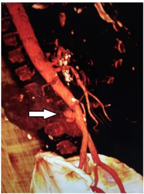
exacerbation of chronic pancreatitis and also revealed evidence suggestive of an abdominal aortic aneurysm. After clinical stabilization and improvement of the acute pancreatitis, he was referred to the vascular surgery service, where angiotomography reveled an
infrarenal abdominal aortic pseudoaneurysm, ive
centimeters from the bifurcation of the iliac arteries (Figure 1).
The decision was taken to perform conventional surgery to repair the pseudoaneurysm, via a xiphopubic incision and transperitoneal access. After clamping the aorta and performing arteriotomy of the anterior
wall, the pseudoaneurysm ostium was identiied in
the posterior wall of the aorta (Figure 2). An aorta to aorta dacron interposition graft was constructed with end-to-end anastomosis (Figure 3).
Figure 1. Angiotomography of the abdominal aorta, showing
a pseudoaneurysm of the posterior wall.
Figure 2. Ostium of the abdominal aortic pseudoaneurysm.
246 J Vasc Bras. 2017 Jul-Set;16(3):244-247
Pseudoaneurysm of the aorta secondary to chronic pancreatitis
The patient recovered well during the postoperative period and was discharged after 5 days in hospital. He is currently attending monthly follow-up consultations with regular assessments of the graft using vascular ultrasonography (Figure 4). To date he has been free from intercurrent conditions and has not suffered further episodes of exacerbation of his pancreatitis.
DISCUSSION
Pancreatitis can be complicated by several vascular disorders, such as bleeding pseudocysts, thrombosis of the venous portal system, varicosities, and pseudoaneurysms.2 This combination results in
elevated morbidity and mortality rates and among these patients survival is directly dependent on diagnosis and early treatment.
During the initial pathophysiologic process of pancreatitis, proteolytic exocrine enzymes such as trypsin are released from acinar cells and activated and these enzymes can cause damage that is not restricted to structures adjacent to the pancreas, but can involve bones, liver, and blood cells and vessels.4 Formation of arterial pseudoaneurysms is
a consequence of enzymatic corrosion of tissues. Many different risk factors contribute to formation of pseudoaneurysms, including necrotizing pancreatitis, multiple organ failure, accumulation of pancreatic
luids, and abscesses.1 The natural progression of this
disease is unpredictable, varying from spontaneous regression to rupture into the abdominal cavity, retroperitoneal space, or gastrointestinal tract. It is known that the risk of rupture is not directly related to pseudoaneurysm size.2
Patients with pseudoaneurysms secondary to pancreatitis may exhibit a variety of clinical presentations, ranging from asymptomatic cases with abdominal pains, through distension, melena, and minor intermittent bleeding, to acute hemorrhage (tachycardia, hypotension).5 The patient in question
remained hemodynamically stable throughout his time in hospital, despite the abdominal aortic pseudoaneurysm.
Imaging exams play a fundamental role in diagnosing pseudoaneurysms, since a large proportion of patients with pancreatitis already have chronic abdominal pains, as in the case reported here. Furthermore, many of them have a history of alcohol abuse and in these cases bleeding may be erroneously attributed to concomitant peptic ulcer disease and esophageal varices, which are common in these cases. Even severe acute hemorrhage is not simple to detect because of the frequency of multiple organ failure in these patients.
Management of abdominal aortic pseudoaneurysm secondary to pancreatitis is driven by the patient’s clinical condition and hemodynamic stability.1
For stable patients, abdominal ultrasonography with
Doppler is generally the irst diagnostic examination
used. It can suggest vascular involvement and identify venous thrombosis, necrotic areas, and abscesses in
the abdominal cavity, but indings are nonspeciic.5
Angiotomography and angiography are more accurate methods that enable diagnosis and therapeutic intervention in selected patients.
There is no well-established screening routine in the literature for secondary pseudoaneurysms. However, Suzuki et al. recommend abdominal Doppler ultrasonography at regular intervals in patients with chronic pancreatitis, as a form of secondary prevention.6
Pseudoaneurysms have a greater propensity to rupture than true aneurysms. Therefore the decision to perform surgery, whether conventional or endovascular, should be arrived at as soon as possible.7
In unstable patients, conventional surgery is
deined as the gold standard treatment for abdominal
aortic pseudoaneurysm associated with pancreatitis.2
In cases in which there is hemodynamic stability, the ideal treatment is controversial because of the small number of case reports that have been published.3
In all of the reports identiied in the literature, the
treatment option chosen was repair with an open technique, with exclusion of the pseudoaneurysm and interposition of a synthetic graft.
Endovascular repair techniques offer an alternative to conventional surgery,8,9 since they eliminate the
surgical traumas of transperitoneal or retroperitoneal access and aorta clamping, with a possible reduction
Figure 4. Longitudinal ultrasonography with Doppler for
247 J Vasc Bras. 2017 Jul-Set;16(3):244-247 Eduardo Carvalho Horta Barbosa, Leonardo Pires de Sá Nóbrega et al.
in the rates of morbidity and mortality associated with the procedure. On the other hand, the open technique is still a safe and effective option, with long term results that are well-established in the literature. It is preferable for younger patients, who have longer life expectancy and physiological reserves compatible with laparotomy and aortic clamping.10,11 These factors
were decisive in our choice of conventional surgery, in view of the age and clinical condition of the patient. Another important factor considered in choice of the therapeutic technique was the incompatibility between the diameter of the patient’s infrarenal aorta, which measured 1.6 centimeters, and the aortic endoprosthesis that was available at our service, which would have obliged us to use an iliac extension endoprosthesis off label.
It can be concluded that abdominal aortic pseudoaneurysm secondary to pancreatitis is a rare
complication that is dificult to diagnose clinically and
involves the possibility of catastrophic progression. It is therefore essential that vascular complications associated with chronic pancreatitis are always included in the list of diagnostic hypotheses, as was the case here.
REFERENCES
1. Barge JU, Lopera JE. Vascular complications of pancreatitis: role of interventional therapy. Korean J Radiol. 2012;13(Supl 1):S45-55. PMid:22563287. http://dx.doi.org/10.3348/kjr.2012.13.S1.S45.
2. Mallick IH, Winslet MC. Vascular complications of pancreatitis. JOP. 2004;5(5):328-37. PMid:15365199.
3. Takagi H, Manabe H, Sekino S, Kato T, Matsuno Y, Umemoto T. Abdominal aortic pseudoaneurysm associated with chronic pancreatitis. Eur J Vasc Endovasc Surg. 2005;9:46-8.
4. He Q, Liu YQ, Liu Y, Guan YS. Acute necrotizing pancreatitis complicated with pancreatic pseudoaneurysm of the superior mesenteric artery: a case report. World J Gastroenterol. 2008;14(16):2612-4. PMid:18442218. http://dx.doi.org/10.3748/ wjg.14.2612.
5. Luciano KS, Souza AR, Erdmann TR, Talamini LT, Cosentino AB, Erdmann AG. Pseudoaneurisma de artéria esplênica como complicação de pancreatite crônica: relato de caso. Arq Catarin Med. 2007;36:82-5.
6. Suzuki T, Ishida H, Komatsuda T, et al. Pseudoaneurysm of the gastroduodenal artery ruptured into the superior mesenteric vein in a patient with chronic pancreatitis. J Clin Ultrasound. 2003;31(5):278-82. PMid:12767023. http://dx.doi.org/10.1002/ jcu.10170.
7. Fankhauser GT, Stone WM, Naidu SG, et al. The minimally invasive management of visceral artery aneurysms and pseudoaneurysms. J Vasc Surg. 2011;53(4):966-70. PMid:21216559. http://dx.doi. org/10.1016/j.jvs.2010.10.071.
8. Giles RA, Pevec WC. Aortic pseudoaneurysm secondary to pancreatitis. J Vasc Surg. 2000;31(5):1056-9. PMid:10805901. http://dx.doi.org/10.1067/mva.2000.102850.
9. Fairman RM, Wang GJ. Abdominal aortic aneurysms: endovascular treatment. In: Rutherford RB, editor. Vascular surgery. Philadelphia: Sauders; 2014. p. 2046-61.
10. Ristow AV, Vescovi A, Massière BV, Correa MP. Aneurisma da aorta abdominal: tratamento pela técnica endovascular. In: Brito CJ, editor. Cirurgia Vascular: cirurgia endovascular, angiologia. Rio de Janeiro: Revinter; 2014. p. 800-71.
11. Coffler GEG, Nascimento RG, Lobato AC. Aneurismas periféricos: tratamento endovascular. In: Brito CJ, editor. Cirurgia Vascular: cirurgia endovascular, angiologia. Rio de Janeiro: Revinter; 2014. p. 941-54.
*
Correspondence
Eduardo Carvalho Horta Barbosa Hospital de Base do Distrito Federal – HBDF SQSW 304, bloco J, apto 611- Sudoeste CEP 70673-410 - Brasília (DF), Brazil Tel.: +55 (61) 98137-5580 E-mail: eduardochbarbosa@gmail.com
Author information
ECHB - Resident physician in Vascular Surgery, Hospital de Base do Distrito Federal. LPSN - Vascular surgeon, Hospital de Base do Distrito Federal; Board-certiied in Vascular eco-Doppler by Colégio Brasileiro de Radiologia; Full member of Sociedade Brasileira de Angiologia e Cirurgia Vascular (SBACV). DAC - Vascular surgeon, Hospital de Base do Distrito Federal. JR - Vascular surgeon, Hospital de Base do Distrito Federal; Full member of Sociedade Brasileira de Angiologia e Cirurgia Vascular (SBACV); Board-certiied in Vascular and Endovascular Surgery by SBACV. CEK - Vascular surgeon, Hospital de Base do Distrito Federal; Full member of Sociedade Brasileira de Angiologia e Cirurgia Vascular (SBACV).
Author contributions
Conception and design: ECHB, LPSN Analysis and interpretation: ECHB, LPSN, DAC Data collection: ECHB, LPSN, CEK Writing the article: ECHB, LPSN, JR Critical revision of the article: LPSN, JR Final approval of the article*: ECHB, LPSN, CEK, DAC, JR Statistical analysis: N/A. Overall responsibility: ECHB
