Original Article
The conventional system of pleural drainage currently in use in the treatment of pleural diseases and in the post-operative period following thoracic surgery is the same as that described by Kenyon in 1916.(1) This method consists
Introduction
After any surgical procedure involving the opening of the pleura, the thoracic cavity must be drained in order to facilitate adequate pulmonary reexpansion and allow the outflow of blood, fluids and air.
Use of a one-way flutter valve drainage system in the
postoperative period following lung resection*
Utilização da válvula unidirecional de tórax como sistema de drenagem no pós-operatório de ressecções pulmonares
Nelson de Araujo Vega1, Hugo Alejandro Vega Ortega2,
Alfio José Tincani3, Ivan Felizardo Contrera Toro4
Abstract
Objective: To evaluate pleural drainage using a one-way flutter valve following elective lung resection. Methods: This was a prospective study, with descriptive analysis, of 39 lung resections performed using a one-way flutter valve to achieve pleural drainage during the postoperative period. Patients less than 12 years of age were excluded, as were those submitted to pneumonectomy or emergency surgery, those who were considered lost to follow-up and those in whom water-seal drainage was used as the initial method of pleural drainage. Lung expansion, duration of the drainage, hospital stay and postoperative complications were noted. Results: A total of 36 patients were included and analyzed in this study. The mean duration of pleural drainage was 3.0 ± 1.6 days. At 30 days after the surgical procedure, chest X-ray results were considered normal for 34 patients (95.2%). Postoperative complications occurred in 8 patients (22.4%) and were related to the drainage system in 3 (8.4%) of those. Conclusions: The use of a one-way flutter valve following elective lung resection was effective, was well tolerated and presented a low rate of complications.
Keywords: Drainage; Postoperative complications; Thoracic surgery.
Resumo
Objetivo: Avaliar a drenagem pleural através de válvula unidirecional de tórax no pós-operatório de ressecção pulmonar eletiva.
Métodos: Foram realizadas 39 ressecções pulmonares, de forma prospectiva e com análise descritiva, em pacientes que utilizaram a válvula unidirecional de tórax (VUT) como o método de drenagem pleural durante o período pós-operatório. Foram excluídos os pacientes com idade inferior a 12 anos, os submetidos à pneumectomia ou a operação de urgência, os que não completaram o seguimento do estudo e os pacientes que utilizaram o sistema de frasco em selo d’água como método inicial de drenagem pleural. Observou-se a expansão pulmonar, o tempo de permanência com o sistema de drenagem, o período de internação e as complicações pós-operatórias. Resultados: Neste estudo, foram incluídos e analisados 36 pacientes. A média de permanência com o sistema de drenagem pleural foi de 3,0 ± 1,6 dias. O laudo da radiografia de tórax, realizado após 30 dias do procedimento cirúrgico foi considerado normal em 34 (95,2%) pacientes. Ocorreram oito (22,4%) casos de complicações pós-operatórias, sendo três (8,4%) relacionadas à VUT. Conclusões: A utilização da VUT no pós-operatório de ressecção pulmonar eletiva foi eficiente, bem tolerada e apresentou baixo índice de complicação.
Descritores: Drenagem; Complicações pós-operatórias; Cirurgia torácica.
* Study carried out at the Santa Casa de Misericórdia de Ribeirão Preto – SCMRP, Santa Casa Hospital at Ribeirão Preto – Ribeirão Preto, Brazil and at Universidade Estadual de Campinas – Unicamp, State University at Campinas – Campinas, Brazil.
1. Professor in the Pulmonology Department. Barão de Mauá University Center, Ribeirão Preto, Brazil.
2. Professor of Thoracic Surgery. Universidade de Ribeirão Preto – UNAERP, Ribeirão Preto University – Ribeirão Preto, Brazil.
3. Assistant Professor in the Department of Head and Neck Surgery. Hospital das Clinicas da Universidade Estadual de Campinas – HC-Unicamp, State University at Campinas Hospital das Clínicas – Campinas, Brazil.
4. Head of the Department of Thoracic Surgery. Faculdade de Ciências Médicas da Universidade Estadual de Campinas – FCM-Unicamp, State University at Campinas School of Medical Sciences – Campinas, Brazil.
Correspondence to: Nelson de Araujo Vega. Av. Saudade, 456, Campos Elíseos, CEP 14085-000, Ribeirão Preto, SP, Brasil. Tel 55 16 3605-0707. E-mail: [email protected]
Financial Support: The authors of this study received no financial support from the manufacturers of the one-way flutter valve. Nor were there any conflicts of interest between any of the authors and the manufacturers of the one-way flutter valve used in the present study or other manufacturers (of pleural drains or of any other thoracic drainage systems).
has since been used in the outpatient treatment of spontaneous pneumothorax.(9)
Methods
The one-way flutter valve is similar to the Heimlich valve and presents the following characteristics:
• It is a silicone rubber device, with a long flat tubular shape, 93 mm long and with a diam-eter of 13 mm. The proximal tubular-shaped extremity is adapted to a connector for the drainage tube. The remainder of the body of the valve, up to the distal extremity, is flat, so that both of its sides remain in touch with each other (closed). Therefore, the valve allows the outflow of air and fluids from the pleural space but prevents their return.
• The valve is inserted into a transparent plastic cylinder, 111 mm long, with a diameter of 22 mm, whose cone-shaped extremities have adequate caliber and grooves for their adap-tation to tubes of various sizes (proximal extremity) and fluid-collection bags or tubes (distal extremity). The device is 172 cm long and weighs 15 g.
In the period from February of 2002 to December of 2003, 39 lung resections were performed in two hospitals in the city of Ribeirão Preto, Brazil: the Santa Casa de Misericórdia de Ribeirão Preto and the Hospital Ribeirânia.
Lung resections included in this study were lobectomy, segmentectomy, bullectomy, wedge resection (excision of a nodule) and lung biopsy. All of the surgical procedures were performed by the same team, who always used similar surgical and anesthetic techniques.
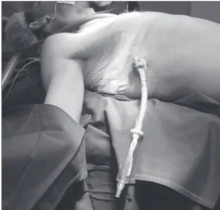
During surgery, right after the insertion and fixation of the drainage tube to the chest wall, the one-way flutter valve was attached to the distal extremity of the tube (Figure 1). Transparent multi-perforated PVC chest tubes with lateral radiopaque lines were used.
Two drainage tubes were used in lobectomies and segmentectomies, whereas a single tube was used in minor resections and bullectomies.
Most of the patients were extubated in the oper-ating room and remained under observation for 6 h in the recovery room. During that period, patients received supplemental oxygen via a Venturi mask and were monitored by means of echocardiography, of inserting the distal extremity of the tube into
a liquid column, contained inside a flask, whose cap has two openings: one for the passage of the drainage tube and one for ventilation (air vent). This is known as a water-seal drainage system. The use of this system in the postoperative period following thoracic surgery was described and disseminated by Lilienthal in 1922.(2)
Most surgeons use water-seal drainage with or without continuous suction in the postopera-tive period following elecpostopera-tive lung resection. This system is efficient, safe and affordable. However, using these flasks might cause risks, disadvantages and inconveniences for patients: they are heavy and large; they restrict the mobility of patients(3,4); frequent clamping performed during transport might cause pulmonary collapse and formation of clots,(3,5) as well as hypertensive pneumothorax; the placement of the flask, always kept in a level below the thorax of the patient, facilitates the disconnec-tion of one of the connecdisconnec-tions(3); and the bubbling inside the flask, when connected to continuous suction, causes an unpleasant sound.(6)
We must emphasize the fact that using this method in a prehospital environment is inappro-priate, because it is not only difficult to keep the flask below the patient, but it is also necessary to perform frequent clamping inside the limited space of an ambulance.(7)
In 1968, Henry Heimlich idealized a device in order to replace water-seal drainage systems, which was initially used in the treatment of American soldiers with thoracic trauma during the Vietnam War. The following advantages of a one-way valve were described: it provides better mobility of patients; clamping is unnecessary during trans-portation; the valve keeps working regardless of its position or level; nursing and medical teams can easily understand how it works; and it is safer and easier to clean.(3)
Since then, interest in developing an alternative and adequate thoracic drainage system has been reported in the literature.(4,6-8)
e) accidental removal of the valve
f) inverted positioning of the valve into the chest tube
• Group 2 - Other complications: a) pneumonia
b) infected surgical wound c) empyema
d) septicemia
e) pulmonary atelectasis f) hypertensive pneumothorax g) respiratory insufficiency h) prolonged air leak (over 7 days)
Patients less than 12 years of age were excluded, as were those submitted to pneumonectomy or emergency surgery, those who were considered lost to follow-up and those in whom water-seal drainage was used as the initial method of pleural drainage.
Chest tubes were never clamped during the postoperative period. We considered using the water-seal drainage system, with or without contin-uous suction, only when the one-way flutter valve method failed.
We defined failure of the drainage system as when patients presented one of the following character-istics: moderate-to-high volumes of intrathoracic fluids within the first 24 h after surgery; pulmonary collapse, higher than 30% of lung expansion by postoperative day 3; hypertensive pneumothorax; and respiratory insufficiency.
The postoperative follow-up period was 90 days, and follow-up evaluation consisted of outpatient visits and chest X-rays.
All participating patients or their legal guardians gave written informed consent.
The study design was approved by the Ethics in Research Committee of the State University at Campinas School of Medical Sciences (Protocol no. 543/2002). This study meets all of the requirements described in Brazilian National Health Council Resolutions 196/96 and 251/97.
This was a prospective study, with descrip-tive analysis of the data stored in a spreadsheet (Microsoft Excel®). We used the nonparametric Kruskal-Wallis test (nonparametric analysis of vari-ance) together with Dunn’s post-test in order to evaluate the difference noted in the duration of drainage in the postoperative period following lung resection. The level of significance was set at 5% (p < 0.05).
pulse oximetry and noninvasive determinations of arterial blood pressure. Following lobectomies and segmentectomies, most patients spent the postop-erative period in intensive care units.
During the postoperative period, lung expan-sion, duration of drainage using the one-way flutter valve, hospital stay and postoperative complications were noted. The patients were submitted to chest X-rays in the immediate postoperative period, after the removal of the drainage system and during the 90-day, postoperative outpatient follow-up period.
The criteria used for tube removal were lung expansion, blood output rate lower than 200 mL/24 h and no air leak.
The postoperative complications considered in this study were subdivided into two groups:
• Group 1 - Complications related to the pleural drainage system using the one-way flutter valve:
a) valve obstruction due to blood clots, preventing gases, blood and fluids from flowing out of the thoracic cavity
b) valve collapse, preventing gases, blood and fluids from flowing out of the thoracic cavity
c) disconnection of the valve and the tube d) gas reflux through the one-way flutter
valve
cystic lung diseases; benign neoplasia; pulmonary tuberculosis; and pleural tumor.
At 30 days after the surgical procedure, the X-rays of 34 patients (95.2%) were considered normal, whereas 2 patients (5.6%) presented residual pleural space (pneumothorax—less than 30% of complete lung expansion).
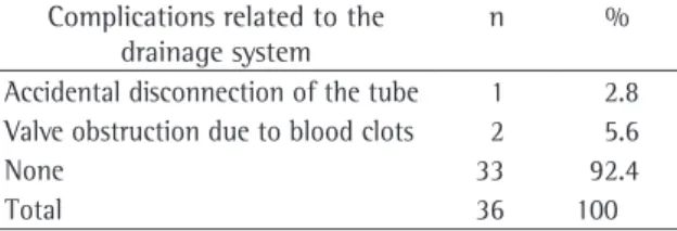
Table 2 shows that 3 (11.2%) of the patients presented postoperative complications related to the one-way flutter valve drainage system. However, the investigation of all postoperative complica-tions revealed 8 cases (22.4%): 3 (8.4%) related to the drainage system; 2 (5.6%) due to pneu-monia; 1 (2.8%) due to dehiscence of the chest wall; 1 (2.8%) due to bronchopleural fistula and empyema; and 1 (2.8%) due to atelectasis and subcutaneous emphysema.
Of the 36 cases, only 1 (2.8%) was classified as a case of failure of the drainage system. In that case, we chose to replace the valve with a water-seal drainage system.
Discussion
The presence of air, blood or fluids in the thoracic cavity counters the negative pressure within the pleural space and causes pulmonary collapse.
Results
Of the 39 patients submitted to lung resection within the period of this study, 3 were excluded for having been submitted to emergency surgery. No deaths occurred in the perioperative period, and 36 patients completed the study.
The mean age was 48.6 years (median, 50 years; range, 13-77 years). Of the 36 patients, 22 (61.6%) were male and 14 (39.2%) were female.
Of the 36 patients, 17 (47.6%) were smokers and 21 (58.8%) presented at least one comor-bidity. Previous diseases and comorbidities, as noted through anamnesis, physical examination and complementary tests, were as follows: cardiac arrhythmia; asthma; cancer of the larynx; diabetes mellitus, chronic obstructive pulmonary disease (COPD), systemic arterial hypertension, hypothy-roidism, mitral insufficiency, symplastic leiomyoma, Hodgkin’s lymphoma, spontaneous pneumothorax, acute myocardial infarction and illicit drug use.
Of the 36 lung resections, 10 were lung biopsies, 10 were wedge resections, 2 were segmentecto-mies, 8 were lobectosegmentecto-mies, and 6 were bullectomies. Conventional thoracotomy was the surgical tech-nique used to gain access to the pleural cavity of 33 patients (92.4%), whereas video-assisted thoracic surgery was performed in 3 patients (8.4%).
All patients were submitted to chest X-rays in the immediate postoperative period. Of the 36 patients, 25 (70%) presented complete lung expansion, whereas 11 (30.8%) revealed some degree of pulmo-nary collapse.
Removal of the chest drainage system ranged from 1 day to 8 days after the surgical procedure (in 19.6% and 2.8%, respectively), with a mean of 3.0 ± 1.6 days and median of 3.0.
The results of the chest X-rays performed after the removal of the drainage system were considered normal in 26 patients (72.8%), whereas incomplete lung expansion was seen in 8 patients (22.4%), small pleural effusion was seen in 1 (2.8%), and 1 (2.8%) developed pneumonia (Table 1).
The mean postoperative hospital stay was 4.5 ± 2.4 days (median, 4 days).
According to the anatomopathological examina-tion results, the diagnoses of patients were classified into eight subgroups: lung cancer; blebs/bullae; benign pulmonary nodule; interstitial lung diseases;
Table 1 - Distribution of patients using the one-way flutter valve system in the postoperative period following lung resection, according to the results of chest X-rays performed after the removal of the drainage system.
Chest X-ray results after the removal of the drainage system
Cases %
Normal 26 72.8
Incomplete expansion 8 22.4
Pleural effusion 1 2.8
Pneumonia 1 2.8
Total 36 100
Table 2 - Frequency of postoperative complications related to the drainage system among the 36 patients included in the study.
Complications related to the drainage system
n %
Accidental disconnection of the tube 1 2.8 Valve obstruction due to blood clots 2 5.6
None 33 92.4
this initiative, there has been interest in developing a drainage method that would replace the water-seal system for the treatment of pleural diseases,(8,17) as well as in the postoperative management following thoracotomies.(4,6,7)
However, the method most commonly used as an alternative to replace the water-seal system is still the Heimlich valve.
Other authors have reported that the outpa-tient treatment of spontaneous pneumothorax using Heimlich valve is safe, efficient and afford-able.(18,19) This valve, attached to a plastic collection bag, has been described as an alternative formula for the treatment of thoracic injuries in prehospital environments,(20) in emergencies,(11,8) and for the treatment of hemothorax.(21)
Using this valve in patients diagnosed with prolonged air leak after undergoing lung volume resection resulted in a decrease in the mean length of the hospital stay.(13,22)
However, this device is not totally free of compli-cations. In 1990, Mainini and Johnson reported two cases of hypertensive pneumothorax due to inverted connection of the valve to the chest tube.(23) Therefore, it is necessary to drain the thoracic cavity
in order to promote adequate lung expansion, as well as to reestablish cardiorespiratory function and negative intrapleural pressure.(10)
Currently, the method most widely used for the treatment of pneumothorax, hemothorax, pleural effusion and empyema is closed water-seal drainage. In the postoperative period following thoracotomy and lung resection, water-seal systems have been widely used. However, controversy remains as to whether one or two chest tubes should be used, as well as to whether continuous suction is required.
Since 1960, most surgeons have preferred to use chest tubes with suction ranging from −10 to
−20 cmH2O in the initial postoperative period.(12,13)
Recent studies have demonstrated that water-seal drainage (without suction) is safe and can promote benefits for patients, since it reduces air leaks, decreasing the duration of drainage and of the hospital stay following lung resection.(14-16)
Between 1962 and 1969, Henry Heimlich described the first studies on the use of a one-way drainage valve in the treatment of pneumothorax, pleural effusion and hemothorax, as well as in the postoperative period following thoracotomy.(3) After
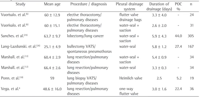
Table 3 - Duration of the drainage following elective thoracotomy reported in various studies (flutter valve drainage bags vs. water-seal drainage system with suction vs. water-seal drainage system without suction vs. one-way flutter valve system).
Study Mean age Procedure / diagnosis Pleural drainage system
Duration of drainage (days)
POC %
n Vuorisalo. et al.(6) 60 ± 12.9 elective thoracotomy/
pulmonary diseases
flutter valve drainage bags
3.3 ± 4.0 - 24 Vuorisalo. et al.(6) 60 ± 15.1 elective thoracotomy/
pulmonary diseases
water-seal + suction
2.6 ± 2.0 - 31
Sanches. et al.(25) 63.7 ± 9.7 lobectomy/lung cancer water-seal +
suction
5.9 ± 4.3 44.0 305 Lang-Lazdunski. et al.(26) 25.1 ± 4.9 bullectomy VATS/
spontaneous pneumothorax
water-seal 5.8 ± 1.2 27.4 167 Marshall. et al.(12) 60.4 ± 2.9 lung resection/pulmonary
diseases
water-seal + suction
5.4 ± 0.9 - 34 Marshall. et al.(12) 66.4 ± 2.6 lung resection/pulmonary
diseases
water-seal 3.3 ± 0.3 - 34
Ponn. et al.(18) 59 lung biopsy VATS/
pulmonary diseases
Heimlich valve 2.5 5.2 19
Vega. et al.a 48.6 ± 16.0 lung resection/pulmonary diseases
one-way flutter valve
3.0 ± 1.6 22.4 36
The only failure of the system occurred in a patient with COPD who was submitted to lobec-tomy due to primary lung cancer. On postoperative day 3, incomplete lung expansion persisted (pneu-mothorax greater than 30%), and the one-way flutter valve was replaced with a water-seal drainage system using continuous suction.
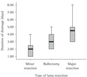
In this study, the use of a one-way flutter valve in the postoperative period following elective lung resection followed the principle that the duration of pleural drainage is generally shorter in minor resections (lung biopsy and wedge resection) and longer in major resections (segmentectomy and lobectomy). Figure 2 shows that this difference was significant (p < 0.001).
The affordability of one-way flutter valves can facilitate their use. The cost of this valve, ready to use, is US$ 5.00.
Table 3 shows the results of this study and those of other studies using various drainage systems in the postoperative period following elective lung resection.(6,12,25,26) The data in Table 3 suggest that the duration of pleural drainage with the one-way flutter valve was similar to or shorter than that of other studies.
The development and learning of new surgical techniques, as well as the development of medical devices and equipment, reduce surgical trauma and air leak. Therefore, some surgeons have been moti-Other authors reported that the one-way flutter
valve system attached to a plastic collection bag is safe and provides better mobility to patients, as well as potentially being more physiological,(4) especially when used in the postoperative period following lung resection.(6,7,24)
During the analysis period of this study (90 days), death occurred in each of the 3 cases (7.6%) that were excluded from the study due to emergency surgery. Two of those deaths occurred in patients hospitalized in an intensive care unit and presenting bilateral opacities of indefinite etiology, according to imaging methods. In those cases, an open lung biopsy was indicated. In both cases, death occurred in the late postoperative period, after the drainage systems had been removed. The third death occurred in a patient who had been diagnosed with severe COPD and presented spontaneous pneumothorax for 10 days and suffered air leak in the drainage system. That patient had been submitted to bullec-tomy. In the first 24 h after surgery, intense air leak was identified, as was subcutaneous emphysema and muscle fatigue. Despite being reintubated, the patient died of respiratory insufficiency.
The results of the postoperative chest X-rays were considered normal when there were minimal or no alterations.
Most of the cases of pulmonary collapse seen on chest X-rays performed in the immediate post-operative period were due to incomplete apical lung reexpansion (less than 3 cm). In those cases, no failure of the drainage system was detected. We identified cases of residual pneumothorax in which there was incomplete lung expansion (less than 30%), with the air leak staunched and drainage outflow of less than 200 mL/day. In those cases, we indicated the removal of the chest tube and the introduction of respiratory physiotherapy. In this sample, only one patient presented bronchopleural fistula and empyema in the postoperative period, being submitted to a new surgical procedure in the thoracic cavity.
We identified three complications related to the drainage system. In two cases, the valve was obstructed by blood clots and it was promptly replaced. In another case, the drainage system disconnected from the chest of the patient after the one-way flutter valve became caught on the bed. In that case, the valve was not replaced.
8.00
7.00
6.00
5.00
4.00
3.00
2.00
1.00
Duration of drainage (day
s)
Minor resection
Major resection Bullectomy
Type of lung resection
9. Ortega HAV, Lima MP, Denadai JO. Válvula unidirecional aplicada ao tratamento ambulatorial do pneumotórax. J Pneumol. 1996;22(4):177-80.
10. Figueiredo Pinto JA, Leite AG, Cavalet D. Drenagem torácica:
Princípios básicos. In: Pinto Filho DR, Cardoso PF, Figueiredo Pinto JA, Scheineider A, editors. Manual de cirurgia torácica. Rio de Janeiro: Revinter;2001. p.109-125.
11. Grégoire J, Deslauries J. Closed drainage and suction systems. In: Pearson FG, Deslauries J, Ginsberg RJ, Hiebert
CA, Mckneally MF, Urschel HC, editors. Thoracic Surgery. New York: Churchill Livingstone; 2002. p. 1281-1297. 12. Marshall MB, Deeb ME, Bleier JI, Kucharczuk JC, Friedberg
JS, Kaiser LR, et al. Suction vs water seal after pulmonary resection: a randomized prospective study. Chest. 2002;121(3):831-5.
13. McKenna RJ Jr, Fischel RJ, Brenner M, Gelb AF. Use of
the Heimlich valve to shorten hospital stay after lung reduction surgery for emphysema. Ann Thorac Surg. 1996;61(4):1115-7.
14. Okamoto J, Okamoto T, Fukuyama Y, Ushijima C, Yamaguchi M, Ichinose Y. The use of a water seal to manage air leaks after a pulmonary lobectomy: a retrospective study. Ann Thorac Cardiovasc Surg. 2006;12(4):242-4.
15. Cerfolio RJ, Bass C, Katholi CR. Prospective randomized trial compares suction versus water seal for air leaks. Ann Thorac Surg. 2001;71(5):1613-7.
16. Antanavicius G, Lamb J, Papasavas P, Caushaj P. Initial chest
tube management after pulmonary resection. Am Surg. 2005;71(5):416-9.
17. Lima AG, Contrera Toro IF, Tincani AJ, Barreto G. A drenagem
pleural pré-hospitalar: apresentação de mecanismo de válvula unidirecional. Rev Col Bras Cir. 2006;33(2):101-6. 18. Ponn RB, Silverman HJ, Federico JA. Outpatient chest tube
management. Ann Thorac Surg. 1997;64(5):1437-40. 19. Campisi P, Voitk AJ. Outpatient treatment of spontaneous
pneumothorax in a community hospital using a Heimlich flutter valve: a case series. J Emerg Med. 1997;15(1):115-9.
20. Williams JG, Riley TR, Moody RA. Resuscitation experience
in the Falkland Islands campaign. Br Med J (Clin Res Ed). 1983;286(6367):775-7.
21. Schweitzer EJ, Hauer JM, Swan KG, Bresch JR, Harmon JW, Graeber GM. Use of the Heimlich valve in a compact
autotransfusion device. J Trauma. 1987;27(5):537-42. 22. Beyruti R, Villiger LE, Campos JR, Silva RA, Fernandez
A, Jatene FB. A válvula de Heimlich no tratamento do pneumotórax. J Pneumol. 2002;28(3):115-9.
23. Mainini SE, Johnson FE. Tension pneumothorax complicating small-caliber chest tube insertion. Chest. 1990;97(3):759-60..
24. Lodi R, Stefani A. A new portable chest drainage device. Ann Thorac Surg. 2000;69(4):998-1001.
25. Sanches PG, Vendrame GS, Madke GR, Pilla ES, Camargo
JJ, Andrade CF, et al. Lobectomy for treating bronchial carcinoma: analysis of comorbidities and their impact on postoperative morbidity and mortality J Bras Pneumol. 2006;32(6):495-504.
26. Lang-Lazdunski L, Chapuis O, Bonnet PM, Pons F, Jancovici R. Videothoracoscopic bleb excision and pleural abrasion for the treatment of primary spontaneous pneumothorax: long-term results. Ann Thorac Surg. 2003;75(3):960-5.
27. Russo L, Wiechmann RJ, Magovern JA, Szydlowski GW,
Mack MJ, Naunheim KS, et al. Early chest tube removal after
vated to modify their approach to the use of chest tubes in the postoperative period. Recent studies have described chest tube removal in the recovery room,(27) as well as surgical procedures in which their use was avoided.(28) In addition, it has been shown that certain types of thoracic surgery can be performed as outpatient procedures.(29)
Thoracic surgeons utilize drainage systems in the postoperative period in various ways and are frequently guided towards a specific approach according to their personal preferences.(16) There are no evidence-based guidelines or consensuses designed to help surgeons determine their approach regarding the postoperative management of drainage systems.(30)
The use of the water-seal drainage system following lung resection is efficacious and has been well established. However, some studies have shown favorable results with the use of one-way flutter valves in the postoperative period following thoracic surgery.
In conclusion, the use of one-way flutter valves in the postoperative period following elective lung resection, in this sample of patients, proved effi-cient, safe and well tolerated. It also presented a low rate of complications, principally following minor resections.
References
1. Kenyon JH. Traumatic Hemothorax: siphon drainage. Ann Surg. 1916;64:728-9.
2. Lilienthal H. Resection of the lung for suppurative infections with a report based on 31 operative cases in which resection was done or intended. Ann Surg. 1922;75(3):257-320. 3. Heimlich HJ. Valve drainage of the pleural cavity. Dis Chest.
1968;53(3):282-7.
4. Waller DA, Edwards JG, Rajesh PB. A physiological comparison
of flutter valve drainage bags and underwater seal systems for postoperative air leaks. Thorax. 1999;54(5):442-3.
5. Lima AG, Rocha ER, Santos NA, Seabra JC, Mussi RK, Santos JG. Avaliação do uso da braçadeira ou “clamp” na
drenagem pleural fechada subaquática. Estudo prospectivo aleatorizado. J Bras Pneumol. 2007;33(Supl 1R):R13. 6. Vuorisalo S, Aarnio P, Hannukainen J. Comparison between
flutter valve drainage bag and underwater seal device for pleural drainage after lung surgery. Scand J Surg. 2005;94(1):56-8.
7. Graham AN, Cosgrove AP, Gibbons JR, McGuigan JA.
Randomised clinical trial of chest drainage systems. Thorax. 1992;47(6):461-2.
29. Molins L, Fibla JJ, Pérez J, Sierra A, Vidal G, Simón C.
Outpatient thoracic surgical programme in 300 patients: clinical results and economic impact. Eur J Cardiothorac Surg. 2006;29(3):271-5.
30. Tang AT, Velissaris TJ, Weeden DF. An evidence-based approach to drainage of the pleural cavity: evaluation of best practice. J Eval Clin Pract. 2002;8(3):333-40.
video-assisted thoracoscopic wedge resection of the lung. Ann Thorac Surg. 1998;66(5):1751-4.