Evaluation of lung volumes, vital capacity and respiratory
muscle strength after cervical, thoracic and lumbar
spinal surgery
Avaliação dos volumes pulmonares, capacidade vital e força dos músculos
respiratórios no pós-operatório de cirurgia na coluna cervical,
torácica e lombar
Marcio Aparecido Oliveira
I, Milena Carlos Vidotto
II, Oliver Augusto Nascimento
III, Renato Almeida
IV, Ilka Lopes Santoro
V,
Evandro Fornias Sperandio
VI, José Roberto Jardim
VII, Mariana Rodrigues Gazzotti
VIIIUniversidade Federal de São Paulo
ABSTRACT
CONTEXT AND OBJECTIVE: Studies have shown that physiopathological changes to the respiratory sys-tem can occur following thoracic and abdominal surgery. Laminectomy is considered to be a peripheral surgical procedure, but it is possible that thoracic spinal surgery exerts a greater inluence on lung func-tion. The aim of this study was to evaluate the pulmonary volumes and maximum respiratory pressures of patients undergoing cervical, thoracic or lumbar spinal surgery.
DESIGN AND SETTING: Prospective study in a tertiary-level university hospital.
METHODS: Sixty-three patients undergoing laminectomy due to diagnoses of tumors or herniated discs were evaluated. Vital capacity, tidal volume, minute ventilation and maximum respiratory pressures were evaluated preoperatively and on the irst and second postoperative days. Possible associations between the respiratory variables and the duration of the operation, surgical diagnosis and smoking status were investigated.
RESULTS: Vital capacity and maximum inspiratory pressure presented reductions on the irst postopera-tive day (20.9% and 91.6%, respecpostopera-tively) for thoracic surgery (P = 0.01), and maximum expiratory pressure showed reductions on the irst postoperative day in cervical surgery patients (15.3%; P = 0.004). The inci-dence of pulmonary complications was 3.6%.
CONCLUSIONS: There were reductions in vital capacity and maximum respiratory pressures during the postoperative period in patients undergoing laminectomy. Surgery in the thoracic region was associated with greater reductions in vital capacity and maximum inspiratory pressure, compared with cervical and lumbar surgery. Thus, surgical manipulation of the thoracic region appears to have more inluence on pulmonary function and respiratory muscle action.
RESUMO
CONTEXTO E OBJETIVO: Estudos têm demonstrado que alterações isiopatológicas no sistema respirató-rio podem ocorrer após cirurgia torácica e abdominal. A laminectomia é considerada uma cirurgia perifé-rica, mas é possível que as cirurgias de coluna torácica exerçam maior inluência sobre a função pulmonar. O objetivo do estudo foi avaliar os volumes pulmonares e as pressões respiratórias máximas em pacientes submetidos a cirurgia de coluna cervical, torácica ou lombar.
TIPO DE ESTUDO E LOCAL: Estudo prospectivo em hospital universitário terciário.
MÉTODOS: Sessenta e três pacientes submetidos a laminectomia com diagnóstico tumor ou hérnia de disco foram avaliados. Foram avaliados, no pré-operatório, no primeiro e no segundo dias de pós-operatório, ca-pacidade vital, volume corrente, volume por minuto e pressões respiratórias máximas. Possíveis associações entre as variáveis respiratórias e duração da cirurgia, diagnóstico cirúrgico e tabagismo foram investigadas.
RESULTADOS: A capacidade vital e a pressão inspiratória máxima apresentaram redução no primeiro dia de pós-operatório (20.9% and 91.6%, respectivamente) nas cirurgias torácicas; P = 0,01), e a pressão expira-tória máxima apresentou redução no primeiro dia de pós-operatório de cirurgia cervical (15.3%; P = 0,004). A incidência de complicações pulmonares foi de 3,6%.
CONCLUSÕES: Houve redução da capacidade vital e das pressões respiratórias máximas no período pós-operatório em pacientes submetidos a laminectomia. A cirurgia na região torácica apresentou asso-ciação com maiores reduções na capacidade vital e na pressão inspiratória máxima em comparação com a cirurgia cervical e lombar. Assim, a manipulação cirúrgica da região torácica parece ter maior inluência na função pulmonar e na ação dos músculos respiratórios.
IPT, MSc. Researcher in the Neurosurgery/ Respiratory Physiotherapy Group of the Respiratory Division, Universidade Federal de São Paulo (Unifesp), São Paulo, Brazil. IIPT, PhD. Associate Professor of the Department of Physiotherapy, Universidade Federal de São Paulo (Unifesp), São Paulo, Brazil.
IIIMD. Attending Physician in the Respiratory Division, Universidade Federal de São Paulo (Unifesp), São Paulo, Brazil.
IVPT. Researcher in the Neurosurgery/Respiratory Physiotherapy Group of the Respiratory Division, Universidade Federal de São Paulo (Unifesp), São Paulo, Brazil.
VMD. Attending Physician in the Respiratory Division, Universidade Federal de São Paulo (Unifesp), São Paulo, Brazil.
VIPT, PhD. Researcher in the Neurosurgery/ Respiratory Physiotherapy Group of the Respiratory Division, Universidade Federal de São Paulo (Unifesp), São Paulo, Brazil. VIIMD. Assistant Professor in the Respiratory Division, Universidade Federal de São Paulo (Unifesp), and Director of the Pulmonary Rehabilitation Center, Unifesp, São Paulo, Brazil. VIIIPT, PhD. Coordinator of the Neurosurgery/ Respiratory Physiotherapy Group of the Respiratory Division, Universidade Federal de São Paulo (Unifesp), São Paulo, Brazil.
KEY WORDS:
Respiratory function tests. Spine.
Muscle strength. Postoperative period. Laminectomy.
PALAVRAS-CHAVE: Testes de função respiratória. Coluna vertebral.
INTRODUCTION
Studies have shown that physiopathological changes to the respiratory system can occur following thoracic and
abdomi-nal surgery.1-4 he most frequently occurring changes are
reduc-tion in lung volume, changes to breathing patterns, altered gas exchange with reduction in partial arterial oxygen pressure
(PaO2), increase in partial arterial carbon pressure (PaCO2) in
the arterial blood and impairment of mucociliary transport.5-7
Recent studies have demonstrated that similar changes also occur in patients who undergo craniotomy, which is consid-ered to be a peripheral surgical procedure. hese patients expe-rience reduction in lung volume, changes to respiratory patterns (from predominantly diaphragmatic to intercostal), hypoxemia
and reduction in mucociliary transport.8,9 Other factors can
contribute to the reduction in pulmonary volumes, such as
gen-eral anesthesia, pain, immobility in bed and supine position.5,6
Up to 95% of patients with normal lungs who undergo general anesthesia may present atelectasis, which persists for more than 24 hours in 50% of the cases. Atelectasis is mainly present in dependent lung regions.
Laminectomy is considered to be a peripheral surgical
pro-cedure and is indicated for treating spinal column conditions.10
To the best of our knowledge, only one previous study has eval-uated lung function in adult patients undergoing laminectomy
to treat disc herniation or tumors.11 he changes to lung
func-tion and the behavior of the respiratory muscles were similar to those seen during the postoperative period following
tho-racoabdominal4 and cranial8,9 surgery. Alterations to
respira-tory function, such as reduction of lung volumes and respi-ratory muscle strength during the postoperative period following elective spinal surgery using a posterior access for tumor removal or herniated disc were correlated with surgical duration ≥ 240 minutes, presence of tumors, or cervicothoracic
surgical access.11 However, these analyses were performed by
pooling cervical surgery and thoracic surgery patients, due to
the small number of patients in the cervical group.11 It is
pos-sible that thoracic spinal surgery exerts a greater inluence on lung function, due to stimulation of relexes that inhibit the action of the diaphragm.
OBJECTIVE
he aim of this study was to evaluate how lung volume and maxi-mum inspiratory and expiratory pressure behave in patients who have undergone spinal (cervical, thoracic, or lumbar) surgery for treatment of disc herniation or tumors.
METHODS
his was a prospective study, conducted at a tertiary-level university hospital, on patients who underwent elective
spinal surgery to correct a herniated disc or remove a tumor. he patients were divided into three groups, based on the loca-tion of the spinal surgery: cervical, thoracic or lumbar. his study received approval from the hospital’s Human Research Ethics Committee (process 1956/07).
he inclusion criteria were that the patients needed to have undergone elective laminectomy under general anesthesia for disc herniation or tumor resection; to be ≥ 18 years of age; and to have signed the informed consent. he exclusion criteria were any presence of preoperative respiratory symptoms or obstruc-tive or restricobstruc-tive lung disease based on self-reports or clinical and radiological analysis; inability to perform spirometric or manometric procedures; postoperative respiratory support last-ing more than 24 hours; pain greater than two points on the visual analog scale (VAS) during evaluations ater the adminis-tration of analgesic medication; and death or brain death during the postoperative period.
Preoperative and postoperative evaluations were performed by the same investigator (MAO). he preoperative evaluation consisted of a clinical examination; investigation of past history of lung disease and smoking status; assessment of any presence of pain; and measurement of ventilation, vital capacity (VC) and respiratory muscle strength. he irst and second-day postoper-ative follow-ups consisted of pain evaluation and measurement of ventilation, VC and maximum respiratory pressures. All the patients received training so that they would be able to carry out the maneuvers during the preoperative period.
Pain was measured using a VAS scale from 0 (no pain) to 10 (worst possible pain), which made use of cartoon faces to depict
pain intensity.12 Based on pain intensity, the patients were
ini-tially medicated orally with an anti-inlammatory drug (tenoxi-cam), followed irst by amitriptyline and then by carbamazepine and amplictil. A narcotic (tramadol) was administered in cases of intense pain. All measurements were performed with the patients in the dorsal decubitus position and experiencing little or no pain (≤ 2 on the pain scale). If pain persisted ater treatment, the patient was excluded from the study, due to the possibility that their pain might inluence the results.
VC, tidal volume (TV) and minute ventilation (VE) were measured using a spirometer (Ohmeda RM 121, Tokyo, Japan), with the patient in the dorsal decubitus position and the bed positioned at zero degrees.
values.13 To measure VE, the patient was instructed to breathe
normally for one minute through a facial mask connected to the spirometer. TV was determined by dividing VE by the respira-tory rate (f).
Maximum inspiratory and expiratory pressures (MIP and MEP, respectively) were measured using a manometer (Comercial Médica, Brazil), using a nasal clip, with the patient in the dorsal decubitus position and the bed at zero degrees. To measure MIP, the patient performed maximal inspiratory efort at residual volume against a closed valve. To measure MEP, the patient performed maximal expiratory efort at total lung capacity against a closed valve. he measurements were repeated ive times or until one measurement was lower than the previous one. he two highest measurements were not to difer by more than 5%, and the highest measurement was used for analysis. Measurements were compared using the predicted
values.14 MIP and MEP were evaluated in order to measure the
respiratory muscle strength.
he duration of the operation (in minutes) was obtained from the anesthesia ile. he following were considered to be postoperative pulmonary complications: acute respiratory infection (pneumonia or purulent tracheobronchitis), atelectasis and/or bronchospasm.
Data analysis
Analysis of variance (ANOVA) with repeated measurements was used to compare two or more dependent samples. Fisher’s exact test or the chi-square test was used for categorical data. Continuous data were expressed as mean ± standard deviation. Statistical signiicance was set at 5% (P ≤ 0.05). he SPSS 17.0 sotware for Windows was used for the analyses.
he fpower macro of the SAS sotware, version 1.2 (Michael Friendly), was used to calculate the sample size, taking into con-sideration an ANOVA model for repeated measurements with the following factor groups: surgery type (cervical, lumbar or thoracic) and evaluation time (preoperative, irst postopera-tive day and second postoperapostopera-tive day). Based on the indings
reported by Di Pietro et al.11 and the preliminary results from
the present investigation, the comparison of interest was based on VC. By taking the signiicance level to be 5% and the test power to be 80%, the minimum sample was determined to be
16 patients in each group.15
RESULTS
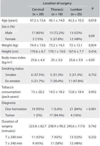
Eighty-four patients fulilled the inclusion criteria. However, twenty-one were excluded: eleven refused to participate; ive were unable to understand the procedures; three had intense pain during the evalu-ation; one was on a ventilator for more than 24 hours; and one died. Among the remaining 63 patients, 20 underwent cervical surgery, 18 underwent thoracic surgery and 25 underwent lumbar surgery.
Regarding tobacco use, 25.4% of the patients were smokers, 36.5% were ex-smokers and 38.1% had never smoked; no signiicant diferences among the groups were found regarding smoking status (P = 0.73) or
total number of pack-years (P = 0.95) (Table 1). Surgery to treat disc
herniation was performed on 41 patients (65.1%) and to treat tumors on
22 patients (34.9%) (Table 1). he median duration of surgery was
240 minutes (mean: 236.7 ± 93.9 minutes). No signiicant diference in the duration of the operation was found among the three groups (P
= 0.74) (Table 1).
he incidence of pulmonary complications was 3.2%. Atelectasis occurred in one patient (5%) who underwent cervical laminectomy, and tracheobronchitis occurred in one patient (5.6%) who under-went thoracic surgery. One death related to hemodynamic instability occurred in the cervical group immediately following surgery.
No diferences in TV (P = 0.06) or VE (P = 0.34) were found among the groups during the postoperative period or within the groups at the diferent evaluation times. Cervical surgery did not result in any signiicant change in VC during the postopera-tive period, whereas thoracic surgery led to a 20.9% reduction in
Location of surgery
P
Cervical
(n = 20)
Thoracic (n = 18)
Lumbar (n = 25)
Age (years) 47.2 ± 15.6 45.1 ± 14.0 43.3 ± 10.3 0.618
Sex n (%)
Male 17 (85%) 13 (72.2%) 13 (52%) 0.09 Female 3 (15%) 5 (27.8%) 12 (48%)
Weight (kg) 74.4 ± 13.6 73.2 ± 14.3 72 ± 12.1 0.834
Height (cm) 170.6 ± 6.7 170.1 ± 10.0 167.6 ± 7.7 0.416
Body mass index
(kg/m2) 25.6 ± 4.4 25 ± 3.0 25.6 ± 3.9 > 0.05
Smoking status
Smoker 6 (37.5%) 5 (31.3%) 5 (31.3%) 0.732
Ex-smoker 5 (21.7%) 7 (30.4%) 11 (47.8%) Tobacco
consumption (pack-years)
13 ± 20.2 14.5 ± 18.2 12.8 ± 18.4 0.952
Diagnosis
Disc herniation 19 (95%) 1 (5.6%) 21 (84%) < 0.001
Tumor 1 (5%) 17 (94.4%) 4 (16%) Duration of
surgery (minutes)
223.8 ± 62.7 238.9 ± 90.2 245.6 ± 117.0 0.742
T ≤ 240 min 11 (55%) 7 (42%) 13 (52%) 0.232
T ≥ 240 min 9 (45%) 11 (58%) 12 (48%)
Table 1. Patients’ characteristics, diagnoses and duration of
surgery according to location of surgery
VC on the irst postoperative day (P = 0.003) and a 17.4% reduc-tion on the second postoperative day (P = 0.01). Lumbar sur-gery led to an 11% reduction in VC on the irst postoperative day (P < 0.001). he reduction in VC was not associated with smok-ing status or the duration of the operation in any of the groups. he VC on the irst and second postoperative days was higher in patients with a diagnosis of herniation than in those with tumors (P = 0.049 and P = 0.019, respectively).
A 47% reduction in MIP occurred in the cervical group on the irst postoperative day (P = 0.03), and this was maintained (43.7% reduction) on the second postoperative day (P = 0.01). In the thoracic group, the reductions were 91.6% and 89.2% on the irst and second postoperative days, respectively (P < 0.001). In the lumbar group, the reductions were 55.7% and 53.4% on the irst and second postoperative days, respectively (P < 0.001) (Table 2). No association was found between reduction in MIP
and smoking status or surgical diagnosis. On the irst postop-erative day, MIP was 29.2% lower (P = 0.04) in patients whose surgery lasted more than 240 minutes than in patients with a duration of surgery of less than 240 minutes. No signiicant dif-ference was found on the second postoperative day.
A signiicant reduction (15.3%) in MEP occurred only in the cervical group in the irst postoperative day (P = 0.02) (Table 2). No associations were found between reduction in
MEP and smoking status or surgery duration. he MEP on the irst and second postoperative days was higher in patients with diagnoses of tumors than in those with herniation (P = 0.024 and P = 0.046, respectively).
DISCUSSION
In the present study, we observed that in patients who under-went spinal surgery to treat herniated discs or tumors, the great-est reduction in VC occurred in thoracic surgery patients with a tumor diagnosis. Inspiratory muscle strength was signiicantly reduced in all groups, with no signiicant association with the surgery site. However, the reduction was greater among patients with a surgical duration of over four hours. he reduction in expiratory muscle strength was greater in patients who under-went cervical or thoracic surgery than in those who underunder-went lumbar surgery. However, there was only a statistical diference in the group that underwent cervical surgery.
here has been increasing interest in the efects of surgi-cal procedures on the respiratory system. horacic and upper abdominal surgical procedures are known to exert a negative
inluence on lung function and respiratory muscle strength.16-18
In upper abdominal surgery, the incidence of pulmonary com-plications ranges from 10% to 81%, whereas the risk of such complications in lower abdominal surgery is inversely propor-tional to the distance between the surgical incision and the umbilicus, and ranges from 0% to 5%. In peripheral surgery (i.e. outside the thoracoabdominal compartment), the incidence of
complications is around 2%.19,20
he incidence of pulmonary complications in the present study was 3.6%, which was similar to the rate expected for lower
abdominal surgery,5 lower than that of upper abdominal
sur-gery21 and higher than that of peripheral surgery.17 Pulmonary
complications have been correlated with anesthesia, supine posi-tioning in the immediate postoperative period, immobility in
bed or postoperative pain.11 However, in this study, pain was
con-trolled with analgesics, and early mobilization was encouraged in order to reduce the possibility of respiratory complications.
No association was found between changes in lung func-tion and smoking status, although 61.5% of the participants were either smokers or ex-smokers. here is no consensus regarding the association between smoking status and decline in lung func-tion during the postoperative period following upper abdominal or neurological surgery. Some studies have found an association between smoking status and the incidence of pulmonary
compli-cations,20 while others have not found this correlation.22,23
Reductions in VC and respiratory muscle strength occurred in all groups in our study, with a greater negative impact (dimin-ished VC) on the thoracic group than on the lumbar group. Several studies have reported pulmonary changes in neurosur-gery patients that potentially increased the risk of
complica-tions.8,9,11 Di Pietro et al.11 were unable to detect changes in lung
function or respiratory strength stemming from surgery in the cervical and thoracic regions, due to their small sample size. However, the anatomical and physiological diferences between
Preoperative 1st PO day 2nd PO day
Vital capacity
Cervical 77.1 ± 16.7 64.4 ± 15.5 71.1 ± 17
Thoracic 79.4 ± 19.8 58.5 ± 15.4* 62 ± 17.5*
Lumbar 84.8 ± 8.6 73.8 ± 8.9* 79.1 ± 8.5
Maximum inspiratory pressure
Cervical 122.5 ± 41.1 75.5 ± 22.1* 78.8 ± 19* Thoracic 148.6 ± 40.9 57 ± 21* 59.4 ± 18.9*
Lumbar 128.7 ± 29.7 73.3 ± 21.1* 75.3 ± 21.5*
Maximum expiratory pressure
Cervical 75.2 ± 17.6 59.9 ± 14.9* 67.7 ± 20 Thoracic 68.4 ± 19 52.8 ± 19 57.8 ± 18
Lumbar 86.2 ± 19 78.6 ± 20.8 80.3 ± 20
Table 2. Vital capacity, maximum inspiratory pressure and
maximum expiratory pressure (% predicted) over time, among patients who underwent cervical, thoracic or lumbar laminectomy
these regions might trigger diferent responses from the respi-ratory system.
In the present study, a 20.9% reduction in VC was found on the first postoperative day in patients who underwent tho-racic surgery (P = 0.003). This was similar to the 25% reduc-tion over the same timeframe following craniotomy that was
found by Sogame et al.22 The reduction in VC following a
craniotomy procedure that these authors found was similar to what had been described in lower abdominal surgery, and they attributed this reduction to the anesthetic effect on the respiratory system, since no thoracoabdominal manipulation had been performed.
A 29.2% reduction in MIP was found on the irst postopera-tive day (P = 0.041) in patients whose duration of surgery was ≥ 240 minutes, in comparison with those whose surgery was < 240 minutes, with no signiicant diferences between the
dif-ferent groups studied. Sogame et al.22 found that duration of
sur-gery ≥ 240 minutes was associated with a reduction in both VC and TV on the irst postoperative day, in patients who underwent
elective craniotomy to correct aneurysm. Di Pietro et al.11 found
that reductions in VC, TV, MIP and MEP were also associated with duration of surgery ≥ 240 minutes. he reduction in inspi-ratory muscle strength suggests that lengthy surgery accentuates the inhibition of the diaphragm relex, independent of the
loca-tion of the surgery.8,9,23
Reduction in respiratory muscle strength is related to diminished lung function and an increase in the incidence of
pulmonary complications in patients who undergo surgery.24 he
overall reductions in MIP of 52.8% and 51.7% on the irst and sec-ond postoperative days, respectively (P < 0.001), were similar to
the MIP reduction found following upper abdominal surgery,24,25
and much higher than the 18% reduction in MIP expected for
laminectomy of the lumbar spine.12 It has been recognized that
inhibition of the diaphragm relex is the main factor for lung vol-ume reduction following thoracic or abdominal surgery, even if direct manipulation of the thoracoabdominal viscera does not
take place.26,27 It is possible that laminectomy may cause
inhibi-tion of this relex, thereby leading to reducinhibi-tions in lung volume and respiratory muscle strength, as found in the present study. he reductions in lung volume and respiratory muscle strength increase the risk of postoperative complications due to atelecta-sis, pneumonia, respiratory failure and exacerbation of underly-ing chronic lung disease.
Reduced respiratory measurements and respiratory muscle strength may be related to pain, fear or lack of cooperation from
the patient.24 However, pain greater than two points on the visual
analog scale (VAS) during evaluations ater administration of analgesic medication was an exclusion criterion in the present study, which suggests that this factor did not exert an inluence
on our indings. hus, it seems that our indings were, in fact, associated with inhibition of the diaphragm relex caused by the surgical procedure.
his study has some limitations that should be noted. Although the pain was controlled, fear and precaution in per-forming maximal respiratory strength evaluations during the postoperative period may have had some inluence on the mea-surements. However, we believe that this is unlikely to have changed our results signiicantly, since the patients were well-oriented regarding pain control through medication. Moreover, they had undergone training to carry out the maneuvers during the preoperative period. Other limitations on this study are that it was conducted in a single center and that the sample size was relatively small.
he study ended ater the second postoperative day because most of the patients were discharged ater the second day. he patients were informed by the physiotherapy team about the changes to the respiratory system inherent to anesthesia and the surgical procedure, but no physiotherapeutic intervention was carried out. Our indings of reductions in VC and respiratory muscle strength justify diferentiated, prophylactic intervention by the entire multidisciplinary team.
CONCLUSIONS
Based on our indings, thoracic laminectomy appears to exert a greater negative inluence on lung function and respiratory mus-cle strength during the postoperative periods, in comparison with cervical and lumbar laminectomy.
REFERENCES
1. Ayoub J, Cohendy R, Prioux J, et al. Diaphragm movement before and after
cholecystectomy: a sonographic study. Anesth Analg. 2001;92(3):755-61.
2. Kim SH, Na S, Choi JS, et al. An evaluation of diaphragmatic movement
by M-mode sonography as a predictor of pulmonary dysfunction
after upper abdominal surgery. Anesth Analg. 2010;110(5):1349-54.
3. Ambrozin ARP, Cataneo AJM. Aspectos da função pulmonary após
revascularização do miocárdio relacionados com risco pré-operatório
[Pulmonary function aspects after myocardial revascularization related
to preoperative risk]. Rev Bras Cir Cardiovasc. 2005;20(4):408-15.
4. Crema E, Benelli AG, Silva AV, et al. Assessment of pulmonary function
in patients before and after laparoscopic and open esophagogastric
surgery. Surg Endosc. 2005;19(1):133-6.
5. Dureil B, Cantineau JP, Desmonts JM. Efects of upper or lower abdominal
surgery on diaphragmatic function. Br J Anaesth. 1987;59(10):1230-5.
6. Shauer PR, Luna J, Ghiatas AA, et al. Pulmonary function after
laparo-scopic cholecystectomy. Surgery. 1993;114(2):389-97; discussion 397-9.
7. Dimopoulou I, Daganou M, Dafni U, et al.Phrenic nerve dysfunction
after cardiac operations: electrophysiologic evaluation of risk factors.
8. Gazzotti MR, Vidotto MC, Sogame LC, Hayashi LY, Jardim JR.
Disminución de la capacidad vital en el período postoperatorio de
la craneotomía electiva [Vital capacity reduction in postoperative of
elective craniotomy]. Rev Neurol. 2008;47(3):124-8.
9. Franceschini J, Sogame LCM, Gazzotti MR, Vidotto MC, Jardim JR.
Pulmonary function and thoraco-abdominal coniguration after
elective craniotomy. Neurosurgery Quarterly. 2008;18(1):22-7.
Available from: http://journals.lww.com/neurosurgery-quarterly/
Abstract/2008/03000/Pulmonar y_Function_and_Thoraco_
abdominal.5.aspx. Accessed in 2015 (Feb 24).
10. Chou R, Loeser JD, Owens DK, et al. Interventional therapies, surgery,
and interdisciplinary rehabilitation for low back pain: an
evidence-based clinical practice guideline from the American Pain Society.
Spine (Phila Pa 1976). 2009;34(10):1066-77.
11. Di Pietro TL, Sogame LM, Vidotto MC, Jardim JR. Study of respiratory
muscle strength, vital capacity, and ventilometry in the postoperative
period of spinal surgery by posterior access. Spine (Phila Pa 1976).
2006;31(12):E367-72.
12. Huskisson EC. Measurement of pain. Lancet. 1974;2(7889):1127-31.
13. Duarte AAO, Pereira CAC, Rodrigues SCS. Validação de novos valores
previstos brasileiros para a espirometria forçada na raça branca e
comparação com os valores previstos obtidos por outras equações
de referência [Validation of new Brazilian predicted values for
forced spirometry in Caucasians and comparison with predicted
values obtained using other reference equations]. J Bras Pneumol.
2007;33(5):527-35.
14. Black LF, Hyatt RE. Maximal respiratory pressures: normal values and
relationship to age and sex. Am Rev Respir Dis. 1969;99(5):696-702.
15. Lui KJ, Cumberland WG. Sample size requirement for repeated
measurements in continuous data. Stat Med. 1992;11(5):633-41.
16. Bigler DR. Aendringer i lungefunktionen ved anaestesi og thoraxkirurgi
[Lung function changes during anesthesia and thoracic surgery].
Ug-eskr Laeger. 2003;165(3):232-5.
17. Hedenstierna G. Mechanisms of postoperative pulmonary
dysfunc-tion. Acta Chir Scand Suppl. 1989;550:152-8.
18. Nomori H, Horio H, Fuyuno G, Kobayaski R, Yashima H. Respiratory
muscle strength after lung resection with special reference to age and
procedures of thoracotomy. Eur J Cardiothorac Surg. 1996;10(5):352-8.
19. Smetana GW. Preoperative pulmonary evaluation. N Engl J Med.
1999;340(12):937-44.
20. Pereira ED, Fernandes AL, da Silva Anção M, et al. Prospective
assessment of the risk of postoperative pulmonary complications in
patients submitted to upper abdominal surgery. Sao Paulo Med J.
1999;117(4):151-60.
21. Soares SM, Nucci LB, da Silva MM, Campacci TC. Pulmonary function
and physical performance outcomes with preoperative physical
therapy in upper abdominal surgery: a randomized controlled trial.
Clin Rehabil. 2013;27(7):616-27.
22. Sogame LC, Faresin SM, Vidotto MC, Jardim JR. Postoperative study
of vital capacity and ventilation measurements following elective
craniotomy. Sao Paulo Med J. 2008;26(1):11-6.
23. Kimball WR, Carwood CM, Chang Y, et al. Efect of efort pain after
up-per abdominal surgery on two independent measures of respiratory
function. J Clin Anesth. 2008;20(3):200-5.
24. Chiavegato LD, Jardim JR, Faresin SM, Juliano Y. Alterações funcionais
respiratórias na colecistectomia por via laparoscópica [Functional
respiratory changes in laparoscopic cholecystectomy]. J Pneumol.
2000;26(2):69-76.
25. Paisani DM, Chiavegato LD, Faresin SM. Volumes, capacidades
pulmonares e força muscular respiratória no pós-operatório de
gastroplastia [Lung volumes, lung capacities and respiratory muscle
strength following gastroplasty]. J Bras Pneumol. 2005;31(2):125-32.
26. Drummond GB. Diaphragmatic dysfunction: an outmoded concept.
Br J Anaesth. 1998;80(3):277-80.
27. Hall JC, Tarala RA, Hall JL, Mander J. A multivariate analysis of the risk of
pulmonary complications after laparotomy. Chest. 1991;99(4):923-7.
Sources of funding: None
Conlict of interest: None
Date of irst submission: November 19, 2014
Last received: January 14, 2015
Accepted: January 26, 2015
Address for correspondence:
José Roberto Jardim
Disciplina de Pneumologia da Unifesp
Rua Botucatu, 740 — 3o andar
Vila Clementino — São Paulo (SP) — Brasil
CEP 04023-062
Tel./Fax. (+55 11) 5572-4301