Cocaine-induced pulmonary changes: HRCT findings*
Cocaine-induced pulmonary changes: HRCT findingsRenata Rocha de Almeida1, Gláucia Zanetti1,2, Arthur Soares Souza Jr.3,
Luciana Soares de Souza4, Jorge Luiz Pereira e Silva5, Dante Luiz Escuissato6,
Klaus Loureiro Irion7, Alexandre Dias Mançano8, Luiz Felipe Nobre9,
Bruno Hochhegger10, Edson Marchiori1,11
Abstract
Objective: To evaluate HRCT scans of the chest in 22 patients with cocaine-induced pulmonary disease. Methods:
We included patients between 19 and 52 years of age. The HRCT scans were evaluated by two radiologists independently, discordant results being resolved by consensus. The inclusion criterion was an HRCT scan showing abnormalities that were temporally related to cocaine use, with no other apparent causal factors. Results: In 8 patients (36.4%), the clinical and tomographic findings were consistent with “crack lung”, those cases being studied separately. The major HRCT findings in that subgroup of patients included ground-glass opacities, in 100% of the cases; consolidations, in 50%; and the halo sign, in 25%. In 12.5% of the cases, smooth septal thickening, paraseptal emphysema, centrilobular nodules, and the tree-in-bud pattern were identified. Among the remaining 14 patients (63.6%), barotrauma was identified in 3 cases, presenting as pneumomediastinum, pneumothorax, and hemopneumothorax, respectively. Talcosis, characterized as perihilar conglomerate masses, architectural distortion, and emphysema, was diagnosed in 3 patients. Other patterns were found less frequently: organizing pneumonia and bullous emphysema, in 2 patients each; and pulmonary infarction, septic embolism, eosinophilic pneumonia, and cardiogenic pulmonary edema, in 1 patient each. Conclusions: Pulmonary changes induced by cocaine use are varied and nonspecific. The diagnostic suspicion of cocaine-induced pulmonary disease depends, in most of the cases, on a careful drawing of correlations between clinical and radiological findings.
Keywords: Cocaine, Cocaine-related disorders; Tomography, X-ray computed; Lung diseases.
*Study carried out at the Universidade Federal do Rio de Janeiro, Rio de Janeiro, Brasil.
1. Programa de Pós-Graduação em Radiologia, Universidade Federal do Rio de Janeiro, Rio de Janeiro, Brasil. 2. Faculdade de Medicina de Petrópolis, Petrópolis, Brasil.
3. Faculdade de Medicina de São José do Rio Preto, São José do Rio Preto, Brasil. 4. Ultra-X, São José do Rio Preto, São José do Rio Preto, Brasil.
5. Departamento de Medicina e Apoio Diagnóstico, Universidade Federal da Bahia, Salvador, Brasil. 6. Departamento de Clínica Médica, Universidade Federal do Paraná, Curitiba, Brasil.
7. Liverpool Heart and Chest Hospital NHS Foundation Trust, Liverpool, United Kingdom. 8. Radiologia Anchieta, Hospital Anchieta, Taguatinga, Brasil.
9. Universidade Federal de Santa Catarina, Florianópolis, Brasil.
10. Universidade Federal de Ciências da Saúde de Porto Alegre, Porto Alegre, Brasil. 11. Universidade Federal Fluminense, Niterói, Brasil.
Correspondence to: Edson Marchiori. Rua Thomaz Cameron, 438, Valparaiso, CEP 25685-120, Petrópolis, RJ, Brasil. Tel.: 55 24 2249-2777. Fax: 55 21 2629-9017. E-mail: [email protected]
Financial support: None.
Submitted: 9 February 2015. Accepted, after review: 7 April 2015.
Introduction
Cocaine is an alkaloid found in the leaves of a bush of the Erythroxylaceae family: the coca bush (Erythroxylum coca).(1) After marijuana, it is
the second most widely consumed and trafficked illicit drug in the world.(2,3) The prevalence of
“lifetime use” of cocaine in the 108 largest cities in Brazil, in 2005, was 2.9%.(3) In 2012,
a survey conducted by the Fundação Oswaldo Cruz involving approximately 25,000 people estimated the number of crack users in Brazil to be 0.81%, i.e., about 370 thousand users.(4)
Cocaine is the most widely consumed illicit drug among patients treated in emergency rooms, as well as being the leading cause of drug abuse-related deaths.(1,5) Several respiratory
problems have been temporally associated with acute or chronic cocaine use.(6,7) Therefore, the
diagnosis of cocaine-induced pulmonary diseases is a challenge for clinicians and radiologists, especially in urban hospitals.
All scans were analyzed for the following: ground-glass opacities, consolidations, interlobular septal thickening, the crazy-paving pattern, nodules, small parenchymal nodules, centrilobular nodules, the tree-in-bud pattern, cavitation, the halo sign, paraseptal emphysema, apical bullae, bullous emphysema, masses, and architectural distortion. The criteria for defining these findings, as well as the terminology used, were those recommended in the Fleischner Society Glossary of Terms(11) and in the consensus guidelines of
the Colégio Brasileiro de Radiologia(12) and the
Departamento de Imagem of the Sociedade Brasileira de Pneumologia e Tisiologia.(13)
In addition, all scans were assessed for the presence of pleural effusion, pneumothorax, pneumomediastinum, and any other associated findings.
The HRCT findings were also analyzed for laterality (bilateral, left, or right), as well as for distribution in the axial plane (central, peripheral, or random) and in the craniocaudal plane (upper, middle, lower, or diffuse). Lesions predominating in the inner third of the lung were defined as central, those predominating in the outer third of the lung were defined as peripheral; and those showing no preferential distribution were defined as random. The craniocaudal distribution of the lesions was characterized as follows: upper, for those located preferably above the level of the aortic arch; middle, for those located from the level of the aortic arch to the level of the carina; lower, for those located below the level of the carina; and diffuse, for those with no apparent predominance.
Results
Clinical and epidemiological aspects
We assessed 22 patients with cocaine-induced pulmonary disease, of whom 18 (81.81%) were male and 4 (18.18%) were female. All patients were adults, and ages ranged from 19 to 52 years (mean age of 32 years). The route of cocaine administration was inhalation (smokers or “snorters”), in 19 cases (86.36%), and i.v. injection, in 3 cases (13.63%). Crack use alone was reported in 9 cases, and other cocaine use, including cocaine hydrochloride and freebase cocaine, was reported in 11 cases. Two patients on chest X-ray (CXR), there have been few studies
describing CT findings.
The objective of the present study was to evaluate, by means of an analysis of HRCT scans of the chest in 22 patients with pulmonary changes that were temporally related to cocaine use, the most common HRCT findings, their morphological characteristics, and the distribution of the lesions in the lung parenchyma. In addition, we studied some epidemiological aspects of those patients.
Methods
The present study was approved by the Research Ethics Committee of the Hospital Universitário Antonio Pedro of the Universidade Federal Fluminense, in the city of Niterói, Brazil. Because the study was retrospective, patient informed consent was not required. This was a descriptive, retrospective observational study of HRCT scans of the chest in 22 patients with pulmonary changes induced by cocaine use, all of which were randomly gathered via personal contacts with radiologists and pulmonologists from seven different institutions, located in six Brazilian states. Eighteen patients were male, and 4 were female. Ages ranged from 19 to 52 years.
Patients were assessed for route of cocaine administration, type of cocaine used, and the presence of AIDS. The diagnosis was based on the association between HRCT findings and their temporal relationship with cocaine use, after excluding other possible causes.
Among the cases studied, we found patients with different types of pulmonary involvement, presenting with different clinical syndromes caused by cocaine use. In order to group patients and their imaging findings efficiently, we defined a subgroup of 8 patients presenting with features of the “crack lung” syndrome, which is characterized by respiratory failure associated with pulmonary opacities that are temporally related to crack use, with no other apparent causal factors, and which resolves rapidly after discontinuation of such use.(8-10)
hemopneumothorax occurred in 1 patient, respectively. Three patients developed talcosis. One of those patients reported using cocaine by inhalation, and the other 2 reported using cocaine by injection. All patients presented with perihilar conglomerate masses associated with architectural distortion and emphysema (Figure 3). In 1 of the injection cocaine users, increased reported both crack and other cocaine use. The
prevalence of AIDS was 22.72% (n = 5).
Tomographic aspects
The clinical and tomographic findings were consistent with the “crack lung” syndrome in 8 cases. Other forms of thoracic involvement included barotrauma (n = 3); talcosis (n = 3); organizing pneumonia (n = 2); bullous emphysema (n = 2); and pulmonary infarction, septic embolism, cardiogenic pulmonary edema, and chronic eosinophilic pneumonia, in 1 patient each (Table 1). Those changes were clinically divided into acute (“crack lung”, barotrauma, pulmonary infarction, septic embolism, and cardiogenic pulmonary edema) or chronic (talcosis, organizing pneumonia, chronic eosinophilic pneumonia, and bullous emphysema).
“Crack lung”
The most common HRCT finding in the 8 patients classified into the “crack lung” subgroup was ground-glass opacities, in 100% of the cases. In addition, consolidations were found in 4 of those cases (50%; Figure 1), and the halo sign was found in 2 (25%). The crazy-paving pattern was identified in 1 case (12.5%). In another case (12.5%), concomitant centrilobular nodules, some with the tree-in-bud pattern, were found. Paraseptal emphysema in the lung apices was identified in 1 case (12.5%; Table 2). Although the association of HRCT patterns was common, ground-glass opacities predominated in all cases analyzed. Regarding laterality, the involvement was bilateral in all 8 cases. The axial plane distribution was predominantly peripheral in 5 cases and predominantly central in the remaining 3. In none of the cases was the distribution random. In the craniocaudal plane, lesions were found to predominate in the upper third of the lung in 2 cases and in the lower third of the lung in 2 cases. In addition, diffuse involvement was seen in 4 cases. No case was found to have lesions predominating in the middle third of the lung.
Less common complications
Barotrauma was found in 3 patients. Two of those patients reported using cocaine by inhalation, and the other one reported using cocaine by inhalation and injection. Pneumomediastinum (Figure 2), pneumothorax, and spontaneous
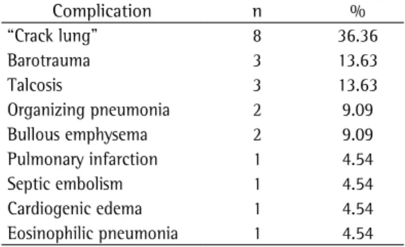
Table 1 - Frequency distribution of the pulmonary complications induced by cocaine use (n = 22).
Complication n %
“Crack lung” 8 36.36
Barotrauma 3 13.63
Talcosis 3 13.63
Organizing pneumonia 2 9.09 Bullous emphysema 2 9.09 Pulmonary infarction 1 4.54
Septic embolism 1 4.54
Cardiogenic edema 1 4.54 Eosinophilic pneumonia 1 4.54
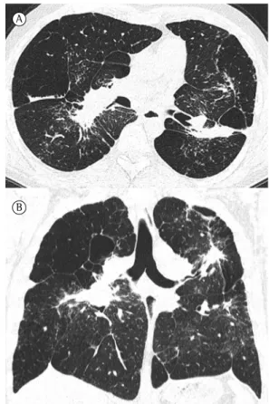
Figure 1 - “Crack lung”. A 24-year-old man with a recent history of crack use. HRCT scan showing consolidations associated with ground-glass opacities.
Table 2 - Frequency distribution of the HRCT findings in the lung parenchyma of the patients with “crack lung” (n = 8).
HRCT finding n %a
Ground-glass opacities 8 100.0
Consolidations 4 50.0
Consolidations with the halo sign 2 25.0 Smooth septal thickening 1 12.5 Crazy-paving pattern 1 12.5 Paraseptal emphysema 1 12.5 Centrilobular nodules 1 12.5 Tree-in-bud pattern 1 12.5
aThe sum of the percentages is greater than 100%, given
Discussion
Cocaine is the second most widely used illicit drug (second only to marijuana) in Brazil and in the world, as well as being associated with numerous health problems, such as those related to the respiratory system.(5,14,15) However, identifying
cocaine use in clinical practice remains difficult, representing a diagnostic challenge. For this reason, few case series have been published on the topic, being primarily limited to the study of the profile of cocaine users and their symptoms, especially those associated with psychological and behavioral changes.(14,16)
Because of the pulmonary impairment observed in cocaine users, chest radiology plays a critical role in the assessment of such patients. Large prospective studies aimed at the radiological investigation of pulmonary changes are scarce density was noted within the masses, whereas, in
the other one, there were also small parenchymal nodules in the adjacent parenchyma.
Organizing pneumonia was identified in 2 patients. Both of them reported using cocaine by inhalation and had HRCT findings of central and peripheral consolidations associated with architectural distortion. The diagnosis was confirmed by lung biopsy. Bullous emphysema was found in 2 patients who smoked cocaine, 1 of whom reported both cocaine and marijuana use. In that patient, HRCT showed large emphysema bullae in the lung apices, associated with architectural distortion. One patient developed pulmonary infarction and reported using cocaine by inhalation. The HRCT scan of that patient showed triangular subpleural consolidation with a pleural base. The diagnosis of pulmonary infarction was based on the clinical condition of the patient in combination with the radionuclide imaging pattern.
The patient with HRCT findings consistent with septic embolism reported using cocaine by injection. In that case, the HRCT findings consisted of predominantly peripheral pulmonary nodules, most of which were cavitated (Figure 4). Cardiogenic edema was identified in 1 patient, who reported using cocaine by inhalation. The HRCT scan of that patient showed ground-glass opacities interspersed with smooth interlobular septal thickening, resulting in a crazy-paving pattern, associated with bilateral pleural effusion and an enlarged cardiac silhouette. The patient with eosinophilic pneumonia reported using crack by inhalation. He presented with peripheral and pulmonary eosinophilia. His HRCT scan showed peripheral areas of ground-glass attenuation.
A
B
Figure 2 - Barotrauma. A 23-year-old man presenting with chest pain and no history of trauma. The HRCT scan shows free air dissecting into the mediastinal structures and the cervical soft tissues.
batteries, sometimes combined with different organic solvents. “Oxi”, or oxidized cocaine, is synthesized by mixing leftovers of cocaine paste with gasoline or kerosene and raw (virgem) lime (CaO). “Oxi” has become popular as an alternative substance that can be sold at a very low price, and its use is spread across Brazil.(23)
There is a relationship between cocaine use and the presence of HIV infection and AIDS(5);
this is due to increased exposure to risky sexual behavior and to transmission via injection drug use.(19) The prevalence of AIDS in our sample
was 22.7%.
The diagnosis of cocaine-induced pulmonary impairment is based primarily on a history of exposure to cocaine, consistent radiological findings, and the exclusion of other apparent causes for those findings.(24) Although knowledge
of whether patients have a history of cocaine use is extremely important for establishing a causal relationship, rarely is this information spontaneously provided by patients or their guardians, which makes the diagnosis difficult. Often, this information is only obtained retrospectively, after direct history taking, and, although 25-60% of crack users exhibit respiratory symptoms after smoking the drug, few seek medical attention.(18,24) In most of the
cases analyzed in our study, cocaine use was mentioned by patients only at a late stage of the investigation.
Certain physical examination findings, such as burned fingertips, resulting from handling the glass pipes typically used to smoke the drug, or the presence of black sputum, characteristic of crack use and attributed to the inhalation of carbon residues from butane or from the alcohol-soaked cotton used for the purpose of cooking the cocaine, can suggest the diagnosis.(5,24)
The frequency of cocaine-induced pulmonary complications is unknown; however, a wide spectrum of changes have been described in literature reviews.(5,8,24-28) Those changes include
“crack lung”, pulmonary edema, alveolar hemorrhage, interstitial disease, pulmonary hypertension, organizing pneumonia, emphysema, barotrauma, infection, lung cancer, pulmonary infarction, eosinophilic disease, aspiration pneumonia, lipoid pneumonia, etc.(5,8,24-28)
In our study, the HRCT scans of 22 patients were evaluated, and the most common finding was “crack lung”, in 8 cases, followed by barotrauma and limited to CXR series.(17,18) Taking only the
literature related to CT into consideration, studies are even scarcer, consisting of case reports and review studies.
Regarding the profile of cocaine users in Brazil and in South America, the incidence of use is higher in males in the 25- to 35-year age group.(19,20) The data found in our sample are
comparable to those from the literature, with the incidence being higher in males (80.95%), especially in young adults (mean age of 32 years).
Currently, the most widely used form of cocaine is crack, mainly because of its intense euphoric effects, which are obtained within a few minutes, and its lower cost.(15,16,21) In our
study, crack also was the most widely used type of cocaine, in 11 cases (50%), and, in 2 of those cases, the patients reported both crack and other cocaine use.
Cocaine can be administered by inhalation (smokers or “snorters”) and/or by i.v. injection. (5)
Currently, the most widely used route of administration is inhalation, especially crack or freebase cocaine smoking.(15) The preference for
inhalation over i.v. injection that has occurred in recent decades is mainly due to the increase in HIV transmission via injection drug use.(22) In
our study, 86.36% of the patients reported using cocaine by inhalation, and only 13.63% reported using i.v. cocaine, which is consistent with data from the national and international literature.
In Brazil, at least two other varieties of freebase cocaine, designated “merla” and “oxi”, are administered by inhalation (smoked). (23)
“Merla” contains acids obtained from car
and 2 cases of pneumothorax, 1 of which was associated with hemothorax.
Talc, silica, cellulose, and other adulterants are added to street cocaine.(6) Inhalation and i.v.
injection of talc-adulterated cocaine each may cause interstitial lung disease.(8) Inhalation talcosis
appears on HRCT as centrilobular or subpleural nodules, conglomerate masses, and lymph node enlargement.(8,31,32) Injection talcosis can manifest
as diffuse small nodules of increased density, areas of ground-glass attenuation, panacinar emphysema predominantly in the lower lobes, and perihilar conglomerate masses, which may contain areas of increased density.(31,33) In our
study, perihilar conglomerate pulmonary masses, associated with architectural distortion and emphysema, were identified in the 3 patients who presented with talcosis. In 1 case, increased density was noted within the masses, and, in another one, there were also small nodules in the adjacent parenchyma.
Organizing pneumonia has been reported in young crack smokers.(5) In our study, the 2
patients with organizing pneumonia, confirmed by lung biopsy, presented with central and peripheral consolidations on HRCT, as well as architectural distortion. Pulmonary bullous emphysema, predominantly in the upper lung region, is reported in 2-4% of injection drug users, typically affecting young men.(5) In our
sample, the two patients with bullous emphysema presented with large emphysema bullae in the lung apices, associated with architectural distortion. Septic pulmonary embolism and community-acquired pneumonia are among the most commonly observed infectious pulmonary complications in i.v. drug users.(5) HRCT findings
in community-acquired pneumonia can vary and are frequently related to the causative agent(34);
whereas septic emboli characteristically appear on HRCT as multiple peripheral pulmonary nodules, in different stages of cavitation, representing areas of septic infarction.(8,35,36) This pattern was
identified in 1 patient in our sample.
Our study had some limitations. First, the study was retrospective. Second, HRCT techniques varied widely, given the multicenter origin of the cases studied. Another important limitation of the present study, as well as of any other study related to drug users, is that, in certain cases, there are difficulties in establishing a causal relationship between cocaine use and HRCT patterns with and talcosis, in 3 cases each. Other findings
included organizing pneumonia and bullous emphysema, in 2 cases each. In addition, pulmonary infarction, septic embolism, cardiogenic edema, and eosinophilic pneumonia were identified in one case each. It should be considered, however, that no radiological finding alone is diagnostic of pulmonary changes induced by cocaine use. Most imaging findings are nonspecific and should be correlated with a history of cocaine use.(24)
The term “crack lung” refers to an acute pulmonary syndrome that occurs after inhalation of freebase cocaine and is associated with fever, hypoxemia, hemoptysis, respiratory failure, and the presence of diffuse alveolar infiltrates rich in eosinophils.(5,14,24,28) Since alveolar hemorrhage,
hypersensitivity pneumonitis, eosinophilic disease, and acute respiratory distress syndrome can be indistinguishable radiologically, the development of respiratory failure related to bilateral opacities, combined with cocaine use and rapid resolution after cessation of such use, has been called “crack lung”.(5,8-10,24)
HRCT findings in patients with “crack lung” include ground-glass opacities, consolidations, airspace nodules, smooth interlobular septal thickening, and, in some cases, the crazy-paving pattern.(5,15,28) In the literature, there are no large
case series investigating the most common HRCT findings in patients with “crack lung” and their distribution in the lung parenchyma. In our study, a bilateral distribution was found in all cases, being predominantly peripheral in the axial plane and diffuse in the craniocaudal plane.
Barotrauma is another complication that is often related to crack smoking and to the inhalation of powdered cocaine.(29) There is an
increase in airway pressure after smoking, either due to episodes of forceful coughing or intentional production of a Valsalva maneuver to increase the absorption and maximize the effect of the drug. (29) Barotrauma can manifest as pneumothorax,
pneumomediastinum, pneumopericardium, or subcutaneous emphysema, and it is usually diagnosed by CXR.(5,29) When CXR is inconclusive,
HRCT can help in the diagnosis.(30) A finding of
pneumomediastinum in young patients with no history of trauma should raise the suspicion of inhaled cocaine use.(27) We found no data on
2002;22 Spec No:S119-35. http://dx.doi.org/10.1148/ radiographics.22.suppl_1.g02oc01s119
9. Kissner DG, Lawrence WD, Selis JE, Flint A. Crack lung: pulmonary disease caused by cocaine abuse. Am Rev Respir Dis. 1987;136(5):1250-2. http://dx.doi.org/10.1164/ ajrccm/136.5.1250
10. Forrester JM, Steele AW, Waldron JA, Parsons PE. Crack lung: an acute pulmonary syndrome with a spectrum of clinical and histopathologic findings. Am Rev Respir Dis. 1990;142(2):462-7. http://dx.doi.org/10.1164/ ajrccm/142.2.462
11. Hansell DM, Bankier AA, MacMahon H, McLoud TC, Müller NL, Remy J. Fleischner Society: glossary of terms for thoracic imaging. Radiology. 2008;246(3):697-722. http://dx.doi.org/10.1148/radiol.2462070712 12. Souza Jr AS, Araujo Neto CA, Jasinovodolinsky D,
Marchiori E, Kavakama J, Irion KL, et al. Terminologia para a descrição de tomografia computadorizada de tórax (sugestões iniciais para um consenso brasileiro). Radiol Bras. 2002;35(2):125-8. http://dx.doi.org/10.1590/ S0100-39842002000200016
13. Silva CI, Marchiori E, Souza Júnior AS, Müller NL; Comissão de Imagem da Sociedade Brasileira de Pneumologia e Tisiologia. Illustrated Brazilian consensus of terms and fundamental patterns in chest CT scans. J Bras Pneumol. 2010;36(1):99-123. http://dx.doi.org/10.1590/ S1806-37132010000100016
14. Duailibi LB, Ribeiro M, Laranjeira R. Profile of cocaine and crack users in Brazil. Cad Saude Publica. 2008;24 Suppl 4:s545-57. http://dx.doi.org/10.1590/ S0102-311X2008001600007
15. Hui P, Walker B, Levy RD. Patient with fever, hypoxemia, and pulmonary consolidations. Chest. 2012;142(5):1348-51. http://dx.doi.org/10.1378/chest.12-0482 16. Oliveira LG, Nappo SA. Characterization of the crack
cocaine culture in the city of São Paulo: a controlled pattern of use [Article in Portuguese]. Rev Saude Publica. 2008;42(4):664-71. http://dx.doi.org/10.1590/ S0034-89102008000400012
17. Eurman DW, Potash HI, Eyler WR, Paganussi PJ, Beute GH. Chest pain and dyspnea related to “crack” cocaine smoking: value of chest radiography. Radiology. 1989;172(2):459-62. http://dx.doi.org/10.1148/radiology.172.2.2748826 18. Leece P, Rajaram N, Woolhouse S, Millson M. Acute and
chronic respiratory symptoms among primary care patients who smoke crack cocaine. J Urban Health. 2013;90(3):542-51. http://dx.doi.org/10.1007/s11524-012-9780-9 19. Osimani ML, Garibotto L, Scarlatta L, Latorre L, Chiparelli
H, Vidal J. VIH, Hepatitis B, Hepatitis C y VDRL en usuarios de cocaína no inyectable en Uruguay. Adicciones. 2005;17(2):157-62.
20. Pascale A, Hynes M, Cumsille F, Bares C. Consumo de pasta base de cocaína en América del Sur: revisión de los aspectos epidemiológicos y médico-toxicológicos [monograph on the Internet]. Washington DC: Organización de los Estados Americanos; 2014 [cited 2014 Oct 15]. [Adobe Acrobat document, 28p.]. Available from: www. cicad.oas.org/oid/pubs/pbc.pdf
21. Pomara C, Cassano T, D’Errico S, Bello S, Romano AD, Riezzo I, et al. Data available on the extent of cocaine use and dependence: biochemistry, pharmacologic effects and global burden of disease of cocaine abusers. Curr Med Chem. 2012;19(33):5647-57. http://dx.doi. org/10.2174/092986712803988811
certainty. Many of those individuals used or use other illicit drugs by inhalation or i.v. injection. Therefore, when crushed and injected into a peripheral vein, oral use tablets can also cause pulmonary talcosis. In other cases, the added use of marijuana can cause pulmonary bullous lesions. Despite these limitations, the present study includes the largest series of patients with cocaine-induced pulmonary changes identified on HRCT scans that has ever been published.
In conclusion, the most frequently found type of pulmonary change was “crack lung”. Other highly prevalent thoracic complications related to cocaine use were barotrauma and talcosis, followed by bullous emphysema and organizing pneumonia, as well as by cases of pulmonary infarction, septic embolism, cardiogenic pulmonary edema, and eosinophilic pneumonia. Pulmonary changes induced by cocaine use are nonspecific and should be temporally correlated with such use, after exclusion of other causes.
References
1. Karila L, Petit A, Lowenstein W, Reynaud M. Diagnosis and consequences of cocaine addiction. Curr Med Chem. 2012;19(33):5612-8. http://dx.doi. org/10.2174/092986712803988839
2. United Nations Office on Drugs and Crime [homepage on the Internet]. Vienna: UNODC. [cited 2013 Aug 2]. World drug report 2012. [Adobe Acrobat document, 112p.]. Available from: http://www.unodc.org/documents/ data-andanalysis/WDR2012/WDR_2012_web_small.pdf 3. Carlini EA, editor. II Levantamento domiciliar sobre o uso
de drogas psicotrópicas no Brasil: estudo envolvendo as 108 maiores cidades do país: 2005. São Paulo: Centro Brasileiro de Informação sobre Drogas Psicotrópicas e Universidade Federal de São Paulo; 2006. 468p. 4. Pimentel J. Estudo da Fiocruz estima alcance do crack
nas capitais brasileiras [monograph on the Internet]. Rio de Janeiro: Portal DSS Brasil; 2013 [cited 2015 Jan 15]. Available from: http://dssbr.org/site/2013/10/estudo-da-fiocruz-estima-alcance-do-crack-nas-capitais-brasileiras 5. Restrepo CS, Carrillo JA, Martínez S, Ojeda P, Rivera
AL, Hatta A. Pulmonary complications from cocaine and cocaine-based substances: imaging manifestations. Radiographics. 2007;27(4):941-56. http://dx.doi. org/10.1148/rg.274065144
6. Tashkin DP. Airway effects of marijuana, cocaine, and other inhaled illicit agents. Curr Opin Pulm Med. 2001;7(2):43-61. http://dx.doi.org/10.1097/00063198-200103000-00001 7. Riezzo I, Fiore C, De Carlo D, Pascale N, Neri M,
Turillazzi E, et al. Side effects of cocaine abuse: multiorgan toxicity and pathological consequences. Curr Med Chem. 2012;19(33):5624-46. http://dx.doi. org/10.2174/092986712803988893
29. Kloss BT, Broton CE, Rodriguez E. Pneumomediastinum from nasal insufflation of cocaine. Int J Emerg Med. 2010;3(4):435-7. http://dx.doi.org/10.1007/ s12245-010-0205-9
30. Alnas M, Altayeh A, Zaman M. Clinical course and outcome of cocaine-induced pneumomediastinum. Am J Med Sci. 2010;339(1):65-7. http://dx.doi.org/10.1097/ MAJ.0b013e3181c371da
31. Marchiori E, Lourenço S, Gasparetto TD, Zanetti G, Mano CM, Nobre LF. Pulmonary talcosis: imaging findings. Lung. 2010;188(2):165-71. http://dx.doi.org/10.1007/ s00408-010-9230-y
32. Marchiori E, Souza Júnior AS, Müller NL. Inhalational pulmonary talcosis: high-resolution CT findings in 3 patients. J Thorac Imaging. 2004;19(1):41-4. http:// dx.doi.org/10.1097/00005382-200401000-00008 33. Siddiqui MF, Saleem S, Badireddi S. Pulmonary talcosis with
intravenous drug abuse. Respir Care. 2013;58(10):e126-8. http://dx.doi.org/10.4187/respcare.02402
34. Sharma S, Maycher B, Eschun G. Radiological imaging in pneumonia: recent innovations. Curr Opin Pulm Med. 2007;13(3):159-69. http://dx.doi.org/10.1097/ MCP.0b013e3280f3bff4
35. Goswami U, Brenes JA, Punjabi GV, LeClaire MM, Williams DN. Associations and outcomes of septic pulmonary embolism. Open Respir Med J. 2014;8:28-33. http:// dx.doi.org/10.2174/1874306401408010028
36. Ye R, Zhao L, Wang C, Wu X, Yan H. Clinical characteristics of septic pulmonary embolism in adults: a systematic review. Respir Med. 2014;108(1):1-8. http://dx.doi. org/10.1016/j.rmed.2013.10.012
22. Baldwin GC, Choi R, Roth MD, Shay AH, Kleerup EC, Simmons MS, et al. Evidence of chronic damage to the pulmonary microcirculation in habitual users of alkaloidal (“crack”) cocaine. Chest. 2002;121(4):1231-8. http:// dx.doi.org/10.1378/chest.121.4.1231
23. Bastos FI, Mendes A, Duarte Pdo C, Bertoni N. Smoked crack cocaine in contemporary Brazil: the emergence and spread of ‘oxi’. Addiction. 2011;106(6):1191-2. http://dx.doi.org/10.1111/j.1360-0443.2011.03427.x 24. Mançano A, Marchiori E, Zanetti G, Escuissato DL, Duarte
BC, Apolinário Lde A. Pulmonary complications of crack cocaine use: high-resolution computed tomography of the chest. J Bras Pneumol. 2008;34(5):323-7. http:// dx.doi.org/10.1590/S1806-37132008000500012 25. Bailey ME, Fraire AE, Greenberg SD, Barnard J, Cagle
PT. Pulmonary histopathology in cocaine abusers. Hum Pathol. 1994;25(2):203-7. http://dx.doi. org/10.1016/0046-8177(94)90279-8
26. Herculiani PP, Pires-Neto RC, Bueno HM, Zorzetto JC, Silva LC, Santos AB, et al. Effects of chronic exposure to crack cocaine on the respiratory tract of mice. Toxicol Pathol. 2009;37(3):324-32. http://dx.doi. org/10.1177/0192623308330790
27. Mangi AA, Mullins ME, McLoud TC. Pneumomediastinum associated with recent cocaine use: Report of two cases and review of the literature. App Radiol. 2000;29(11):50-2. 28. de Almeida RR, de Souza LS, Mançano AD, Souza AS