Arq Bras Cardiol volume 72, (nº 1), 1999
Vacanti e col Relapsing thromboembolism in paraplegic
75 Sancor – Pronto Socorro Cardiológico de Santos
Mailing adress: Luciano J. Vacanti – Av. Cel. Joaquim Montenegro, 81/22 – 11035-001 – Santos, SP - Brazil
Luciano J. Vacanti, João J. Rodrigues, Cláudio H. Fisher, Sandra F. Delsin, Hermes T. Xavier
Santos, SP - Brazil
Relapsing Pulmonary Embolism in a Patient
with a Spinal Cord-Injure
Case Report
Spinal cord injures are associated with a high inciden-ce of deep vein thrombosis (DVT), and, in consequeninciden-ce, associated also with a greater frequency of pulmonary emboli (PE), reported in the literature to occur in 70 to 100% of those cases 1. The extent of the pulmonar vascular
obs-truction is probably the most important determinant of the right ventricular dysfunction, which in turn leeds to worse prognosis and higher risk of death and reccurence 2. These
patients are therefore good condidates for thrombolytic therapy. After clinical suspicion of PE, if there are unequi-vocally visible thrombi and signs of hight ventricular dys-function at echocardiography, treatment, including throm-bolysis, can be started 3. We report the case of a paraplegic
patient with unusual echocardiographic findings in whon two different therapeutic approaches for PE could be evaluated.
Case Report
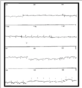
A 52-year-old male had been paraplegic for 17 months due to spinal cord injury resulting from an automobile accident. He had underwent surgery for medullary decom-pression, without success 9 months prior. Approximately two months after surgery (February 97), he experienced sudden dyspnea and syncope, and was admitted to another hospital with the diagnosis of probable pulmonary embo-lism. Enzymatic evaluation was then performed and an increased LDH was observed. The electrocardiogram showed secondary abnormalities of ventricular repo-larization in the anterior wall, probably due to right ventri-cular overload. These abnormalities were not present in his prior ECG performed six months earlier (fig. 1). Chest radiography revealed elevation of the left diaphragm with obliteration of the costophrenic angle due to pleural reaction. The transthoracic echocardiogram showed significant right ventricular and moderate right atrium enlargement, an important right ventricular dysfunction and dilatation of the inferior vena cava. The echocardiogra-phic image suggested a large, multilobular, highly mobile right atrial thrombus with an apparent free pedicle, partially
protruding into the right ventricle during diastole (fig. 2). Doppler echocardiography disclosed a slight tricuspid reflux. The highest estimated systolic pressure at the pulmonary artery was 70mmHg. There was also a moderate left ventricular hypertrophy with slight ectasia of the discending aorta. The ejection fraction was 74%, and the shortening fraction was 36% (table I).
The patient had low blood pressure that regressed with the infusion of crystalloid fluids. Full heparinization was started, maintaining activated partial thromboplastin time (APTT) at 1.5 to 2.5 times the basal value. On the fifth day, oral anticoagulation treatment was started maintaining prothrombin time (PT) at 1.5 to 2.0 times the basal value. On the eighth day of treatment, echocardiography was repeated. The image of the thrombus could no longer be seen. The pulmonary flow showed a mesosystolic notch, suggesting pulmonary hypertension, which could not be estimated. A moderate right ventricular deficit was also
76
Vacanti e col
Relapsing thromboembolism in paraplegic
Arq Bras Cardiol volume 72, (nº 1), 1999
present (table I). The patient was discharged, and the international normalization ratio (INR) was maintained at 2 to 2.5 times the basal value.
Two months after the onset of clinical findings (April 97), echocardiography was performed revealing only a slight left ventricular hypertrophy, normal contractile function in both ventricles, and no pulmonary hyperten-sion nor thrombi (table I and fig. 2).
Six months after hospital admission (August 97), the oral anticoagulant treatment was suspended due to a new surgery at the lumbar level. Three weeks later, the patient presented again with chest pain, dyspnea, and signs and symptoms of low cardiac output. Electrocardiography showed the same pattern as before, and echocardiography revealed a thrombus in the main pulmonary artery with an estimated pulmonary hypertension of 75mmHg, and invol-vement of the right ventricle (table 1 and fig. 2).
Initially, streptokinase was administered at a dosage of 250,000 IU in thirty minutes. It was then maintained at 100,000 IU/h for 24 hours. The oral anticoagulant treatment was restarted after thrombolytic infusion, which had an uneventful course. After seven days of hospitalization, the patient was discharged, and the INR was maintained at 2 to 2.5 times the basal value.
The echocardiography performed on the tenth day showed no thrombus, and the estimated pulmonary artery pressure was 37mmHg. One month later (October 97), the patient had mild right ventricular hypokinesis and no signs of pulmonary hypertension nor thrombi (table I and fig. 2).
Discussion
The majority of pulmonary emboli result from thrombi that originate in the deep veins of the upper and lower limbs
Arq Bras Cardiol volume 72, (nº 1), 1999
Vacanti e col Relapsing thromboembolism in paraplegic
77
and the pelvis. When a thrombus detaches from its original site, it moves through the venous system to the pulmonary circulation and most often obstructs a major branch of the pulmonary artery. The spinal cord lesion is responsible for a high occurrence of deep venous thrombi and, consequen-tly, a higher frequency of pulmonary thromboembolism, with a reported incidence in 70% to 100% of cases 1.
The extension of the pulmonary vascular obstruction might be the main determining factor of the right ventricular dysfunction. The larger the obstruction, the higher the pulmonary artery pressure, the amount of vasoconstrictive substances released, and the hypoxemia. These in turn in-crease vascular resistance resulting in pulmonary hyper-tension. The sudden increase in pulmonary artery pressure causes increased afterload and tension in the right ventri-cular wall. These are followed by distension and dilatation of this chamber, leading to an interventricular septum shift to-wards the left ventricle, reducing chamber filling and, consequently cardiac output 2. These findings were
documented by echocardiography and, after treatment, they disappeared on both occasions.
Pulmonary thromboembolism must be suspected in hypotensive patients when there is evidence of or predis-posing factors to deep venous thrombosis and in the pre-sence of signs of right ventricular failure. These signs inclu-de distension of the jugular veins, gallop rhythm, tachy-cardia, and tachypnea, especially when associated with electrocardiographic abnormalities. These electrocardio-graphic abnormalities include complete or incomplete block of the right branch, S wave in D1 and aVL >1.5 mV, Qs in D3 and aVF but not in D2, QRS > 90º or undetermined, low vol-tage and inverted T wave in D3 and aVF or from V1 to V4 3. In
a series of 49 consecutive patients with pulmonary throm-boembolism, at least three of these echocardiographic signs were present in 76% of the patients 4. The
electrocar-diogram in this case showed QS in D3 and aVF, but not in D2, incomplete block of the right branch, and inverted T wave from V1 to V4.
In the patient here reported, besides the electrocardio-graphic abnormalities, dyspnea, arterial hypotension, and syncope were observed, suggesting a massive
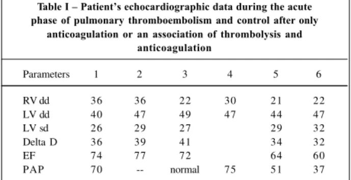
thromboem-Table I – Patient’s echocardiographic data during the acute phase of pulmonary thromboembolism and control after only
anticoagulation or an association of thrombolysis and anticoagulation
Parameters 1 2 3 4 5 6
RV dd 36 36 22 30 21 22
LV dd 40 47 49 47 44 47
LV sd 26 29 27 29 32
Delta D 36 39 41 34 32
EF 74 77 72 64 60
PAP 70 -- normal 75 51 37
RV dd - right ventricle diastolic diameter; LV dd - left ventricle diastolic diameter; LV sd - left ventricle systolic diameter; Delta D - shortening fraction; EF - ejection fraction; PAP - pulmonary artery pressure.
bolism, probably with an obstruction superior to 50% of the pulmonary arterial tree. Generally, the presence of dyspnea, syncope, and cyanosis reflects a more threatening episode of pulmonary embolism.
The echocardiographic findings of diagnostic useful-ness include thrombus direct visualization (rare), right ventricle dilatation, anomalous septal movement, tricuspid valve regurgitation, pulmonary artery dilatation, and reduc-tion of the inspiratory collapse of the inferior vena cava 5. In
the present case, all these echocardiographic signs could be observed, including the thrombus visualization at both hospital admissions. Patients with right ventricular failure due to pulmonary embolism, as in the present case, have a poorer prognosis and might be at a greater risk of relapse and death, when compared with those with normal right ventricular function 6.
Even though perfusion studies and angiography were not performed, this might have been a case of massive pul-monary embolism, according to clinical and laboratory findings of right ventricular failure, low cardiac output, and a quick and positive hemodynamic answer to volume infu-sion without the necessity of vasoactive drugs. As these patients seem more predisposed to relapsing and fatal pul-monary embolisms, in spite of anticoagulation, the current practice is the primary treatment of pulmonary embolism with thrombolytics or mechanical intervention 6. The
primary therapy reduces the thrombus and the hemodyna-mic impact of embolism, preventing the continuous release of serotonin and other neurohumoral factors that aggravate the pulmonary hypertension. It may also dissolve the embo-lic sources located in the pelvis and in the deep veins, and when combined with anticoagulation helps in preventing relapse 7.
There is a tendency to use thrombolysis in cases diag-nosed using noninvasive techniques, without angiogra-phy, which reduces the incidence of bleeding complications, particularly hematomas at the catheterization sites. In the two services where this patient was hospitalized, ventila-tion and perfusion slans were not available. It would have been necessary to remove him to a radiological clinic in a mobile intensive care unit to perform these tests. In case pulmonary embolism is clinically suspected and if there is unequivocal visualization of the thrombus in the atrium, in the right ventricle, or inside the pulmonary artery, and also echocardiographic signs of right ventricle overload, then the treatment should be started. It should include throm-bolysis or surgery, especially if other tests are not available or not advisable 3. As there was no diagnostic doubt and the
onset of appropriate therapy was imminent, full heparini-zation was chosen during the first occasion of treatment of this patient. Due to a prior extensive surgery, to the descri-bed hypothetical risk of thrombus fragmentation, and the occurrence of hemodynamic stabilization with only volume infusion, the classic treatment was chosen using no throm-bolytic agent.
78
Vacanti e col
Relapsing thromboembolism in paraplegic
Arq Bras Cardiol volume 72, (nº 1), 1999
1. Green D, Lee MY, Lim AC et al - Prevention of thromboembolism after spinal cord injury using low-molecular-weight heparin. Ann Intern Med 1990; 113: 571-4. 2. Lualdi JC, Goldhaber SZ - Right ventricular dysfunction after acute pulmonary embolism: Pathophysiologic factors, detection, and therapeutic implications. Am Heart J 1995; 130: 1276-80.
3. Goldhaber SZ, Morpurgo M – Diagnosis, treatment and prevention of pulmonary embolism. JAMA 1992; 268: 1727-33.
4. Sreeram N, Cheriex EC, Smeets JLRM et al – Value of 12-lead electrocardiogram at hospital admission in the diagnosis of pulmonary embolism. Am J Cardiol 1994; 73: 298-303.
thrombolytic agent was used, without performing ventila-tion and perfusion scans or angiography, for the reasons already explained.
Comparing the use of heparin alone to its association with thrombolysis, Goldhaber et al 7, in a study of 101
patients, concluded that in the second group there was an improvement of right ventricular function, a decrease of the final diastolic diameter of the right ventricle and an improve-ment in pulmonary perfusion. All these parameters were statistically significant. The authors stressed that the sub-group of patients with right ventricular hypokinesis, due to the high risk of adverse events with the isolated use of heparin, seemed to be excellent candidates for thrombolytic therapy. Contrary to thrombolysis for acute myocardial
infarction which is limited to the first 12 hours, in pulmonary embolism the patients within 6-14 days of symptom onset benefite equally from this treatment.
Despite of the current recommendation of thrombo-lysis in cases of moderate or massive pulmonary embolism and considering the contraindications, such as cerebral vascular disease, recent surgery or trauma, we had the op-portunity to evaluate two different therapeutic approaches for the same clinical presentation, in the same patient, on different occasions. Although the results were similar, it is not possible to compare the two treatments because our pa-tient was an isolated case. In conclusion, with the available data and in the absence of contraindications, thrombolysis should be, whenever possible, the treatment of choice.
References
5. Kasper W, Geibel A, Tiede N et al – Distinguishing between acute and subacute massive pulmonary embolism by conventional and Doppler echocardiography. Br Heart J 1993; 70: 352-6.
6. Wolfe MW, Lee RT, Feldstein ML et al – Prognostic significance of right ventricular hypokinesis and perfusion lung scan defects in pulmonary embolism. Am Heart J 1994; 127: 1371-5.