ISSN 1806-3713 © 2017 Sociedade Brasileira de Pneumologia e Tisiologia
http://dx.doi.org/10.1590/S1806-37562017000000005
A storm of stones in the lungs: an
uncommon sequela of varicella pneumonia
Gaetano Rea1, Adriano Costigliola2, Cecilia Calabrese21. Dipartimento di Radiologia, A.O. dei Colli, Ospedale Monaldi di Napoli, Napoli, Italia.
2. Dipartimento di Scienze Cardio-Toraciche e Respiratorie, Seconda Università degli Studi di Napoli, Napoli, Italia.
A 29-year-old female patient, never smoker, restorer
by profession, presented with slight malaise and the lu
that lasted for a few days. An X-ray of the chest showed
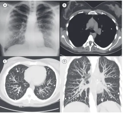
various nodules of varying sizes in random distribution and with high density in both lungs, similar to calciications (Figure 1A). The laboratory test results were unremarkable. Pulmonary function tests and blood gases were normal:
FEV1 = 3.23 L (97% of predicted); FVC = 3.81 L (98% of predicted); DLCO = 98%; pO2 = 97 mmHg; and pCO2 = 39 mmHg. An HRCT of the chest was performed to clarify the chest X-ray indings, showing that the totality of nodules was calciied, but there were no calciied lymph nodes (axial HRCT scan with mediastinal window; Figure 1B). The HRCT scans also showed numerous small bilateral
nodules—sharply deined and randomly distributed in both lungs—but no interstitial thickening or any other pathologic indings (Figures 1C and 1D). Parathyroid function, autoantibodies, and quantiFERON-TB (Cellestis, Ltd., Carnegie, Australia) testing was negative, and blood calcium levels were normal. The patient conirmed a severe varicella infection in childhood (at age 4 years), and antibody testing for the varicella-zoster virus showed positive results (IgG = 348 mIU/mL and IgM = 0.34 mIU/ mL). Varicella (chickenpox) is a contagious viral disease transmitted by respiratory droplets. The development of multiple, small, diffuse nodular calciications in both lungs with noncalciied lymph nodes is an uncommon sequela of varicella pneumonia.
Figure 1. In A, anteroposterior chest X-ray showing innumerable, coarse and punctate micronodules with high calciic density throughout both lungs; in B, axial HRCT scan (mediastinal window) showing no mediastinal calciied lymph nodes. Axial (in C) and coronal (in D) HRCT scans of the chest (thickness of maximum intensity projection: 14 mm and 30 mm, respectively) showing scattered, randomly distributed calciied nodules in both lungs, with no calciied mediastinal lymph nodes, suggestive of healed varicella pneumonia in the appropriate clinical setting.
RECOMMENDED READING
1. Zanetti G, Hochhegger B, Marchiori E. Calciied multinodular lung
lesions: a broad differential diagnosis. Clin Respir J. 2015 Jun 16. [Epub ahead of print] https://doi.org/10.1111/crj.12338
2. Marchiori E, Souza AS Jr, Franquet T, Müller NL. Diffuse high-attenuation pulmonary abnormalities: a pattern-oriented diagnostic approach on
high-resolution CT. AJR Am J Roentgenol. 2005;184(1):273-82. https:// doi.org/10.2214/ajr.184.1.01840273
3. Amin SB, Slater R, Mohammed TL. Pulmonary calciications: a pictorial
review and approach to formulating a differential diagnosis. Curr Probl Diagn Radiol. 2015;44(3):267-76. https://doi.org/10.1067/j.cpradiol.2014.12.005
A B
C D
J Bras Pneumol. 2017;43(3):246-246
246
