An Analysis of Anatomic Variations of the
Sphenoid Sinus and Its Relationship to the
Internal Carotid Artery
Myrian Marajó Dal Secchi
1Ricardo Landini Lutaif Dolci
1Reginaldo Teixeira
2Paulo Roberto Lazarini
11Department of Otorhinolaryngology, Faculdade de Ciências Médicas
da Santa Casa de São Paulo, São Paulo, SP, Brazil
2Department of Radiology, Irmandade da Santa Casa da Misericórdia
de Santos, Santos, SP, Brazil
Int Arch Otorhinolaryngol 2018;22:161–166.
Address for correspondence Myrian Marajó Dal Secchi, PhD, Department of Otorhinolaryngology, Faculdade de Ciências Médicas, Santa Casa de São Paulo, Av. Ana Costa 254, CJ 72, Santos, SP 11060-000, Brazil (e-mail: [email protected]).
Introduction
Surgical access to the anterior region of the skull base via transsphenoidal surgery is commonly used by otorhinolar-yngologists. Radiologic assessment of this region pre- and intraoperatively is fundamental for surgeons to identify the neurovascular structures and their anatomic relationships, thereby reducing possible complications during and after surgical intervention. Computed tomography (CT) of the
paranasal sinus is the best method for studying the anatomy and variation of this region.1–3
The sphenoid sinus (SS) may show varying degrees and directions of pneumatization. Its various extensions and critical relationship to neurovascular structures make intimate knowl-edge of the anatomy of the region and its variations something essential. The internal carotid artery (ICA) is the anatomic structure that must be assessed in the region of the skull
Keywords
►
carotid artery
►
internal
►
paranasal sinuses/
surgery
►
skull base
►
sphenoid sinus
►
sphenoid bone
►
tomography
►
X-ray computed
Abstract
Introduction
The sphenoid sinus (SS) has a high variability; its anatomical relations
and variations must be well understood prior to the expanded endoscopic surgery (EES)
at the skull base via the endonasal transsphenoidal approach. A feared complication is
injury to the internal carotid artery (ICA).
Objective
To evaluate the anatomic variations of the SS and its relationship to the ICA
using computed tomography (CT).
Methods
Cross-sectional retrospective study. Analysis of 90 patients
’
CT scans on
axial, coronal and sagittal planes with 1 mm slices, evaluating lateral and posterior
extensions of pneumatization of the SS, deviation of the sphenoid septum, presence of
septations and their relationship to the parasellar and paraclival segments of the
internal carotid artery (psICA and pcICA, respectively).
Results
The association between the protrusions of the psICA and the pcICA was
statistically signi
fi
cant (
p
<
0.001), as was the association between the lateral extension
of pneumatization of the SS and the protrusion of the psICA (
p
¼
0.014). The presence of
the posterior extension of pneumatization of the SS and protrusion of the pcICA occurred in
46% of the cases. Deviation of the sphenoid septum in the direction of the pcICA was
present in 14% and dehiscence of the pcICA was seen in 3.6% of the cases.
Conclusion
Using the CT scan to recognize the type of extensions of pneumatization
of the SS, the deviation of the sphenoid septum, and the presence of septations is
bene
fi
cial to identify accurately the ICA and to reduce the risk of injury to it.
received April 3, 2017 accepted
September 3, 2017 published online October 25, 2017
DOIhttps://doi.org/ 10.1055/s-0037-1607336. ISSN 1809-9777.
Copyright © 2018 by Thieme Revinter Publicações Ltda, Rio de Janeiro, Brazil
base before and during the surgery. Axial and coronal CT scans are indispensable for disclosing the location of the ICA.4–6
In the clinical practice, CT assessment prior to surgery allows the relationships between the SS and the ICA, and between the lesion and the ICA, to be established. This is fundamental, for example, for planning the endoscopic surgical approach and for safe removal of the lesion with ICA control.6,7 The objective of the present study is to use the CT to analyze the lateral and posterior extensions of pneumatiza-tion of the SS and their relapneumatiza-tionship with parasellar and paraclival segments of the internal carotid artery (psICA and pcICA, respectively). The study also aims to determine the frequency of possible local anatomic variations of the SS in relation to these segments of the ICA adjacent to it.
Methods
This study was approved by the Research Ethics Committee of the Institution (CAAE 14725713.7.0000.5479). A cross-sectional retrospective study of 90 paranasal sinus CT scans was performed in patients clinically indicated for this sup-plementary exam. Inclusion criteria: tomography scans of individuals older than 18 years of age, with rhinosinusal symptoms and request from physician ordering tomography assessment of the nose and paranasal sinus. Exclusion cri-teria: tomography scans identifying individuals with facial bone fractures, rhinosinusal neoplasms, or rhinosinusitis of the posterior paranasal sinus.
Tomography scans were performed using an electro spin resonance (ESR) Hispeed helicoidal tomography manufac-tured by GE, model CT/e (Computed Tomography/helicoidal), series 1676 HMS (GE, Boston, MA, USA). The analysis of the variables was performed using the eFilm 2.1 computer soft-ware program (Merge Healthcare Inc., Chicago, IL, USA).
The exam protocol included the acquisition of images using the helicoidal technique on axial, coronal and sagittal planes, with reconstruction of 1.0/1.0 mm to 1.0/1.0 mm slices (slice thickness/increment). Thefirst image on coronal or axial plane, which identifies the structures of the SS and the ICA, was used for the tomographic analysis in the anterior to posterior and superior to inferior directions, respectively. Based on the images acquired by the tomography scans, the following variables were assessed: the type of tization of the SS, the extensions of the sellar type pneuma-tization, the anatomic variations of the ICA, the presence of septations, and the position of the sphenoidal septum.
Classification of Anatomic Variations of the SS
- Classification of the type of pneumatization of the SS in relation to the sella turcica into: conchal, presellar and sellar. Analysis of the sagittal plane.
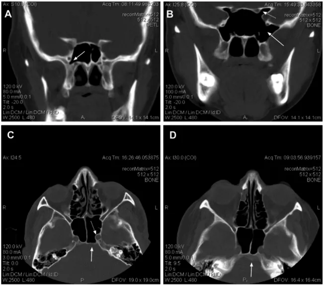
- Classification of the extensions of sellar-type pneumatiza-tion into: lateral (lesser wing, greater wing, pterygoid and complete (greater wing and pterygoid process) (►Fig. 1A,
►Fig. 1B) and posterior (critical and non-critical) (►Fig. 1C, ►Fig. 1D). Analysis of the lateral extensions
on coronal plane and the posterior extensions on axial plane. Two types of posterior extension were considered according to the width between the posterior boundary of the SS and the clivus identified on the axial slices. The type was defined as critical for thickness<2 mm and non-critical for clivus thickness2 mm.
Classification of Anatomic Variations of the Parasellar and Paraclival Segment of the ICA in Relation to the Lateral and Posterior Extensions of the SS
– Absence of protrusion, with protrusion, and presence of
dehiscence. Analysis of the psICA on coronal plane (►Fig. 2A) and the pcICA on axial plane (►Fig. 2B). Protru-sion was defined as>50% projection of the structure into the sphenoid sinus and dehiscence as the absence of visible bone separating the ICA from the SS along its path.
Classification Regarding the Presence or Absence of Septations
Analysis on Axial and Coronal Planes (►Fig. 3A,►Fig. 3B)
– Classification of the sphenoidal septum: on the sagittal
plane, with deviation to the right or left and in the direction of the ICA. Analysis of axial plane (►Fig. 3C,►Fig. 3D).
The chi-square test was used for statistical analysis of the data, and a 5% level of significance was adopted.
Results
Total 90 tomographic imaging exams were studied in pa-tients whose ages ranged from 19 to 84 years with a mean of 46 years (SD¼16.4) and median of 44 years. The sample comprised 33% males and 67% females.
The analysis of the pneumatization of the SS in relation to the sella turcica revealed a predominance of the sellar type in 98% of the patients, the presellar type in 2% of patients and no cases of the conchal type. Lateral extension of pneumatiza-tion of the SS for the lesser wing of the sphenoid was found in 13% of the individuals, with the greater wing type in 47%, the pterygoid type in only 1%, the complete type in 23% and absent in 16%. The presence of posterior extension was found in 78% of the individuals, comprising critical type pneuma-tization in 42%, non-critical type in 36%, and absent in 22%. Protrusion of the psICA was found in 26% of the indivi-duals and it was absent in 74%, whereas protrusion of the pcICA was noted in 35% and absent in 64% of the patients. Dehiscence of the pcICA was observed in four cases (3.6%), two of which were right side and two were left sides.
The statistical analysis using the chi-square test for the association between protrusions of the pcICA and psICA was statistically significant (p<0.001), as was the association between the lateral extension of pneumatization of the SS and protrusion of the psICA (p¼0.014) (►Table 1). A pre-sence of protrusion of the pcICA in individuals of the poster-ior extension of pneumatization of the SS was present in 46% and absent in 54% of the cases.
was observed between individuals exhibiting septations with or without posterior extension (p¼0.908).
The deviation of the sphenoid septum either to the right or left was found in 60% of the individuals; on the sagittal plane, in 26%; deviation in the direction of the ICA, in 14%, and in one or more septum, in 5% of the CT scans. In those individuals exhibiting posterior extension, the position of the sphenoid septum most commonly observed was devia-tion. No significance was reached on the chi-square test (p¼0.289) (►Table 2).
Discussion
Extensive pneumatization of the SS can be associated with irregularities in the sinus wall featuring protrusions and
recesses, such as prominences of the ICA. Computed tomo-graphy can help to identify the relationship of the ICA with the lateral and posterior extensions of pneumatization of the SS, and alert the surgeons to regions for possible injury occurrences.8,9
The present study evaluated the degree and types of pneumatization of the SS and their relationship with the paraclival (pcICA) and parasellar (psICA) segments of the ICA. These segments of the ICA are adjacent to the SS and can be damaged during expanded endoscopic approaches (EEA) to the skull base.5,7,10,11
In the literature, protrusion of the ICA ranges from 8 to 70%.12In the present study, however, the presence of psICA protrusion occurred in 26% of the individuals, while the presence of protrusion of the pcICA was detected in 35%. In this study, a statistically significant association was found between protrusion of the psICA and pcICA (p<0.001). Dehiscence in the pcICA was found in 3.6% of the individuals compared with 5% of cases identified by Meloni et al,11.5% by Kazkayasi et al,314.4% by Johnson et al,4and 5.3% of cases by Unal et al.13In the current study, the pcICA and psICA were evaluated separately and no cases of dehiscence in the psICA was detected. Axial and coronal CT scans are indispensable for disclosing the location of these segments adjacent to the SS.4–6 Assessment of pneumatization of the SS helps surgeons to elect the optimum approach and determine the impact of anatomic variations on the surgery.9,14,15 In this study, the Fig. 3 (A) Axial plane, presence of septations (arrows), (B) Coronal plane, presence of septation to the left (arrow), (C) Axial plane, sphenoid septum on sagittal plane (arrow) and protrusion of paraclival segment (pcICA) and (D) Axial plane, deviation of the sphenoid septum in the direction of the paraclival segment of the internal carotid artery (pcICA).
Table 1 Percentage distribution of lateral extension of pneumatization of sphenoid sinus in relation to protrusion of the psICA
Extension Protrusion of psICA
Total p
absent present
Lateral absent 46 54 100 0.014
present 17 83 100
sellar type was the most frequent, found in 98% of the cases, the presellar type occurred in 2% of cases, while no instances of the conchal type were detected, mirroring the results of the study by Wang et al.8On CT, the configuration of the SS can be identified on sagittal plane, where the sellar type facilitates EEAs, with lower risk of injury to the neurovascular structures surrounding the SS, although there is less bone to remove during exposure of the operative region. However, it becomes easier to identify the landmarks, whereas for the presellar type, the SS walls tend to be thicker and more difficult for transsphenoidal approaches. Lateral and posterior extension may occur in sellar-type pneumatization, and a classification of these extensions is important as a guide to the approaches extending beyond the sella turcica.
In the present study, the greater wing lateral type exten-sion was the most frequently found, in contrast to Wang et al,8who predominantly found the complete type when the extension is to the greater wing and the pterygoid process. Both types facilitate access to the cavernous sinus, middle cranial fossa and petrous apex because there is less bone to remove. These disparities may be explained by individual variations in the population. The posterior extension, in turn, was identified in 78% of all CTs, comprising 42% critical and 36% non-critical types, demonstrating the high incidence of this anatomic variation. Haetinger et al16detected posterior extension in 69% of the cases and 44% of these were of the critical type. The critical type facilitates access to lesions located in the clivus and petrous apex, but there is an associated risk of perforation of the clivus due to the bone thickness of this region.16This classification of the lateral and the posterior extensions in the CT provides a guide to evaluate the degrees and directions of the pneumatization of the SS and its relationship with the psICA and the pcICA. In this study, extensions of pneumatization into the lesser wing were considered lateral type extensions because this definition betterfits the classification of the lateral extensions on CT scans, given the location of this extension in the superior and lateral region of the SS and its relationship with both the optic nerve and psICA. On the other hand, Wang et al7classified the lesser wing type separately from the lateral extensions. This extension was found in 13% of the current cases.
A statistically significant association was found between pneumatization of the lateral extension and the presence of protrusion of the psICA (p¼0.014). Pneumatization of the posterior extension and presence of protrusion of the psICA were relevant in 46% of the cases. This association disclosed
on CT alerts surgeons of the impact of these variations on the surgery, whose outcome is linked to the increase of the SS to beyond normal anatomic limits. Thus, preoperative CT can identify areas with potential risk of complication, such as ICA injury in the SS featuring these extensions.
The local variations of the SS identified on CT include deviations of the sphenoid septum and the presence of septa-tions in the SS.2The computed tomography provides preo-perative anatomic information regarding the course of the ICA to these local variations of the SS. Injury to the ICA can thus be avoided during surgery. In this study, for those individuals exhibiting posterior extension, the position of the septum most commonly observed was deviation, found on 60% of scans, although this association was not statistically significant (p¼0.289), 14% of the individuals had deviation of the septum in the direction of the pcICA, and septations were identified in 39% of the cases. No statistically significant difference between individuals exhibiting septations with and without posterior extensions was found (p¼0.908).
The sphenoidal septum is often deflected to one side or the other, and can attach to the bone wall that protects the ICA, where fracture of the septum in order to gain access to the ICA can damage this artery.2,3,9,13,17There is a potential risk of injury when septations are located near the promi-nence of the ICA. Therefore, identifying the artery is crucial to keep a safe distance during tumor resection.9,18–20
An analysis of the anatomic variations of the SS observed on CT scans and their relationships with the segments of the ICA adjacent to the SS allows for more specific descriptions by the radiologists regarding patients who require EEA to the skull base, where key anatomic features are highlighted such as: the types of pneumatization of the SS and their extensions; irregularities in the walls of the psICA and pcICA; position of the septum in relation to the ICA and presence of septations in the SS. This information acquired, in conjunction with a preoperative assessment of imaging exams by the surgeon and their team, alert to all the aspects outlined above. There-fore, identifying the anatomic boundaries of this region for surgery can be done with more accuracy and reliability.
Conclusion
The evaluation of the SS preoperatively using a CT scan can aid surgeons to be prepared before performing an endo-scopic skull base surgery. Particularly, it provides essential tools to know what will be found at the surgery, avoiding any Table 2 Percentage distribution of the position of sphenoid septum, confirmed by paranasal sinus CT, according to the presence or absence of posterior extension of pneumatization of sphenoid sinus
Extension Sagittal
plane
Deviation ICA path deviation
Total p
Posterior present 23 60 17 78 0.289
absent 35 60 5 22
Total 26 60 14 100
iatrogenic lesion, such as damage to the ICA or noble struc-tures, and thus allowing to better plan the surgery. The anatomic variations and their relationship should be re-ported by the radiologist.
Preoperative recognition of the type of extensions of pneumatization of the SS, deviation of the sphenoid septum and presence of septations are beneficial to identify safely the psICA and pcICA adjacent to the SS. This information may be helpful if intraoperative surgical navigation is used.
Conflict of Interest
No conflicts of interest have been declared by the authors.
Informed Consent
This study was retrospective; thus, the informed consent was not required.
References
1 Meloni F, Mini R, Rovasio S, Stomeo F, Teatini GP. Anatomic variations of surgical importance in ethmoid labyrinth and sphe-noid sinus. A study of radiological anatomy. Surg Radiol Anat 1992;14(01):65–70
2 Sirikci A, Bayazit YA, Bayram M, Mumbuç S, Güngör K, Kanlikama M. Variations of sphenoid and related structures. Eur Radiol 2000; 10(05):844–848
3 Kazkayasi M, Karadeniz Y, Arikan OK. Anatomic variations of the sphenoid sinus on computed tomography. Rhinology 2005;43 (02):109–114
4 Johnson DM, Hopkins RJ, Hanafee WN, Fisk JD. The unprotected parasphenoidal carotid artery studied by high-resolution com-puted tomography. Radiology 1985;155(01):137–141
5 Kennedy DW, Zinreich SJ, Hassab MH. The internal carotid artery as it relates to endonasal sphenoethmoidectomy. Am J Rhinol 1990;4(01):7–12
6 Labib MA, Prevedello DM, Carrau R, et al. A road map to the internal carotid artery in expanded endoscopic endonasal ap-proaches to the ventral cranial base. Neurosurgery 2014;10 (Suppl 3):448–471, discussion 471
7 Gardner PA, Tormenti MJ, Pant H, Fernandez-Miranda JC, Snyder-man CH, Horowitz MB. Carotid artery injury during endoscopic
endonasal skull base surgery: incidence and outcomes. Neuro-surgery 2013;73(2, Suppl Operative)ons261–ons269, discussion ons269–ons270
8 Wang J, Bidari S, Inoue K, Yang H, Rhoton A Jr. Extensions of the sphenoid sinus: a new classification. Neurosurgery 2010;66(04): 797–816
9 Hamid O, El Fiky L, Hassan O, Kotb A, El Fiky S. Anatomic variations of the sphenoid sinus and their impact on trans-sphenoid pitui-tary surgery. Skull Base 2008;18(01):9–15
10 Amin SM, Nasr AY, Saleh HA, Foad MM, Herzallah IR. Endoscopic orientation of the parasellar region in sphenoid sinus with ill-defined bony landmarks: an anatomic study. Skull Base 2010;20 (06):421–428
11 Anusha B, Baharudin A, Philip R, Harvinder S, Shaffie BM. Anato-mical variations of the sphenoid sinus and its adjacent structures: a review of existing literature. Surg Radiol Anat 2014;36(05): 419–427
12 Kantarci M, Karasen RM, Alper F, Onbas O, Okur A, Karaman A. Remarkable anatomic variations in paranasal sinus region and their clinical importance. Eur J Radiol 2004;50(03):296–302 13 Unal B, Bademci G, Bilgili YK, Batay F, Avci E. Risky anatomic
variations of sphenoid sinus for surgery. Surg Radiol Anat 2006;28 (02):195–201
14 Vaezi A, Cardenas E, Pinheiro-Neto C, et al. Classification of sphenoid sinus pneumatization: relevance for endoscopic skull base surgery. Laryngoscope 2015;125(03):577–581
15 Hammer G, Radberg C. The sphenoidal sinus. An anatomical and roentgenologic study with reference to transsphenoid hypophy-sectomy. Acta Radiol 1961;56:401–422
16 Haetinger RG, Navarro JA, Liberti EA. Basilar expansion of the human sphenoidal sinus: an integrated anatomical and compu-terized tomography study. Eur Radiol 2006;16(09):2092–2099 17 Aksoy F, Yenigun A, Goktas SS, Ozturan O. Association of accessory
sphenoid septa with variations in neighbouring structures. J Laryngol Otol 2017;131(01):51–55
18 Fernandez-Miranda JC, Prevedello DM, Madhok R, et al. Sphenoid septations and their relationship with internal carotid arteries: anatomical and radiological study. Laryngoscope 2009;119(10): 1893–1896
19 Poirier J, Duggal N, Lee D, Rotenberg B. Sphenoid sinus septations: unpredictable anatomic landmarks in endoscopic pituitary sur-gery. J Otolaryngol Head Neck Surg 2011;40(06):489–492 20 Dasar U, Gokce E. Evaluation of variations in sinonasal region with