Are Histologic Studies of Adenotonsillectomy
Really Necessary?
Giseli Rebechi
1Thiago Euênio Pontes
1Elias Lobo Braga
1Willian Maduel Matos
1Fernando Rebechi
1Cícero Matsuyama
11Setor de Otorrinolaringologia, Instituto CEMA, São Paulo/SP, Brasil
Int Arch Otorhinolaryngol 2013;17:387–389.
Address for correspondence Giseli Rebechi, Avenida Indianópolis, 740, Bairro, Moema, CEP 04062-001, São Paulo/SP, Brasil (e-mail: [email protected]).
Introduction
Adenotonsillectomy is surgery to remove the tonsils and adenoids, and it is one of the most common and most frequently performed surgical procedures in the world.1,2 Its different indications can be divided into therapeutic, diagnostic, as well as access to other surgeries. Sleep apnea syndrome, hypertrophic tonsils and adenoids, chronic tonsil-litis, halitosis, and suspicion of malignancy are indications for adenotonsillectomy, but the most frequent indication is recurrent tonsillitis.
Most otolaryngology services routinely send adenotonsil-lectomy specimens for histopathologic examination, whether
for malignancy investigation, analysis of suspect material, or medicolegal documentation of surgical removal.1 Recent studies have shown that routine histopathologic analysis of the tonsil is dispensable, because they have a very low probability of diagnosing occult malignancies. Unfortunately, this risk is still not zero, so the need for routine histopatholo-gy is still controversial.
Objective
Define the real need for routine histopathologic examination of adenotonsillectomy specimens and perform a cost–benefit anal-ysis of its use in patients without risk factors for malignancy. Keywords
►
tonsillar neoplasms
►
tonsillectomy
►
adenoidectomy
Abstract
Introduction
In most ear, nose, and throat services, it is routine to send the material
extracted from tonsillectomy for histologic study to research malignancy, to analyze
suspect material, or to provide medical-legal documentation. Recent studies have
shown that this routine analysis is dispensable.
Objective
To evaluate the actual need and perform a cost
–
bene
fi
t analysis of routine
histopathologic examination in tonsillectomy with no signs or symptoms of malignancy.
Methods
A retrospective observational study evaluated the charts of patients
under-going adenotonsillectomy, tonsillectomy, or adenoidectomy from January 2008 to
September 2009 at the Institute of Otorhinolaryngology CEMA-SP. Costs of this test for
the public health system were analyzed and the literature reviewed.
Results
We studied 281 patients between 2 and 22 years of age; 142 (50.5%) were
male and 139 (49.5%) were female. Of the surgeries, 201 were adenotonsillectomies
(71.5%), 41 were tonsillectomies (14.5%), and 39 were adenoidectomies (14%). The
most common indication for surgery was recurrent infection (63.3%). None of study
patients had clinical suspicion of malignancy. The tests showed a cost of R$20.03 per
tonsil analyzed.
Conclusion
Routine histopathologic examination in patients undergoing
adenoton-sillectomy with no signs or symptoms of malignancy is dispensable and increases the
cost of the surgeries.
received June 8, 2013 accepted July 11, 2013
Copyright © 2013 by Thieme Publicações Ltda, Rio de Janeiro, Brazil
DOI http://dx.doi.org/ 10.1055/s-0033-1353441. ISSN 1809-9777.
Materials and Methods
This retrospective observational study evaluated the records of all patients who had adenotonsillectomy, tonsillectomy, or adenoidectomy from January 2008 to September 2009 at the CEMA Institute of Otorhinolaryngology, São Paulo, Brazil. This project was approved by the Ethics and Research Institution under the protocol 17.205/2009. Patients or their guardians signed an informed consent form, shown inAppendix 1. Data analysis was performed using descriptive statistics, and the results are presented in absolute numbers.
We excluded patients with malignancy symptoms and signs. All patients underwent general anesthesia and tonsil-lectomy by extraction dissection technique. The pharyngeal tonsils were removed with a Beckmann curette. Specimens were immediately placed in sterile glass with 10% formalin and sent for histologic analysis. The tonsils werefixed in 10% formalin and embedded in paraffin, and sections were stained with hematoxylin-eosin. We studied the results of the histopathologic examinations, regardless of age, sex, and indication for surgery. We also analyzed the cost of this test for the public health system, and we reviewed the literature.
Results
A total of 281 patients were recruited, between 2 and 22 years old. Of them, 142 (50.5%) were male and 139 (49.5%) were female. Most patients had tonsil hypertrophy grade III accord-ing to the Brodsky classification and pharyngeal tonsil hyper-trophy documented by nasofibrolaryngoscopy.
Of the surgeries, 201 were adenotonsillectomies (71.5%), 41 were tonsillectomies (14.5%), and 39 were adenoidecto-mies (14%). The most common surgical indications were recurrent tonsillitis (63.3%) and obstructive sleep apnea syndrome (38.7%). None of the patients in our study had clinical suspicion of malignancy.
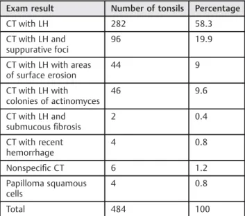
Results of the pathologic examinations of tonsils and adenoids are shown in ►Tables 1 and 2. The tests cost R$20.03 per piece analyzed; the adenotonsillectomy surgery has three pieces, reaching a total of R$60.09 per surgery.
Discussion
Tonsils with malignant processes present different aspects than benign lesions on clinical examination; physical exami-nation and medical history can be important guides. These lesions may start with nonspecific symptoms and insidious symptoms, and they are usually diagnosed in patients over 60 years old. Smoking and alcoholism are risk factors.
According Beaty et al,3risk factors for tonsillar malignancy include history of head and neck cancer, tonsillar asymmetry, visible lesion, ulcerated or hard consistency on palpation of the tonsil, unexplained weight loss or constitutional symp-toms, and cervical lymphadenopathy. Usually the patient starts with dysphagia without signs of acute infection with ipsilateral otalgia, difficulty in mobility of the tongue, nasal voice, halitosis, and nasal reflux. Symptoms such as changes in tone of voice, drooling, bloody saliva, and trismus indicate deep infiltration of the tumor.
Many authors have reported that tests with positive results for malignancy had a suspected diagnosis before surgery. The result of histopathologic exams on tonsil specimens correlates well with preoperative clinical impressions, and theirfindings rarely change the management of the patient.4
Randall et al reported a prevalence of malignancy of 0.087% in routine examinations. In these patients, 88% had preoperative suspicion of malignancy. Among the tests, 0.011% had a positive result but no risk factor; the authors concluded that routine examination was unnecessary if there was no suspicion of malignancy.1Garavello et al noted a 0.18% incidence of positive histopathologic analysis without clinical suspicion in children, concluding that routine examinations were unnecessary.5 Felix et al found a 0.19% incidence of positivity; however, all of the patients had some risk factor for tonsillar malignancy. The examinations did not locate any hidden malignancies.6 Williams and Brown found 4,070 tonsils with malignancy on histopathology, and all of them had been diagnosed during preoperative evaluations.7 A study by DellAringa et al showed no malignancy in the patients analyzed,finding a negative cost–benefit ratio for routine histopathologic exams.8 Younis et al presented Table 1 Results of histopathologic examination of the tonsils
Exam result Number of tonsils Percentage
CT with LH 282 58.3
CT with LH and suppurative foci
96 19.9
CT with LH with areas of surface erosion
44 9
CT with LH with colonies of actinomyces
46 9.6
CT with LH and submucousfibrosis
2 0.4
CT with recent hemorrhage
4 0.8
Nonspecific CT 6 1.2
Papilloma squamous cells
4 0.8
Total 484 100
Abbreviations: CT, chronic tonsillitis; LH, lymphatic hyperplasia.
Table 2 Results of histopathologic examination of the pharyngeal tonsils
Exam result Number of tonsils Percentage
Luschka pharyngeal adenoid hypertrophy
237 98.75
Lymphoid tissue of reactive pattern
2 0.84
Malpighian mucosa with vascular ectasia
1 0.41
Total 240 100
International Archives of Otorhinolaryngology Vol. 17 No. 4/2013
research that showed none of the 2,099 pediatric patients undergoing tonsillectomy had malignancy found on histo-pathologic exam, but this incidence differed from the adult population.9Mohamad et al also found in their study that routine examination is not necessary in the pediatric popu-lation.10 Many authors have showed that histopathologic exam of specimens from children is superfluous11and that increasing age is a risk factor to be considered.12This fact is due to very different indications for tonsillectomy in different age groups; in adults, the incidence of excisional biopsies and symptoms of malignancy are much higher than in pediatric patients.
Spending with microscopic examinations by tonsil varies greatly in the studies, ranging from US $12.85 to US $90.00. Annual spending in the United States is approximately US $35,467,080.00. For the Brazilian government, spending on each piece is R$20.03.
Conclusion
Routine histopathologic examination in pediatric adenotonsil-lectomy specimens is dispensable, with a negative cost–benefit ratio. Despite all these studies showing that such tests burden the public purse as well as private health systems, surgeons are required to apply for the pathologic study because health plans require its result for the payment of the surgery.
We emphasize the importance of a thorough history and clinical examination in all patients undergoing adenotonsil-lectomy. In children, the chance of occult malignancy is very low. In patients with risk factor present in clinical ear, nose, and throat examination, this exam is indispensable. With more careful preoperative assessment, we could save millions of dollars per year.
References
1 Randall DA, Martin PJ, Thompson LDR. Routine histologic exami-nation is unnecessary for tonsillectomy or adenoidectomy. Laryn-goscope 2007;117:1600–1604
2 Cinar F. Significance of asymptomatic tonsil asymmetry. Otolar-yngol Head Neck Surg 2004;131:101–103
3 Beaty MM, Funk GF, Karnell LH, et al. Risk factors for malignancy in adult tonsils. Head Neck 1998;20:399–403
4 Ikram M, Khan MAA, Ahmed M, Siddiqui T, Mian MY. The histopathology of routine tonsillectomy specimens: results of a study and review of literature. Ear Nose Throat J 2000;79:880–882
5 Garavello W, Romagnoli M, Sordo L, Spreafico R, Gaini RM. Inci-dence of unexpected malignancies in routine tonsillectomy speci-mens in children. Laryngoscope 2004;114:1103–1105
6 Felippe F, Gomes GA, de Souza BP, Cardoso GA, Tomita S. Evaluation of the utility of histopathologic exam as a routine in tonsillecto-mies. Braz J Otorhinolaryngol 2006;72:252–255
7 Williams MD, Brown HM. The adequacy of gross pathological examination of routine tonsils and adenoids in patients 21 years old and younger. Hum Pathol 2003;34:1053–1057
8 Dell’Aringa AR, Juares AJ, Melo Cd, Nardi JC, Kobari K, Perches Filho RM. Histological analysis of tonsillectomy and adenoidectomy specimens–January 2001 to May 2003. Braz J Otorhinolaryngol 2005;71:18–22
9 Younis RT, Hesse SV, Anand VK. Evaluation of the utility and cost-effectiveness of obtaining histopathologic diagnosis on all routine tonsillectomy specimens. Laryngoscope 2001;111:2166–2169
10 Mohamad I, Hassan S, Salim R. The routine histopathological examination of tonsillectomy specimens at hospital universiti sains Malaysia—retrospective study and its implication. Malays J Med Sci 2007;14:19–21
11 Strong EB, Rubinstein B, Senders CW. Pathologic analysis of routine tonsillectomy and adenoidectomy specimens. Otolaryngol Head Neck Surg 2001;125:473–477
12 Erdag TK, Ecevit MC, Guneri EA, Dogan E, Ikiz AO, Sutay S. Pathologic evaluation of routine tonsillectomy and adenoidec-tomy specimens in the pediatric population: is it really necessary? Int J Pediatr Otorhinolaryngol 2005;69:1321–1325
Appendix 1 Model of the term of consent
CEMA Institute (Department of Otorhinolaryngology) Term of consent
We invite the Sr _______________________________________ to participate in the survey“Are histologic studies of adenotonsillectomy really necessary?”under the responsibility of the researcher Dra Giseli Rebechi, to analyze the need for routine histopathologic studies on the products of adenotonsillectomy in patients without risk factors for malignancies and to analyze the costs and benefits of the studies. Your participation is voluntary and will be through the authorization and release of medical records. If you agree to participate, you will contribute to analysis of the real need for routine checkups and lower expenditures of public and private health care.
If after consenting to participation you decide to stop participating, you have the right and freedom to withdraw consent at any stage of the research, either before or after the collection of data, regardless of the reason, and without any prejudice. You will not have expense and you will not receive any remuneration. The search results will be displayed, but your identity will not be disclosed and will be kept secret. For any other information, you can may contact the researcher at the address Indianapolis Avenue, 740, Moema, São Paulo/SP, Brazil, by phone (11) 5082 3420, or you may contact the Committee on Ethics in Research-Institute of Otorhinolaryngology CEMA, Pascoal Moreira street, 450, Mooca, São Paulo/SP, Brazil, phone (11) 2602 4000.
Informed consent
I, ___________________________________________________________, was informed about what the researcher wants to do and why she need my collaboration, and I understand the explanation. Therefore, I agree to participate in the project, knowing that I will not gain anything and I can leave whenever I want. This document is issued in two copies to
be signed both by me and the researcher, providing a copy to each of us.
_________________________________ Signature of Participant
_________________________________
Signature of Researcher Principal