C&ar A. Cuba Cuba,3 Philip
D. Marsden,
Air C. Baneto,
Roxicler
Rocha,3 Raymunda
R. Sampaio,3 and Laura Patzlaff4
The purpose of the work reported here is to evaluate a group of parasitologic and immunologic techniques used to diagnose American (mucocutaneous) tishmnn- iasix One of the chief di#iculties encountered is that of detecting parasites in the lesions. This makes it necessary to ure serologic methodc that are less than ideally sensitive and specific, a shortcoming con$rmed by the findings of this study.
Introduction
A marked increase in the number of muco- cutaneous leishmaniasis cases coming to the dermatology service of Brasilia University’s teaching hospital, together with study of an area endemic for this disease in the state of Bahia, led us to focus our attention on the problem of laboratory diagnosis of this proto- zoan disease.
For routine diagnosis, the usual procedure consists of checking the patient’s lesions for
Leishmania amastigotes and administering the Montenegro intradermal test (27). Of the other diagnostic methods available, the in- direct immunofluorescence test (24 22) has not been thoroughly evaluated, while the di- rect agglutination and immunodiffusion tests (I, 6,9) are not used for routine diagnosis.
With regard to the etiologic agent of the dif- ferent clinical forms of mucocutaneous leish- maniasis found in South America, researchers
‘Also published in Spanish in the Boleth de la Oficina SanitrwiaPanameTica~ 89(3):195-208, 1980.
ZThis study was conducted with the assistance of the National Scientific and Technological Development Council of Brazil and the Superintendency of Public Health Campaigns (SUCAM) of the Brazilian Ministry of Health.
3Professor, Health Sciences Faculty, University of Bra&a, Bra&a, D.F., Brazil.
4Professor, Institute of Exact Sciences, University of Brasilia, Brasilia, D.F., Brazil.
have become increasingly interested in iso- lating the responsible parasites-so as to in- vestigate their biological (3,23,2#, 25), bio- chemical (8,17), and ultrastructural (18) char- acteristics for purposes of identification.
This article presents a number of observa- tions concerning mucocutaneous leishmania- sis patients who received parasitologic and im- munodiagnostic examinations, and discusses the sensitivity of the tests employed.
Materials and Methods
Selection of Patients
A total of 56 patients with mucocutaneous leishmaniasis were observed at the Brasflia University teaching hospital’s dermatology service and at various places in the municipal- ities of Cravolandia, Wenceslau Guimarzes, and Ubaira in the Brazilian state of Bahia. Each patient was given a physical examina- tion; and, as a result, 29 were classified clini- cally as having an active cutaneous form of the disease, while the other 27 were classified as having an active mucosal form. A sample of venous blood was taken from each patient for serologic testing. The length of time that the lesions had been present ranged from 3 months to 26 years. The possibility that any patient might have Chagas’ disease or kala- azar was ruled out.
250 PAHO BULLETIN . vol. 15, no. 3, 1981
Diagnostic Laboratory Methods
Insofar as possible, nearly all the selected patients were given the same diagnostic tests in a parallel and systematic manner. Biopsy specimens taken with a skin punch were used to prepare contact smears and histological specimens, and to provide a source of tritu- rated tissue for injection into hamsters. In ad- dition, each patient was given the Montene- gro intradermal test, and patient sera were tested for Leishmania antibody by indirect im- munofluorescence and direct agglutination tests.
Parasitologic tests. Three contact smears were procured from each patient and were stained with Giemsa (pH 7.5) for 1 hour.
For purposes of histologic examination, part of each biopsy specimen was placed in Zenker-form01 fixative for up to 6 hours and was then stored in 70 per cent alcohol to await processing. Subsequently, sections 5 microns thick were prepared and stained with hema- toxylin-eosin for examination.
Triturates prepared from the biopsy speci- mens were inoculated into the snouts and hind feet of hamsters (Mesocricetus aura@) obtained from Brasilia University’s central vivarium. The animals were observed periodically so as to obtain a systematic record of the evolution of their infections. Material from infected hamsters was subsequently inoculated into other hamsters and Swiss 44 white mice. The isolated parasites were classified biologically according to the recommendations of Lainson and Shaw (23,24,25). Cultures were produced in NNN and LIT5 media as described by Ca- margo (7), were maintained at 22’C, and were examined and transferred every 15 days. Immunologic tests. All the patients were given the Montenegro intradermal test. That is, all were tested intradermally with antigen (leish- manin) made from promastigotes of Leishma-
‘NNN = Navy, Nicolle, and McNeil medium; LIT = liver infusion tryptose medium.
nia mexicana amazonensis (Josefa strain) isolated from a human case with a cutaneous lesion ob- served at the University of Brasilia. These had been maintained in NNN and LIT culture media, with periodic reinoculation into ham- sters. The protein nitrogen (N) content of ap- proximately 5 x lo6 flagellates per ml was determined in 30 pg N per ml, using the Goa microburet method (19).
A tenth of a milliliter of antigen was inocu- lated intradermally into each patient’s right forearm, and an equal volume of phenolated physiologic solution was inoculated into the left forearm as a control. The reading was made in millimeters of induration perceptible to the touch 72 hours after inoculation.
Antigen for the indirect immunofluores- cence test was prepared from L. mexicana ama- zonensis promastigotes following the general guidelines of the method recommended by Guimarzes et al. (20). The reactivity of the 56 patients’ sera was then tested using this an- tigen.
In addition, amastigote antigen was pre- pared from the same strain, following the pro- cedure described by Shaw and Lainson (31), and was used to test sera from 24 patients with parasitologically confirmed cases. This pro- vided a means of comparing the relative sensi- tivity of the results when promastigotes and amastigotes were used as antigen.
Both indirect immunofluorescence methods employed human anti-gamma-globulin la- beled with fluorescein isothiocyanate (F/P 0.64) from the Wellcome Research Labora- tories, England (Lot K 0403); a Zeiss-Jena dark-field fluorescent microscope; a HBO-50 light source and ultraviolet lamp; and barrier filters and exciters OGl and BB 224, respec- tively. Interpretation of the findings obtained with our stra took into account the criteria adopted by Chiari (IO), Guimarzes et al. (201, and Shaw and Lainson (31).
mexicana amazonensis. A total of 46 patients were tested using this procedure.
Statistical Analysis
The data obtained were assessed by calcula- tion of the reciprocals of geometric mean ti- ters, tests of variance, and Student’s t test of averages of the distribution frequency of the patients’ serum titers. The levels of the tests were ~~0.1 and p<O.O5.
Results
Parasitologic Tests
The three parasitologic tests (involving con- tact smears, histologic examination, and ham- ster inoculation) revealed the presence of parasites in 24 of the 56 patients tested. Four- teen patients (25 per cent) yielded parasite- positive smears, and histologic examination of biopsy specimens from 15 of 55 patients (27.3 per cent) revealed amastigotes. Table 1 shows the clinical features of the 24 parasite-positive cases, as well as the results of these and other tests.
Of the 56 patients, 29 had the active cuta- neous form of American leishmaniasis while 27 had the mucosal form of the disease. Six- teen cases (55.1 per cent) of the group with cutaneous lesions showed evidence of amasti- gotes in the lesions, but only 8 cases (29.6 per cent) in the group with mucosal lesions could be parasitologically confirmed. Of the 24 parasitologically confirmed cases (both cuta- neous and mucosal), three (12.5 per cent) were confirmed on the basis of smears alone; one (4.1 per cent) was confirmed on the basis of histologic examination alone; and six (25 per cent) were confirmed on the basis of ham- ster inoculation alone. A relatively high densi- ty of amastigotes was found in another six pa- tients with a long history of chronic lesions go- ing back anywhere from 7 to 20 years, the parasites being detected in both smears and histologically examined biopsy specimens
from the members of this group. The remain- ing eight patients all yielded positive results in at least two of the three parasitologic tests.
Four patients with recent ulcers (compatible with American leishmaniasis) only yielded positive parasitologic results through hamster inoculation-when the inoculated animals de- veloped nodules containing numerous amas- tigotes. (The other two patients yielding para- sitologically positive results only via hamster inoculation had chronic lesions.)
Of the 18 patients whose lesions yielded parasitologically positive results by other methods, 17 provided infected material that was inoculated into hamsters. Nine of these 17 animals became infected. Five showed no signs of cutaneous infection over periods of observation ranging between 5 and 9 months from the time of inoculation. Two were sacri- ficed because they were on the verge of dying but showed no signs of being infected 7 months after inoculation. One died unob- served. And one Leishmania strain (from pa- tient 21) produced a visceral infection.
The isolates from patients 1, 2, and 3 were subsequently reinoculated into other hamsters between six and eight consecutive times. Although the incubation period was shorter for these latter passages-35-40 days, prob- ably due to the fact that the inoculum became progressively richer in parasites-the evolu- tion of the infection retained its basic char- acteristics (rapid growth of the lesion, early ulceration, intense cutaneous parasitization, and minimal inflammatory infiltrate). Cul- tures were easily obtained, and growth of the promastigote forms was exuberant. The three isolates adapted easily to Swiss 44 white mice; and the organisms from patient 3 produced metastasis in both hamsters and mice.
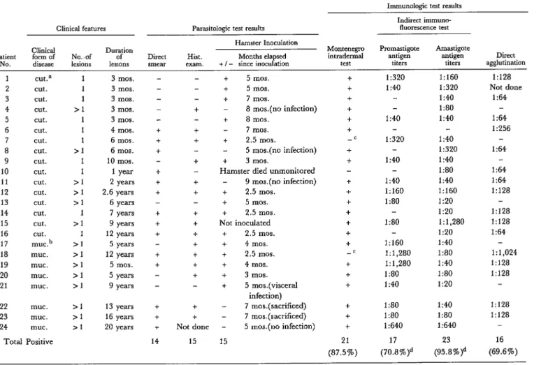
Table 1. Clinical features, parasitologic findings, and immunologic test results of 24 cases of parasitologically confirmed American (mucocutaneous) leishmaniasis.
Clinical features Parasitologic test results
Immunologic test results Indirect immuno-
fluorescence test Hamster Inoculation
Clinical Montenegm Pmmastigote Amastigote
Patient form of No..of D”LPn Direct Hist. Months elapsed intradermal antigen antigen Direct
NO. disease lesions lewns smear exam. + I - since inoculation test titers titers agglutination
1 2 3 4 5 6 7 8 9 10 11 12 13 14 15 16 17 18 19 20 21 22 23 24 clKa cut. cut. cut. cut. cut. cut. cut. cut. cut. cut. cut. cut. cut. cut. cut. muc.b muc. muc. UIUC. muc. muc. muc. muc.
Total Positive
1 1 1 11 1 1 1 >l 1 1 >l >l >l 1 >l 1 11 >l >l >l 11 >l >l >1 3 mm. 3 mos. 3 mos. 3 mos. 3 mos. 4 mm. 6 mm. 6 mm. 10 mos. 1 year 2 years 2.6 years 6 years 7 years 9 years 12 years 5 years 12 years 5 mos. 5 years 9 years 13 years 16 years 20 years + + + + + + + + + + + + + + 14 + + + + + + + +
Not done 15
+ 5 mos.
+ 5 mos. + 7 mos.
- 8 mos.(no infection) + 8 mos.
- 7 mos. + 2.5 mos.
- 5 mos.(no infection) + 3 mos.
Hamster died unmonitored - 9 mos.(no infection) + 2.5 mos.
+ 5 mos. + 2.5 mos. Not inoculated
+ 2.5 mos. + 4 mos. + 2.5 mos. + 4 mos. + 3 mos. + 5 mos.(visceral
infection) 7 mos.(sacrificed) 7 mos.(sacrificed) - 5 mos.(no infection) 15 + + + + + + -c + f f + + + + + + -c + + + + + +
21 17 23 16
(87.5%) (70.8%)d (95.8%)d (69.6%)
1:320 l:# 1:40 1:160 1:320 1:40 1:80 1:40 1:320 1:40 1:40 1:160 1:80 1:80 1:160 1:1,280 1:1,280 1:80 1:40 1:40 1:320 1:40 1:80 1:40 1:160 1:20 1:20 1:1,280 1:20 1:40 1:80 1:40 1:80 1:20
1:80 1:40
1:80 1:80
1:640 1:640
1:128 Not done
1:64 1:64 1:256 1:64 1:64 1:64 1:128 1:128 1:128 1:64 1:1,024 1:128 1:128 1:128 1:128 aCutaneous lesion(s). bMucosal lesions.
‘Patients giving no cutaneous response to PPD, candidin, or trichophytin.
biological and biochemical study of these and other isolates is now being prepared for pub- lication. (Recently we were able to identify some of these strains isolated two years ago with the following results: L. mexicana ama- zonensis in patients 2 and 3 and L. braziliensis braziliensis in patient 20. We used: (a) bio- logical parameters: morphologic growth in cultures, behavior in hamsters, and develop- ment in the digestive tract of Luttomyia longi- pa&s, and (b) isoenzyme patterns-Cuba, C., in preparation.)
Immunologic Tests
Montenegro intmdermal test. Of the 56 original patients, 53 (94.6 per cent) gave a positive response to the Montenegro intradermal test. Repeated tests of the other three patients at various times failed to produce a positive response. Two of these patients also failed to react when tested intradermally with such an- tigens as PPD tuberculin, candidin, and trich- ophytin, and also yielded a negative response to the DNCB6 sensitization test, suggesting cellular immunodeficiency. All three of these patients were among the 24 yielding positive parasitologic results (see Table 1).
The indirect immunojluorescence test (promastigote antigen). Our records show that this test eli- cited positive titers for circulating fluorescent antibody from 35 (62.5 per cent) of the 56 pa- tients. When the distribution of these positive
6DNCB = 2,4 dinitrochlorobenzene.
titers among different types of patients was ex- amined, the following results were obtained:
(a) With respect to the proven presence of parasites, 17 (70.8 per cent) of the 24 patients whose smears or tissues yielded amastigotes gave a positive response, with titers ranging from I:40 to 1: 1,280 and the geometric mean titer being 1:78. Most of the titers were dis- tributed in the range 1:40-l: 160. Somewhat fewer (18 of 32 or 59.4 per cent) of the patients without positive parasitologic findings yielded a positive response. Titers for these patients ranged from 1:40 to 1: 160; the geometric mean titer was 1:43.
(b) With regard to the number of lesions present, Student’s t test showed a significant statistical difference (p <0.05) between the average of the serologic titers from patients with one lesion and that from those with mul- tiple lesions (Table 2). This suggests that the number of lesions had an influence on circu- lating antibody levels.
(c) Regarding the duration of infection (the time of development of the initial lesion), 23 patients whose initial lesions apparently de- veloped within the preceding year were com- pared with patients whose lesions were more than a year old. In general, the distribution of positive titers (between 1:40 and 1: 160) was similar in the two groups, and no statistically significant differences were found.
(d) Regarding the two clinical forms of the disease, a statistically significant difference (p< 0.1) was found between the mean fluores- cent antibody titers for patients with cuta- neous and mucosal forms of the disease (Table
Table 2. Fluorescent antibody titers obtained with sera from 54 American (mucocutaneous) leishmaniasis patients with single or multiple lesions, using promastigote antigen.
No. of lesions
No. of sera yielding the recipmcal titers shown % positive No. of No. of (reciprocal s 20 40 80 160 320 640 1,280 2,560 sera sera titers
positive tested 240)
Geometnc mean reciprocal
titer
Single 12 8 3 4 1 - - - 16 28 57.1% 42=
Multiple 73 8 4 1 1 2 - 19 26 73.1% 8Oa
254 PAHO BULLETIN . vol. 15, no. 3, 1981
3))
suggesting a relationship between the clinical form of the disease and these titers.The direct agglutination test. Forty-six patient sera were tested by this method, field-work limitations precluding procurement of sera from the rest of the 56 patients studied. In all, 60.8 per cent of these sera yielded titers of 1:64 or greater that were considered positive for American leishmaniasis. The distribution of the positive responses was then analyzed in terms of the parasitology and indirect im- munofluorescence test results, as well as the number of patient lesions, duration of the le- sion(s), and the clinical form of the disease.
As Table 4 indicates, sera from parasitolog- ically negative patients yielded a relatively low rate of positive response. On the other hand, 69.6 per cent of the patients with amastigotes present in their lesions produced serologic titers of 1:64 or greater.
Comparison of the geometric mean titers for fluorescent antibodies (1:78) and aggluti- nating antibodies (1:79) revealed no statis- tically significant difference in the two tests’ results with sera from parasitologically posi- tive patients. On the other hand, the geomet- ric mean titers for fluorescent antibodies
(1:43) and agglutinating antibodies (1:62) among parasitologically negative patients did show a statistically significant difference (p ~0.1). Overall, however, the two tests (in- direct immunofluorescence and direct agglu- tination) showed virtually the same levels of diagnostic sensitivity when promastigotes were used as antigen in the indirect immuno- fluorescence test.
The number of lesions and the time it took the initial lesion to develop had no statistically significant effect (p< 0.05) on the titers of cir- culating agglutinating antibodies; nor did Student’s t test reveal any significant differ- ence (p< 0.05) between the titers of aggluti- nating antibodies obtained from the sera of patients with cutaneous forms of the disease ) and those with mucosal forms.
The 46 patients whose sera were tested for both fluorescent and agglutinating antibody yielded the following results:
1) No. responding positively to at least one test:
40 (87.0 per cent)
2) No. responding positively to the indirect im- munofluorescence test: 29 (63.0 per cent)
3) No. responding positively to the direct agglu- tination test: 28 (60.9 per cent)
Table 3. Fluorescent antibody titers obtained with sera from 56 American (mucocutaneous) leishmaniasis patients with cutaneous or mucosal lesions, using promastigote antigen.
Clinical form of disease
No. of sera yielding the reciprocal titers shown 9% positive No. of No. of
920 40 80 160 320 640 1,280 2,560 sera sera (reciprocal titers positive tested 2 40)
Geometric mean reciprocal
titer
Cutaneous 13 8 5 1 2 - - - 16 29 55.2% 40=
Mucosal 8 3 6 7- 1 2 - 19 27 70.4% 7ea
%&dent’s t test indicates a statistically significant difference (piO.05) between these two figures.
Table 4. Agglutinating antibody titers obtained with sera from 46 American (mucocutaneous) leishmaniasis patients with or without parasitologically confirmed cases. All the cases were diagnosed positive
on the basis of clinical observations and the response to the Montenegro intradermal test. Presence of
amastigotes parasitologically confirmed
No. of sera yielding the reciprocal titers shown
No. of No. of 532 64 128 256 512 1,024 2,048 sera sera
positive tested
% positwe (reciprocal
uters 2 64)
Geometric mean reciprocal
titer
Yes 7 6 8 l- l- 16 23 69.6 79
In all, the indirect immunofluorescence test detected 12 cases not detected by the direct ag- glutination test, and the latter detected 11 cases not detected by the former. The remain- ing 17 cases responded positively to both tests. The individual responses of parasitologically positive patients to both tests are shown in Table 1. With regard to indirect immunofluo- rescence tests using amastigotes as antigen, the results showed this test to be very sensitive for diagnosing mucocutaneous leishmaniasis. That is, 23 (95.8 per cent) of the 24 sera tested by this method yielded a positive response, as compared to 70.8 per cent of those tested using promastigote antigen and 69.6 per cent tested for agglutinating antibody. Overall, it appears that the test using amastigote antigen is significantly more sensitive (p< 0.1) than the latter two. However, the distribution range of the titers in both fluorescent antibody tests was similar; the geometric mean titers were 1: 136 (with amastigote antigen) and 1:80 (with promastigote antigen).
Discussion
It is not always easy to find the amastigote form of Leishmania in the characteristic ulcers of patients who have been clinically diagnosed as positive for mucocutaneous leishmaniasis. It is generally accepted that parasites are abundantly present in recent lesions and that amastigotes can be found with relative ease in smears from such lesions. However, when the lesions are chronic, whether mucosal or cuta- neous, it is difficult and frequently impossible to find the parasite (2,16). Zeledon and Ponce (34) have reported finding parasites in the le- sions of 36 per cent of a group of 75 patients with the clinically diagnosed cutaneous form of leishmaniasis by using direct smears. In our study, contact smears from 14 of the 56 pa- tients (25 per cent) yielded positive results, and the evidence points to the presence of par- asites belonging to both the L. braziliensis and L. mexicana complexes. It is possible that the differences in the percentages of patients whose stained smears showed positive results
could be partly due to the incidence of para- sites belonging to the L. braziliensis complex.
Another matter that has not been fully re- solved is how material to be examined should best be removed from the lesion or lesions of a parasitized individual and how the smear should be prepared. This question appears to be important, since there are a number of dif- ferent criteria and procedures-such as scrap- ing the ulcer’s inner rim, excising a wedge- shaped fragment from the edge of the lesion, using a skin punch, and so forth. It should be noted that Araggo, Pessoa, and Pestana, who pioneered research on mucocutaneous leish- maniasis in Brazil, laid particular stress on the need for care and patience in obtaining and examining specimens for parasitologic diag- nosis of the disease (15).
Regarding microscopic examinations, it was generally difficult or almost impossible to identify parasitic forms of Leishmania in the histologic tissue specimens, even when the presence of such forms in the smears could be demonstrated easily. Overall, during the course of our study we observed amastigotes in tissue specimens from 27.3 per cent of the 55 patients whose specimens were subjected to histologic examination.
The use of hamsters in leishmaniasis re- search has resulted in noteworthy progress in the fields of pathology (12,14) and epidemiol- ogy (23,24,25) and has improved our under- standing of Leishmania transmission (11,26). In general, the authors whose works are cited have found the hamster very susceptible to Leishmania infection. To date we have not found a culture method that is satisfactory for primary isolation.
256 PAHO BULLETIN . vol. 15, no. 3, 1981
marked clinical and immunological evidence of active Leishmania infections-including the presence of parasites in smears and tissue specimens- developed no signs of parasitized lesions, despite prolonged periods of observa- tion. Such negative results probably had to do with the density of the parasites in the pa- tients’ lesions, the viability of the parasite forms in the inoculum, or the absence of par- asites in the injected inoculum, among other things. But it could also relate to the theoreti- cal possibility that the hamster is not the ani- mal of choice, or is not sufficiently sensitive to become infected with some strains of Leish-
mania capable of infecting man.
In any event, and despite these shortcom- ings, the hamster is the only laboratory animal we have at present for isolating Leishmania from mucocutaneous leishmaniasis patients. Obviously, inoculation of susceptible animals is of secondary importance in laboratory diag- nosis of the disease (28). Nevertheless, when one is attempting to confirm a diagnosis, es- tablish a basis for treatment, or evaluate the action of a drug used in a patient, demonstra- tion of the parasite’s presence is of impor- tance-and often this can only be done by using the hamster.
The usefulness of hamsters, despite their limitations, may be seen by examining the data on patients 1, 2, 3, 5, 13, and 20 that are shown in Table 1. All of these patients gave a positive response to the Montenegro intrader- mal test and also to two or all three of the sero- logic tests. Though neither smears nor histo- logic examinations revealed the parasites, hamsters inoculated with material from the patients’ lesions developed parasite-infested nodules between 3 and 8 months after inocu- lation.
In this vein, Medina and Belfort (29) have recommended that material from suspected human lesions be inoculated into hamsters whose immune systems have been partly sup- pressed by administration of steroids. They have found this method invaluable for diag- nosing mucocutaneous leishmaniasis. In addi- tion, Hommel et al. (21) have succeeded in
growing Leishmania bmziliensis braziliensis in immunosuppressed hamsters.
Not long ago, Melo et al. (30) reported using the Montenegro test on patients who had been clinically and parasitologically diag- nosed as having mucocutaneous leishman- iasis. Using antigens with varying concen- trations of protein nitrogen (N) per ml, they demonstrated the existence of a linear rela- tionship between the average areas covered by the skin reactions and the antigen concentra- tions used. On this basis, they concluded that a higher percentage of positive reactions was obtained when 40 pg N per ml was used. The antigen we used for our study contained 3Opg N per ml, and 94.6 per cent of our patients reacted positively; of those patients with para- sitologically confirmed infections, 87.5 per cent reacted positively to the intradermal test. Leishmaniasis serology has recently seen wider use of the indirect immunofluorescence technique for detection of circulating an- tibodies (10,20,33). In our study, when this technique was applied using antigen prepared from promastigote leishmanias, the method detected only 62.8 per cent of the clinically diagnosed and Montenegro-positive cases, and only 70.8 per cent of the parasitologically confirmed cases. These results were obtained despite utilization of techniques known to be effective, and despite the fact that modifica- tions which might interfere with the results were held to a minimum.
Aside from the acknowledged subjectivity involved in reading slides, various factors af- fecting the immunologic responses triggered by the infective agent lead to qualitative and quantitative variations in those responses as the disease runs its course. This is suggested by the information reported in Table 1, which shows that the levels of fluorescent antibody in sera from seven patients with active lesions containing parasites was so low as to elude de- tection when antigen prepared from promas- tigotes was used.
atively small number of positive cases tested, our findings show the test using amastigote antigen to be highly sensitive in diagnosing this disease- results agreeing with those of Shaw and Lainson (31)-and also show it to be significantly more sensitive than the same test when promastigote antigen is used. Thus, comparison of the antigenicity of the promas- tigote and amastigote forms of Leishmania shows that the intracellular (amastigote) forms apparently afford earlier and more effi- cient detection of circulating antibodies for purposes of diagnosing the disease. As Table 1 indicates, of eight patients with relatively re- cent lesions (3 to 12 months’ development), sera from seven yielded positive titers when amastigote antigen was used, while only three yielded positive titers with promastigote an- tigen. Generally speaking, however, the average titers obtained with the amastigote antigen tended to be low, a finding previously reported by other authors (.5,13).
Our study failed to find any correlation be- tween the fluorescent antibody titers obtained with amastigote antigen and the clinical mani- festations of the disease, the duration of the le- sions, or the number of lesions involved. The observed titers ranged from 1:40 to 1: 1,280. Some of the relatively high ones were obtained with sera from patients with chronic forms (both cutaneous and mucosal) of the disease, despite a low incidence of very recent lesions in these patients. Before attempting to draw conclusions about this matter, we feel addi- tional observations are needed.
Despite the relatively small number of cases tested, our findings suggest that mucosal in- volvement may have a marked influence on circulating antibody titers. This is contrary to the findings reported by Bittencourt et al, (4), who feel there is no relation between the level of circulating antibodies and the clinical form of the disease.
The discovery that circulating fluorescent antibody titers are influenced by the number of the patient’s lesions was reported earlier by Chiari (lo), after studying a group of muco-
cutaneous leishmaniasis patients with recent and exclusively cutaneous lesions. Chiari maintains, however, that the length of time involved in development of the initial lesion, parasitologic confirmation of the infection, and the patient’s age have no significant effect on the circulating antibody titers.
In regard to the indirect immunofluores- cence test, we feel there is every reason to use antigen prepared from amastigote forms of the parasite, to test such antigen’s efficacy in epi- demiologic surveys of the prevalence of infec- tions, to confirm its usefulness in follow-up of patients under treatment, and to use it in as- sessing the efficacy of new therapeutic drugs (32).
Our experience with the direct agglutina- tion test showed it to be reasonably useful (its diagnostic performance was basically similar to the indirect immunofluorescence test using promastigote antigen), and we believe that its speed and simplicity would make it even more valuable for performing field tests. Its diag- nostic sensitivity in our study failed to equal that obtained by Allain and Kagan (I), but we have been unable thus far to determine why. It should be noted, however, that our expe- rience showed simultaneous use of these two serologic tests can lead to substantially better immunodiagnostic results (86.9 per cent de- tection of positive cases) than use of either test alone.
258 PAHO BULLETIN . vol. 15, no. 3, 1981
SUMMARY This article reports on the sensitivity of various tests for detecting mucocutaneous (American) leishmaniasis, as indicated by the results obtained with 56 patients diagnosed as having the disease. Each patient received a physical examination and was classified as having either the cutaneous or the mucosal form of American leishmaniasis.
Biopsy specimens were then obtained for prep- aration of direct smears, slides for histologic ex- amination, and triturated tissue for inoculation into hamsters. In the case of 24 patients, at least one of these procedures revealed the presence of Leishma- nia parasites. All three tests detected the parasite in six cases; two of the three yielded positive results in eight cases; and only one test provided parasitologic confirmation of the disease in 10 cases. Overall, each of the three methods detected parasites in about the same number of cases.
Also, patients’ sera were used to perform indirect
immunofluorescence and direct agglutination tests. Promastigote forms of Leishmania nexicana amazonen- sis were used in both tests, and amastigote forms were used in additional indirect immunofluores- cence tests. When promastigotes were used, both tests obtained relatively low rates of positive response (63 and 61 per cent, respectively). How- ever, when amastigote antigen was used in the in- direct immunofluorescence test, a positive response was obtained from 95.6 per cent of the sera tested. Among other things, these results reconfirm the desirability of using antigen prepared from the parasite’s amastigote forms in the indirect immu- nofluorescence test. It would also appear appro- priate to evaluate the efficacy of this procedure in epidemiologic surveys of the prevalence of leish- maniasis infections, to confirm its usefulness in pa- tient follow-up, and to use it in seeking to deter- mine the efficacy of new therapeutic drugs.
REFERENCES (1) Allain, D. S., and I. G. Kagan. A direct ag- glutination test for leishmaniasis. Am J Trob Med
Hyg 24(2):232-236, 1975.
(2) Azulay, R. D. Leishmaniose tegumentar.
Jornal Brasileiro de Medicina, pp. 50-70, 1974. (3) Barbosa, W., M. do C. Moreira de Souza, J. M. de Souza, D. M. Rassi, B. B. Gerais, and R. L. de Oliveira. Note on the classification of the
Leishmania sp. responsible for cutaneous leishman- iasis in the East Central Region of Brazil. Ann Trap
Med Parasitol70(4):389-399, 1976.
(4) Bittencourt, A. L., A. Sod&, and Z. A. An- drade. Pesquisa de anticorpos circulantes pelo m& todo de immunofluorescencia na leishmaniose te- gumentar. Rev lnst Med Trap Siio Paul0 10(4):247- 252, 1968.
(5) Bray, R. S., and R. Lainson. The immu- nology and serology of leishmaniasis: I. The fluo- rescent antibody staining technique. Trans R Sot Trap Med Hyg 59(5):535-544, 1965.
(6) Bray, R. S. Immunodiagnosis of Leishman- iasis. In S. Cohen and E. Sadun (eds.). Immunology
of Parasitic Infections. Blackwell Scientific Publica- tions, London, 1976, pp. 65-76.
(7) Camargo, E. P. Growth and differentiation in Ttypanosoma cnrzi: I. Origin of metacyclic trypa- nosomes in liquid media. Rev Inst Med Trap Sio
Paul0 6:93-100, 1964.
(8) Chance, M. L., W. Peters, and L. Shchory. Biochemical taxonomy of Leishmania: I. Observa-
tions on DNA. Ann Trap Med Pam&to1 68:307-316,
1974.
(9) Chaves, J. Immuno-diffusion reactions among various samples of Leishmania. Acta Cientz$ca
Venezolana 21:68-70, 1970.
(10) Chiari, C. A. Pesquisa de anticorpos circu- lantes na leishmaniose tegumentar americana pela rea$o de immunofluorescfncia indireta. Thesis. Federal University of Minas Gerais, Belo Horizon- te, Brazil, 1971.
(11) Coelho, M. de V. Recent researches on the transmission of South American cutaneous Leish- maniasis. Proceedings of Ihe 7th International Congress on
Tropical Medicine and Malaria 2:320-323, 1964.
(1.2) Coelho, M. de V., and E. Coutinho-Abath. Experimental cutaneous leishmaniasis: I. Infection of albino mice and Syrian hamsters by Leishmania
mexicana Rev Inst Med Trap S&o Paul0 7: 136-144,
1965.
(13) Convit, J., and M. E. Pinardi. Applying the indirect immunofluorescence test to the study of American cutaneous leishmaniasis. Derm Int 8: 17- 20, 1969.
(14) Coutinho-Abath, E., and M. de V. Coelho. Experimental cutaneous leishmaniasis: II. The pathology of leishmaniasis by Leishmania mexicana. Rev Znst Med Trap S&o Paul0 7~145-155, 1965.
(16) Furtado, T. A. DiagnBstico laboratorial de leishmaniose tegumentar americana. Anais Bra- sileiras de Dmnatologia 471211-218, 1972.
(17) Gardener, P. J., M. L. Chance, and W. Peters. Biochemical taxonomy of Leishmania: II Electrophoretic variations of malate dehydroge- nase.Ann Trap Med Parasitol68:317-325, 1974.
(18) Gardener, P. J., L. Shchory, and M. L. Chance. Species differentiation in the genus Leish-
mania by morphometric studies with the electron microscope. Ann Trap Med Pa&to1 71(2):147-155, 1977.
(19) Goa, J. A micro buret method for protein determination; determination of total protein in cerebrospinal fluid. Stand J Clin Lab Invest 5:218- 222, 1953.
(20) Guimarzes, M. C. S., V. L. Giovannini, and M. E. Camargo. Antigenic standardization for mucocutaneous leishmaniasis immunofluorescence test. Rev Inst Med Trap Sco Paul0 16~145-148, 1974.
(21) Hommel, M. W., W. Peters, and M. L. Chance. Leishmania braziliensis braziliensis in immu- nosuppressed hamsters and nude mice. Tram R Sot
Trap Med Hyg 69(1):9-10, 1975.
(22) Kagan, I. G. Advances in the immunodiag- nosis of parasitic infections. Z Parasitenkd 45: 163- 195, 1974.
(23) Lainson, R., and J. J. Shaw. Leishmaniasis in Brazil: V. Studies on the epidemiology of cuta- neous leishmaniasis in Mato Gross0 State and Ob- servations on two distinct strains of Leishmanin
isolated from man and forest animals. Trans R SOG Trap Med Hyg 64(5):654-667, 1970.
(24) Lainson, R., and J. J. Shaw. Epidemiolog- ical Considerations of the Leishmanias with Partic- ular Reference to the New World. In A. Fallis (ed.). Symposium on Ecolou and Physiology of Parasites.
University of Toronto, 1971,
(25) Lainson, R., and J. J. Shaw. Leishmaniasis of the New World: Taxonomic problems. Br Med Bull 28144-48, 1972.
(26) Lainson, R., R. D. Ward, and J. J. Shaw. Experimental transmission of Leishmania chagasi,
causative agent of neotropical visceral leishmania-
sis, by the sandfly Lutzomya longipalpis. Nature 266:628-630, 1977.
(27) Marsden, P. D. Leishmaniasis. N Engl J Med 300:350-352, 1979.
(28) Marsden, P. D., and R. R. Nonata. Muco- cutaneous leishmaniasis: A review of clinical as- pects. Rev&a Sociedade Brasileira Medicina Tropical
6:309-326, 1975.
(29) Medina, R., and E. Belfort. Leishmaniasis de la mucosa naso-bucal. Archives Venezolanos a? Medicina Tropical y Parasitologia Mtdica 1~307-316, 1973.
(30) Melo, M. N., W. Mayrink, C. A. da Costa, P. A. Magalhges, M. Dias, P. Williams, F. G. Araujo, M. de V. Coelho, and S. M. Batista. PadronizaGzo do antigen0 de Montenegro. Rev Inst Med Trap Silo Paul0 19:161-164, 1977.
(31) Shaw, J. J., and R. Lainson. A simply pre- pared amastigote leishmanial antigen for use in the indirect fluorescent antibody test for leishmaniasis.
J Parasitol63:384-385, 1977.
(32) Walton, B. C. The indirect fluorescent anti- body test for evaluation of effectiveness of chemo- therapy in American leishmaniasis. J Parasitol 56
(section II): 480-481, 1970.
(33) Walton, B. C., W. H. Brooks, and I. Ar- jona. Serodiagnosis of American leishmaniasis by indirect fluorescent antibody test. Am J Trap Med Hyg 21:296-299, 1972.