online | memorias.ioc.fiocruz.br
In the New World, cutaneous leishmaniasis presents in forms with different clinical, histological and immune characteristics and differing grades of severity (Convit et al. 1993). The most common clinical form is localised cutaneous leishmaniasis (LCL), in which lesions usu-ally heal spontaneously and predominantly exhibit a T-helper (Th)1 immune response associated with a delayed hypersensitivity reaction. Some infected individuals de-velop diffuse cutaneous leishmaniasis (DCL), which is characterised by multiple non-healing lesions and a pre-dominant Th2 immune response associated with the pro-duction of non-protective antibodies (Caceres-Dittmar et al. 1993, Convit et al. 1993). Chronic cutaneous leishma-niasis, also called intermediate cutaneous leishmaniasis (ICL), is a persistent form that presents with extensive cutaneous lesions containing plaques and ulcers and is characterised by an exacerbated cell-mediated immune response that forms a granuloma containing many acti-vated T cells and produces a variety of cytokines (Cace- res-Dittmar et al. 1993, Diaz et al. 2002).
The regulation of cellular migration from lymphoid to peripheral tissues plays an important role in the im-mune response and chemokines and chemokine recep-tors are critical to the molecular mechanisms driving in this process. The recruitment of naïve and memory
T cells to peripheral tissue is mediated by a combina-tion of adhesion molecules and chemokine receptors (Laudanna et al. 2002). It is also known that cytokines are directly involved in chemokine production and may precede the expression of chemokines (Ohmori et al. 1993). In fact, interleukin (IL)-12 is required for the in-duction of Th1-related chemokines such as XCL1 (also known as lymphotactin), CXCL10 [induced protein-10 (IP-10)] and CCL2 [monocyte chemoattractant protein (MCP)1] (Zaph & Scott 2003) and interferon (IFN)-γ selectively induces CXCL10 and CXCL9 (monokine in-duced by IFN-γ) (Farber 1997). Other chemokines, such as CXCL5 (RANTES) and CCL11 (eotaxin), have been associated with a Th2 response. Although chemokine receptors are not exclusively expressed on specific T cell subsets (Kim et al. 2001), CXCR3 appears to be ex-pressed by most Th1 cells, whereas CCR3 is exex-pressed primarily by Th2 cells (Bonecchi et al. 1998, Sebas-tiani et al. 2001).
The role of chemokines in Leishmania infection has been exhaustively studied and these findings indi-cate that chemokines and chemokine receptors play a crucial role in determining the outcome of the disease (Teixeira et al. 2006).
During early Leishmania infection in mice, the in-flammatory infiltrate is composed of polymorphonuclear cells (PMNs) and macrophages (Silva et al. 2005, Peters et al. 2008). Humans PMNs containing Leishmania para-sites produce CXCL8 (IL-8) and CCL4 [macrophage inflammatory protein (MIP)1β], which are essential for recruiting macrophages and PMNs to the site of infection (van Zandbergen et al. 2004). Adaptive immunity is trig-gered by dendritic cells (DCs) in the skin that internalise Leishmania parasite antigens and transporting them from doi: 10.1590/0074-0276108042013008
Financial support: CDCH-UCV (PG-09-74292008/1) + Corresponding author: nilkadiaz@gmail.com Received 19 November 2012
Accepted 27 March 2013
Chemokines and chemokine receptors expression in the lesions
of patients with American cutaneous leishmaniasis
Nilka Luisa Díaz/+, Olga Zerpa, Félix Jacobo Tapia
Instituto de Biomedicina, Universidad Central de Venezuela, Caracas, Venezuela
American cutaneous leishmaniasis (ACL) presents distinct active clinical forms with different grades of sever-ity, known as localised (LCL), intermediate (ICL) and diffuse (DCL) cutaneous leishmaniasis. LCL and DCL are associated with a polarised T-helper (Th)1 and Th2 immune response, respectively, whereas ICL, or chronic cuta-neous leishmaniasis, is associated with an exacerbated immune response and a mixed cytokine expression profile. Chemokines and chemokine receptors are involved in cellular migration and are critical in the inflammatory re-sponse. Therefore, we evaluated the expression of the chemokines CXCL10, CCL4, CCL8, CCL11 and CXCL8 and the chemokine receptors CCR3, CXCR3, CCR5 and CCR7 in the lesions of patients with different clinical forms of ACL using immunohistochemistry. LCL patients exhibited a high density of CXCL10+, CCL4+ and CCL8+ cells, indicating an important role for these chemokines in the local Th1 immune response and the migration of CXCR3+ cells. LCL patients showed a higher density of CCR7+ cells than ICL or DCL patients, suggesting major dendritic cell (DC) migration to lymph nodes. Furthermore, DCL was associated with low expression levels of Th1-associated chemokines and CCL11+ epidermal DCs, which contribute to the recruitment of CCR3+ cells. Our findings also suggest an important role for epidermal cells in the induction of skin immune responses through the production of chemokines, such as CXCL10, by keratinocytes.
the skin to the draining lymph node for antigen presenta-tion to naïve T cells. This migratory ability depends on the expression of CCR7, the receptor for CCL21 that is expressed in lymphoid tissues (Förster et al. 1999).
Despite the numerous studies describing chemokine participation in the immune response to Leishmania, very little is known about the chemokine and chemokine receptor expression patterns in human cutaneous leish-maniasis. Ritter et al. (1996) found that CCL2 and CCL3 (MIP1α) appear to be responsible for macrophage acti-vation in skin lesions. Similarly, LCL patients infected with Leishmania mexicana exhibited high CCL2, CX-CL9 and CXCL10 expression levels that were associ-ated with a concentrassoci-ated dermal infiltrate comprising macrophages and large numbers of CD4-positive T cells. In contrast, higher levels of CCL3 and lower lev-els of CXCL10 expression were present in non-healing DCL lesions, which were characterised by a more dif-fuse infiltrate with fewer CD4-positive T cells (Ritter & Körner 2002). Recently, high expression of CXCR3 has been observed in the early stages of LCL lesion, whereas a higher expression level of CCL7 is typically observed in DCL lesions, suggesting that different types of cells are recruited to the site of infection for different clinical forms (Campanelli et al. 2010).
In this study, we evaluated the local production of chemokines related to Th1/Th2 responses, including CXCL10, CCL4, CCL8, CCL11 and CXCL8, as well as the expression of the receptors CCR3, CXCR3, CCR5 and CCR7 in the lesions of patients with different clini-cal forms of American cutaneous leishmaniasis (ACL) using immunohistochemistry. Patients exhibited differ-ent chemokine expression patterns related to the type of adaptive response that was observed. CXCR3-positive lymphocyte recruitment was increased in LCL and ICL patients, whereas DCL and ICL patients showed in-creased expression of CCL11 in epidermal DCs.
PATIENTS, MATERIALS AND METHODS
Patients - This study included patients with LCL (n = 20), ICL (n = 5) and DCL (n = 10) who were treated at the Institute of Biomedicine, Caracas, Venezuela. Before starting the study, the project was approved by the Ethical Committee of the Institute of Biomedicine, which grant-ed the corresponding certification. Patients voluntarily participated in this study after being informed in detail about the research project and signing the informed con-sent form, according to the declaration of Helsinki issued by the World Medical Association (Williams 2008).
Patients were diagnosed using established clinical, epidemiological, histopathological and parasitological criteria (Convit et al. 1993). LCL patients with one or two ulcerated lesions were not receiving treatment at the time of the study. ICL patients were identified by the pro-longed natural history of their disease and the presence of single or multiple lesions with verrucous, sarcoidal or vegetative development and by the presence of plaques with numerous ulcers and an overall poor response to treatment. DCL patients presented with multiple lesions disseminated throughout the body, with one-40 years of parasitic evolution and several relapses after treatment
with pentavalent antimonials. Parasitological confirma-tion of the clinical diagnosis was based on demonstraconfirma-tion of Leishmania amastigotes in Giemsa or haematoxylin-eosin (H&E) staining of smears from biopsies.
Skin biopsies were obtained from all subjects after they signed the informed consent form. The specimens were embedded in Cryomatrix™ resin (Shandon Pitts-burgh, USA), snap-frozen and stored in liquid nitrogen until examination. Frozen sections (5 μm) were cut at -30ºC with a cryostat (Shandon, Pittsburgh, USA) and air-dried overnight prior to the immunostaining procedure.
Antibodies - All antibodies used were diluted in phosphate-buffered saline (PBS), pH 7.2. For chemokine characterisation of frozen sections, affinity-purified goat polyclonal antibodies directed to the following hu-man chemokines were used: CXCL10 (IP-10) (sc-1406, Santa Cruz Biotechnology Inc, California, USA) (2 μg/ mL), CCL8 (MCP2) (sc-1307, Santa Cruz Biotechnol-ogy Inc, California, USA) (2 μg/mL), CCL4 (MIP1β) (AF-271-NA, R&D Systems Inc, USA) (5 μg/mL) and CXCL8 (IL-8) (AF-208-NA, R&D Systems, Inc, USA) (2.5 μg/mL). For CCL11 (eotaxin) detection, a rat mono-clonal antibody (MAB-320, R&D Systems Inc, USA) (5 μg/mL) was used. Mouse monoclonal antibodies against the following human chemokine receptors were pur-chased from R&D Systems Inc (Minneapolis, USA) (5 μg/mL): CXCR3 (MAB-160), CCR3 (MAB-155), CCR5 (MAB-181) and CCR7 (MAB-197). Biotinylated horse anti-mouse IgG, biotinylated horse anti-goat IgG or bi-otinylated rabbit anti-rat secondary antibodies were used at 15 μg/mL (all Vector Laboratories, Burlingame, USA). All antibodies were diluted in PBS.
Immunohistochemistry - For immunohistochemistry, we used a previously described immunostaining proce-dure (Diaz et al. 2002). Briefly, the samples were hydrated in PBS and sequentially incubated for 60 min at room tem-perature with a primary mouse monoclonal antibody or for 120 min at 37ºC with a goat polyclonal antibody. Then, the samples were incubated with a biotinylated horse anti-mouse IgG, a biotinylated horse anti-goat IgG or a bioti-nylated rabbit anti-rat IgG diluted 1:100 (15 μg/mL, all from Vector Laboratories, Burlingame, USA) for 30 min. Next, the samples were treated with the Vectastain®Elite
ABC kit (Vector Laboratories, Burlingame, USA) for 30 min. Incubations were performed in a Shandon Cover-plate™ system (Pittsburgh, USA) and 5-min washes with PBS were performed between steps. Reactions were de-veloped for 3 min with Vector®NovaRed™ substrate. The
sections were then washed, counterstained with Harris haematoxylin, dehydrated and mounted with DPX (BDH Chemicals Ltd, Pool, England). Omission of the primary antibody served as the control.
Leukocyte quantification - Cells were counted using a light microscope (Leica, Wetzlar, Germany) connected to a colour video monitor that was calibrated to deter-mine the number of cells/mm2. Only cells with a visible
inter-est were counted in each section at a magnification of 400X, resulting in 2-4x10⁴ counted cells per section. The
percentage of each phenotype was calculated. Previous counts of the nucleated cells in H&E-stained sections in-dicated that there were approximately 6,000 cells/mm2
in the infiltrate of LCL and ICL patients and approxi-mately 4,000 cells/mm2 in DCL patients.
Statistical analysis - The distribution of the data was analysed with the Kalmogorov-Smirnov normality test. Because not all variables from the leukocyte quantifica-tion had a normal distribuquantifica-tion and the size of the patient groups was small, the comparison between groups was performed using the nonparametric Mann-Whitney U test. The data are represented as the numbers of posi-tive cells/mm2 (medians, range and interquartile range)
and p values less than 0.05 were considered significant. Correlations between the variables were analysed using the Spearman rank coefficient. Moreover, clinical data depicting the leishmanin test values and the time of evo-lution showed a normal distribution and the comparison between groups was therefore performed using an un-paired t test. All tests were performed using GraphPad InStat 3.02 (GraphPad Software, San Diego, California USA) (graphpad.com).
RESULTS
Three groups of patients with different clinical forms of cutaneous leishmaniasis were studied (LCL, n = 20; ICL, n = 5; DCL, n = 10). These patients were diagnosed using established clinical, epidemiological, histopatho-logical and parasitohistopatho-logical criteria (Convit et al. 1993).
The clinical examination showed that LCL patients had one or two localised ulcers with infiltrated bor-ders in exposed areas; the mean evolution period was 2.2 months at the time of the first medical consultation. ICL patients were identified according to the presence of single or multiple lesions with verrucous, sarcoidal or vegetative plaques with 6 ± 2.3 months of evolution and DCL patients presented with a prolonged natural history (118 ± 132 months) and multiple disseminated lesions such as papules, plaques and nodules. The leish-manin skin test was strongly positive in LCL (21.2 ± 8.3, mean ± standard deviation) and ICL patients (15.3 ± 4.8), whereas DCL patients were negative (0.7 ± 2.4) (Table). The histological analysis of skin biopsies confirmed the diagnosis of LCL, ICL or DCL, with the variable pres-ence of Leishmania parasites in LCL and ICL patients and large numbers of parasites in DCL patients.
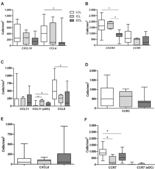
The immunohistological analysis showed a ten-dency for a higher density of CXCL10-positive cells in the granulomas of patients with LCL (median 250 cells/ mm2, ranging from 0-888 cells/mm2) and ICL (263,
0-525 cells/mm2) as compared to DCL patients (138,
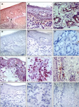
0-488 cells/mm2) (Fig. 1A). All patients with LCL and
ICL and 70% of those with DCL showed CXCL10 ex-pression in the epidermis, sweat glands and nerve fibres (Fig. 2A-C). The analysis of CCL4 and CCL8 expression showed similar results for infiltrating granuloma cells and an augmented density of CCL4- and CCL8-positive cells in patients with LCL (406, 0-1,050 CCL4+ cells/
mm2 and 487, 0-1,238 CCL8+ cells/mm2, respectively)
as compared to DCL (75, 0-925 CCL4+ cells/mm2 and
100, 0-825 CCL8+ cells/mm2) (both p< 0.05) (Fig. 1A,
C). Although no differences were observed regarding CCL11 expression in the infiltrate between groups, 35% of patients with LCL, 80% of those with ICL and 50% of those with DCL exhibited CCL11-positive DCs in the epidermis (Fig. 2G). The density of CCL11-positive epi-dermal DCs was also higher in ICL (150, 0-225 cells/ mm2) and DCL (38, 0-138 cells/mm2) patients than in
LCL patients (0, 0-188 cells/mm2, p< 0.05) (Fig. 1C).
Furthermore, when we evaluated the chemokine re-ceptors expressed on the infiltrating cells in the lesion, we found higher numbers of CXCR3-positive cells in pa-tients with LCL (1,637, ranging from 425-2,750 cells/mm2)
and ICL (1,722, 1,400-2,000 cells/mm2) as compared to
those with DCL (950, 125-1,313 cells/mm2, p < 0.05) (Fig.
1B), indicating a positive correlation with the expression of its ligand CXCL10 (r = 0.3984; p= 0.0239). Although these differences were not significant, we observed a ten-dency for an increased density of CCR5-positive cells in LCL patients (575, 0-1,763 cells/mm2) compared to DCL
patients (238, 0-1,163 cells/mm2) (Fig. 1B). However,
no correlation between CCR5 and its ligand CCL4 was observed. Moreover, no significant differences were ob-served regarding the density of CCR3+ cells in the differ-ent clinical forms of leishmaniasis evaluated (LCL, 500, 0-1,765 cells/mm2; ICL, 638, 0-1,025 cells/mm2; DCL,
307, 0-11,253 cells/mm2) (Fig. 1D) and CCR3 expression
was not correlated with that of its ligand CCL11.
In addition, we evaluated CXCL8 expression, which plays a role in the recruitment of PMN cells. The ex-pression of this chemokine was not significantly differ-ent between the groups of patidiffer-ents evaluated (Fig. 1E). However, we found clear differences in CCR7 expression in granuloma cells from patients with different clinical forms; lesions from patients with LCL showed a higher density of CCR7-positive cells (887, 288-1,763 cells/mm2)
as compared to patients with ICL (173, 0-775 cells/mm2, p
< 0.05) or DCL (536, 100-1,300 cells/mm2, p< 0.05) (Fig.
1F). In addition, 30% of patients with LCL and 20% of patients with ICL, but none of the DCL patients, showed CCR7-positive epidermal DCs (Figs 1F, 2I).
TABLE
Clinical characteristics of different groups of patients
Diagnostic
Age (mean ± SD)
Leishmanin (mm) (mean ± SD)
Time of evolution (months) (mean ± SD)
LCL (n = 20) 34 ± 16 21.2 ± 8.3a 2.2 ± 1.8a ICL (n = 5) 36 ± 20 15.3 ± 4.8a 6 ± 2.3a DCL (n = 10) 17 ± 13 0.7 ± 2.4 118 ± 132
a: p ≤ 0.05 compared with diffuse cutaneous leishmaniasis
DISCUSSION
Chemokines play an important role during innate and adaptive immunity in cutaneous leishmaniasis. It is known that macrophages produce CCL4, an inflamma-tory chemokine that recruits CD4+ lymphocytes (Schall et al. 1993) and is increased in the serum of patients with cutaneous leishmaniasis (Vargas-Inchaustegui et al. 2010). In lesions, we observed elevated levels of CCL4 and CCL8 in LCL patients and these chemokines con-tribute to the recruitment of macrophages and DCs acti-vated by CCL2 in synergy with IFN-γ (Ritter et al. 1996, Jimenez et al. 2010). In contrast, the expression of CXCL8 was similar in LCL, ICL and DCL patients. It has been demonstrated that human monocytes infected with Leish-mania major demonstrate chemotactic activity for neutro-phils and monocytes due to CXCL8 production (Badolato et al. 1996). Moreover, contact with Leishmania induces the release of CXCL8 by PMNs (van Zandbergen et al. 2002), which indicates a potential role for neutrophils in
the pathogenesis of active forms of leishmaniasis, as pre-viously demonstrated by Boaventura et al. (2010) in mu-cosal leishmaniasis associated with a Th17 response.
Immature DCs are able to internalise parasites and migrate to the regional lymph node for antigen presenta-tion. This migratory ability depends on the expression of CCR7, the receptor for CCL21 and CCL19 produced in secondary lymphoid tissues (Förster et al. 1999). These results suggest that patients with LCL have im-proved migration of DCs to lymph nodes. In addition, epidermal DCs may be activated to migrate in a CCR7-dependent manner in patients with LCL and ICL but not DCL and this may contribute to the development of a more efficient adaptive response against the parasite in LCL than in other active clinical forms of the disease. It has also been demonstrated that reduced expression of CCR7 and defective DC migration play a major role in the pathogenesis of visceral leishmaniasis in mice (Ato et al. 2002, 2006). Furthermore, the expression level of
CCR7 and the DC response to the CCR7 ligand CCL21 was shown to be increased after contact with L. major in both resistant and susceptible mice (Steigerwald & Moll 2005). Thus, it is possible that the modulation of chemokine receptor expression on DCs depends upon the infecting Leishmania species.
The chemokines produced at the site of infection are critical for determining the composition of the infiltrat-ing cells. In the lesions of LCL and ICL patients, elevat-ed levels of CXCL10 are producelevat-ed by the epidermis and stromal cells in the dermis, which attracts CXCR3+ cells, including Th1 cells (Sebastiani et al. 2001) and the major-ity of the infiltrating cells in LCL and ICL lesions (Con-vit et al. 1993). These data confirm our previous result indicating that a Th1 response occurs in LCL, whereas a mixed Th1/Th2 response occurs in patients with ICL (Diaz et al. 2002). Our results also coincide with those obtained by Geiger et al. (2010), who concluded that CX-CR3+ Th1 lymphocytes are important for the resolution of cutaneous lesions in leishmaniasis. Furthermore, LCL patients with a high density of CXCR3+ cells respond well to treatment and may resolve lesions spontaneously, whereas ICL patients develop more severe and persistent lesions following primary infection. Thus, it is possible
that patients with ICL and other relapsed forms of leish-maniasis mount a specific type of immune response. In addition, recent evidence demonstrates the involvement of a Th17 response in the pathogenesis of cutaneous, mu-cosal and post-kala-azar dermal leishmaniasis (Bacellar et al. 2009, Lopez Kostka et al. 2009, Boaventura et al. 2010, Katara et al. 2012).
Following early natural killer cell stimulation by IFN-γ, resident cells release elevated levels of CXCL10. Subsequently, CXCR3+ Th1 cells are recruited and more IFN-γ is released and keratinocytes are stimulated to produce more CXCL10 and CCL2 (Albanesi et al. 2001). In LCL and ICL patients, this continuous feedback loop leads to the elimination of most parasites by activated macrophages. However, the exacerbated immune re-sponse in ICL lesions leads to extensive tissue damage and this exacerbated immune response is likely due to failures in the regulation of the T cell response. For ex-ample, Rodriguez-Pinto et al. (2012) demonstrated that CD4+CD25+ cells from asymptomatic individuals had a higher capacity to suppress IFN-γ production by CD4+ effector T cells than those from patients with chronic cu-taneous leishmaniasis.
An association between CCL11 and the Th2 response in allergic dermatitis has been demonstrated (Yawalkar et al. 1999), as well as the expression of its receptor CCR3 on a subset of Th2 cells (Sebastiani et al. 2001). In the pres-ent study, no significant differences in CCL11 or CCR3 expression were observed between groups of patients and this result agrees with recent work showing a similar ex-pression pattern for CCL11 in patients with LCL and dis-seminated leishmaniasis in Brazil (Machado et al. 2011). However, we observed in LCL lesions that CXCL10+ cells were twice as abundant as CCL11+ cells, whereas DCL lesions expressed similar levels of both chemokines. Furthermore, CCL11 is expressed on epidermal DCs in ICL and DCL patients, suggesting a role for these cells in the recruitment of CCR3+ cells in these patients.
This study detected expression of the chemokines CCL11 and CXCL10 in epidermal cells, suggesting the participation of the epithelium in the immune response against Leishmania. In support of this notion, growing evidence has demonstrated that chemokines released from epidermal cells control inflammatory skin diseas-es. Keratinocytes secrete both Th1 and Th2-associated chemokines, although the former type is more abundant-ly produced than the latter. In fact, decreasing keratino-cyte production of chemokines is one of the therapeutic approaches to treat cutaneous inflammatory disorders (Tokura et al. 2008).
In conclusion, for a protective immune response against Leishmania, the recruitment of appropriate leu-kocyte subpopulations resulting from the expression of chemokines and chemokine recognition by the appropri-ate receptors is essential. Our data suggest that the im-mune response in the lesions of LCL patients is associ-ated with a CXCL10-dominassoci-ated pattern of chemokine expression, which serves to attract CXCR3+ Th1 cells and promote efficient DC migration to lymph nodes via CCR7 expression. In contrast, DCL patients show low expression levels of chemokines associated with a Th1 Fig. 2: immunolabelling of chemokine in lesions American
response and instead express CCL11 in epidermal DCs, which contributes to the recruitment of CCR3+ cells. In both of these immune processes, the epidermis plays an important role as the inducer of the immune response via chemokine production by keratinocytes and epidermal DCs, which can be activated and migrate to lymph nodes in LCL patients, but not in DCL patients.
ACKNOWLEDGEMENTS
To the patients who participated in this study.
REFERENCES
Albanesi C, Scarponi C, Sebastiani S, Cavani A, Federici M, Sozzani S, Girolomoni GA 2001. Cytokine-to-chemokine axis between T lymphocytes and keratinocytes can favor Th1 cell accumulation in chronic inflammatory skin diseases. J Leukoc Biol 70: 617-623.
Ato M, Maroof A, Zubairi S, Nakano H, Kakiuchi T, Kaye PM 2006. Loss of dendritic cell migration and impaired resistance to Leish-mania donovani infection in mice deficient in CCL19 and CCL21. J Immunol 176: 5486-5493.
Ato M, Stager S, Engwerda CR, Kaye PM 2002. Defective CCR7 expression on dendritic cells contributes to the development of visceral leishmaniasis. Nat Immunol 12: 1185-1191.
Bacellar O, Faria D, Nascimento M, Cardoso TM, Gollob KJ, Dutra WO, Scott P, Carvalho EM 2009. Interleukin 17 production among patients with American cutaneous leishmaniasis. J Infect Dis 200: 75-78.
Badolato R, Sacks DL, Savoia D, Musso T 1996. Leishmania major: infection of human monocytes induces expression of IL-8 and MCAF. Exp Parasitol 82: 21-26.
Boaventura VS, Santos CS, Cardoso CR, de Andrade J, dos Santos WL, Clarêncio J, Silva JS, Borges VM, Barral-Netto M, Brod-skyn CI, Barral A 2010. Human mucosal leishmaniasis: neutro-phils infiltrate areas of tissue damage that express high levels of Th17-related cytokines. Eur J Immunol 40: 2830-2836.
Bonecchi R, Bianchi G, Bordignon PP, D’Ambrosio D, Lang R, Bor-satti A, Sozzani S, Allavena P, Gray PA, Mantovani A, Siniga-glia F 1998. Differential expression of chemokine receptors and chemotactic responsiveness of type 1 T helper cells (Th1s) and Th2s. J Exp Med 187: 129-132.
Caceres-Dittmar G, Tapia FJ, Sanchez MA, Yamamura M, Uyemura K, Modlin RL, Bloom BR, Convit J 1993. Determination of the cytokine profile in American cutaneous leishmaniasis using the polymerase chain reaction. Clin Exp Immunol 91: 500-505.
Campanelli AP, Brodskyn CI, Boaventura V, Silva C, Roselino AM, Costa J, Saldanha AC, de Freitas LA, de Oliveira CI, Barral-Net-to M, Silva JS, Barral A 2010. Chemokines and chemokine re-ceptors coordinate the inflammatory immune response in human cutaneous leishmaniasis. Hum Immunol 71: 1221-1227.
Convit J, Ulrich M, Fernandez CT, Tapia FJ, Caceres-Dittmar G, Castes M, Rondon AJ 1993. The clinical and immunological spectrum of American cutaneous leishmaniasis. Trans R Soc Trop Med Hyg 87: 444-448.
Diaz NL, Zerpa O, Ponce LV, Convit J, Rondon AJ, Tapia FJ 2002. Intermediate or chronic cutaneous leishmaniasis: leukocyte im-munophenotypes and cytokine characterisation of the lesion. Exp Dermatol 11: 34-41.
Farber JM 1997. Mig and IP-10: CXC chemokines that target lympho-cytes. J Leukoc Biol 61: 246-257.
Förster R, Schubel A, Breitfeld D, Kremmer E, Renner-Muller I, Wolf E, Lipp M 1999. CCR7 coordinates the primary immune response
by establishing functional microenvironments in secondary lym-phoid organs. Cell 99: 23-33.
Geiger B, Wenzel J, Hantschke M, Haase I, Ständer S, von Stebut E 2010. Resolving lesions in human cutaneous leishmaniasis predominantly harbour chemokine receptor CXCR3-positive T helper 1/T cytotoxic type 1 cells. Br J Dermatol 162: 870-874.
Jimenez F, Quinones MP, Martinez HG, Estrada CA, Clark K, Gara-vito E, Ibarra J, Melby PC, Ahuja SS 2010. CCR2 plays a critical role in dendritic cell maturation: possible role of CCL2 and NF-kappa B. J Immunol 184: 5571-5581.
Katara GK, Ansari NA, Singh A, Ramesh V, Salotra P 2012. Evidence for involvement of Th17 type responses in post kalaazar dermal leishmaniasis (PKDL). PLoS Negl Trop Dis6: e1703.
Kim CH, Rott L, Kunkel EJ, Genovese MC, Andrew DP, Wu L, Butcher EC 2001. Rules of chemokine receptor association with T cell polarization in vivo. J Clin Invest 108: 1331-1339.
Laudanna C, Kim JY, Constantin G, Butcher E 2002. Rapid leukocyte integrin activation by chemokines. Immunol Rev 186: 37-46.
Lopez Kostka S, Dinges S, Griewank K, Iwakura Y, Udey MC, von Stebut E 2009. IL-17 promotes progression of cutaneous leishma -niasis in susceptible mice. J Immunol 182: 3039-3046.
Machado PR, Rosa ME, Costa D, Mignac M, Silva JS, Schriefer A, Teix-eira MM, Bacellar O, Carvalho EM 2011. Reappraisal of the immu-nopathogenesis of disseminated leishmaniasis: in situ and systemic immune response. Trans R Soc Trop Med Hyg 105: 438-444.
Ohmori Y, Wyner L, Narumi S, Armstrong D, Stoler M, Hamilton TA 1993. Tumor necrosis factor-alpha induces cell type and tissue-specific expression of chemoattractant cytokines in vivo. Am J Pathol 142: 861-870.
Peters NC, Egen JG, Secundino N, Debrabant A, Kimblin N, Kam-hawi S, Lawyer P, Fay MP, Germain RN, Sacks D 2008. In vivo imaging reveals an essential role for neutrophils in leishmaniasis transmitted by sandflies. Science 321: 970-974.
Ritter U, Körner H 2002. Divergent expression of inflammatory der-mal chemokines in cutaneous leishmaniasis. Parasite Immunol 24: 295-301.
Ritter U, Moll H, Laskay T, Brocker E, Velazco O, Becker I, Gillitzer R 1996. Differential expression of chemokines in patients with localized and diffuse cutaneous American leishmaniasis. J Infect Dis 173: 699-709.
Rodriguez-Pinto D, Navas A, Blanco VM, Ramírez L, Garcerant D, Cruz A, Craft N, Saravia NG 2012. Regulatory T cells in the patho-genesis and healing of chronic human dermal leishmaniasis caused by Leishmania (Viannia) species. PLoS Negl Trop Dis 6: e1627.
Schall TJ, Bacon K, Camp RD, Kaspari JW, Goeddel DV 1993. Hu-man macrophage inflammatory protein alpha (MIP-1 alpha) and MIP-1 beta chemokines attract distinct populations of lympho-cytes. J Exp Med 177: 1821-1826.
Sebastiani S, Allavena P, Albanesi C, Nasorri F, Bianchi G, Traidl C, Sozzani S, Girolomoni G, Cavani A 2001. Chemokine receptor expression and function in CD4+ T lymphocytes with regulatory
activity. J Immunol 166: 996-1002.
Silva F, Gomes R, Prates D, Miranda JC, Andrade B, Barral-Netto M, Barral A 2005. Inflammatory cell infiltration and high antibody production in BALB/c mice caused by natural exposure to Lutzo-myia longipalpis bites. Am J Trop Med Hyg 72: 94-98.
Steigerwald M, Moll H 2005. Leishmania major modulates chemokine and chemokine receptor expression by dendritic cells and affects their migratory capacity. Infect Immun 73: 2564-2567.
Tokura Y, Kobayashi M, Kabashima K 2008. Epidermal chemokines and modulation by antihistamines, antibiotics and antifungals. Exp Dermatol17: 81-90.
van Zandbergen G, Hermann N, Laufs H, Solbach W, Laskay T 2002. Leishmania promastigotes release a granulocyte chemotactic fac-tor and induce interleukin-8 release, but inhibit gamma interfer-on-inducible protein 10 production by neutrophil granulocytes. Infect Immun 70: 4177-4184.
van Zandbergen G, Klinger M, Mueller A, Dannenberg S, Gebert A, Solbach W, Laskay T 2004. Cutting edge: neutrophil granulocyte serves as a vector for Leishmania entry into macrophages. J Im-munol 173: 6521-6525.
Vargas-Inchaustegui DA, Hogg AE, Tulliano G, Llanos-Cuentas A, Arevalo J, Endsley JJ, Soong L 2010. CXCL10 production by hu-man monocytes in response to Leishmania braziliensis infection. Infect Immun 78: 301-308.
Williams JR 2008. The Declaration of Helsinki and public health. Bull World Health Organ 86: 650-652.
Yawalkar N, Uguccioni M, Scharer J, Braunwalder J, Karlen S, Dewald B, Braathen LR, Baggiolini M 1999. Enhanced expression of eo-taxin and CCR3 in atopic dermatitis. J Invest Dermatol113: 43-48.