Mother and daughter with adolescent-onset
severe frontal lobe dysfunction and epilepsy
Giordani Rodrigues dos Passos1, Alonso Cuadrado Fernández1,
Adriana Machado Vasques2, William Alves Martins1,3, André Palmini1,3
ABSTRACT. Familial cases of early-onset prominent frontal lobe dysfunction associated with epilepsy have not been reported to date. We report a mother and her only daughter with incapacitating behavioral manifestations of frontal lobe dysfunction and epilepsy of variable severity. The possibility of a hitherto undescribed genetic condition is discussed.
Key words: frontal lobe, genetics, behavioral, neuropsychiatry, epilepsies, partial, heredity.
MÃE E FILHA COM DISFUNÇÃO SEVERA DO LOBO FRONTAL E EPILEPSIA COM INÍCIO NA ADOLESCÊNCIA
RESUMO. Casos familiares de disfunção proeminente do lobo frontal associada a epilepsia com início precoce ainda não foram relatados. Nós descrevemos uma mãe e sua filha única com manifestações comportamentais incapacitantes de disfunção do lobo frontal e epilepsia de severidade variável. A possibilidade de uma condição genética ainda não descrita é discutida.
Palavras-chave: lobo frontal, genética comportamental, neuropsiquiatria, epilepsias parciais, hereditariedade.
INTRODUCTION
F
rontal lobe dysfunction is nonspeciic and may be the presenting feature of many diferent brain insults. Some are obviously acquired and indeed classical texts in Neu-rology based the description of the ‘frontal lobe syndrome’ on patients with extensive traumatic, neoplastic, vascular or degenera-tive destruction of the frontal lobes.1How-ever, a number of genetically-determined conditions have been increasingly reported, mostly associated with degenerative or meta-bolic diseases, often presenting in late adult-hood and having unequivocal progression.2
Furthermore, when disorders predominantly involving the frontal lobes present early in life, they usually have additional neurologi-cal features, such as white matter progres-sive abnormalities and motor dysfunction.2
hus, earlier-onset prominent frontal lobe syndrome unaccompanied by overt motor or imaging abnormalities is apparently rare and even more so with a familial presentation.
Genetic factors are being increasingly rec-ognized as underlying many types of epilepsy. Some constitute severe epileptic encepha-lopathies and others focal forms of epilepsy, previously considered cryptogenic.3 Some of
the latter may involve the frontal lobes with-out overt imaging abnormalities, but are not speciically associated with severe frontal lobe-related behavioral and emotional dys-regulation. For instance, autosomal dominant nocturnal frontal lobe epilepsy (ADNFLE), a type of frontal lobe epilepsy with proven genetic origin, is in fact a relatively benign entity, whose seizures can be easily controlled using antiepileptic medication and usually associated with normal cognition and behav-ior.4,5 Other types of frontal-predominant
epilepsies of genetic origin include gross malformations of cortical development, such as bilateral frontal polymicrogyria, which can be inherited and occur in families.6 Hence,
genetic forms of frontal lobe epilepsy without gross imaging abnormalities and with
inca-This study was conducted at the Neurology Service, São Lucas Hospital, Pontifical Catholic University of Rio Grande do Sul, Porto Alegre RS, Brazil.
1Neurology Service, São Lucas Hospital, Pontifical Catholic University of Rio Grande do Sul, Porto Alegre RS, Brazil. 2Institute of Geriatrics and Gerontology, Pontifical
Catholic University of Rio Grande do Sul, RS, Porto Alegre, Brazil.3Porto Alegre Epilepsy Surgery Program, Neurology Service, São Lucas Hospital, Pontifical Catholic
University of Rio Grande do Sul, Porto Alegre, RS, Brazil.
Giordani Rodrigues dos Passos. Avenida Ipiranga, 6690 / sala 220 – 90610-000 Porto Alegre RS – Brazil. E-mail: [email protected]
Disclosure: The authors report no conflicts of interest
Received August 13, 2016. Accepted in final form August 16, 2016.
pacitating behavioral manifestations consistent with frontal lobe dysfunction are apparently rare and have not been reported to date.
Here we report a mother and daughter with a largely similar, challenging incapacitating non-degenerative frontal lobe syndrome associated with nonlesional, pharmacoresistant frontal lobe epilepsy. heir condition is apparently unique in that severe frontal lobe dysfunc-tion is associated with epilepsy, normal imaging and a likely autosomal dominant genetic substrate. Further-more, more than 20 years´ follow-up of both patients have provided us the rare opportunity of a longitudinal assessment. Because the patients were deemed legally incapable, written informed consent for case reporting was obtained from their legal guardian.
CASE REPORTS
The mother. his is a 49-year-old Caucasian woman with no history of parental consanguinity or family history of neuropsychiatric disorders. Pregnancy, delivery and early development were uneventful and there was no history of febrile seizures. When she entered school, agitation, learning diiculties and relationship prob-lems became apparent. At age 10 she began having weekly diurnal seizures with an aura of “feeling as if I am about to lose control”, followed by hemiclonic move-ments in alternating sides and secondary generaliza-tion. Seizures were diicult to control with medication, but the most striking aspect of her adolescence was a marked behavioral worsening. Despite a good socio-cultural background, she started to frequently engage in promiscuous relationships, mostly with men she met in the streets. his later deteriorated into bouts of verbal aggression, threats to relatives and neighbors with a knife and stealing objects, leading to problems with the law. At age 20 years, she fell pregnant and abandoned her parents’ house to live with her partner in a setting involving extreme poverty and the need for begging. She soon demonstrated negligent and aggres-sive behavior towards her daughter, resulting in a court decision deeming her incapable of raising a child, which led to sterilization.
In parallel with this severe frontal lobe dysfunction and psychopathic social functioning, seizure control progressively worsened. She was initially seen at our Center at age 26 and referred for presurgical assess-ment at age 29, following unequivocal docuassess-mentation of seizure refractoriness. Scalp electroencephalography (EEG) showed intense epileptiform discharges over the right frontotemporal regions, with much less intense discharges in the homologous area of the left
hemi-sphere. Presurgical neuroimaging with magnetic reso-nance imaging (MRI) was negative, leading to invasive investigation with intracranial electrodes. Invasive EEG recordings showed bilateral frontal epileptiform activity, much more pronounced in the right side, where seizures were recorded. She had resection of the right superior frontal gyrus and anterior cingulate cortex (Figure 1), guided by intraoperative electrocorticography. Histo-pathological examination of the resected brain sample showed only astroglial proliferation and relative reduc-tion in neuronal populareduc-tion, with no evidence of focal cortical dysplasia or inlammation.
Following the surgery, there was marked improve-ment in seizures, which were completely controlled over the years. In contrast, behavioral problems continued to worsen and also proved refractory to all medica-tion attempts. he most dramatic episode was when she threw boiling water into her 8-year-old daughter’s face, which led to loss of custody of the child. At this time, and on two other occasions, she was admitted to psychiatric wards. Due to exhaustion of all medication and behavioral therapeutic strategies, she underwent simultaneous bilateral posteromedial hypothalamotomy (Figure 1) at age 37, with the aim of decreasing violent behavior. Despite improvement in aggressiveness, she remained incapable of social functioning, being trans-ferred to a long-term nursing care facility one year later.
Currently, she is on carbamazepine 1200 mg/day and has been seizure-free for one year. She is also on quetiapine (50 mg/day and increasing), but serious non-adherence to social norms and persistent aggressiveness remain as major concerns.
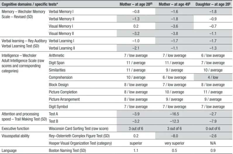
Neuropsychological tests performed at age 28 and again at age 49 were compared (Table 1). On the irst assessment (before epilepsy surgery), intelligence was rated as average to low average; there were signiicant deicits in attention and speed of processing, memory, learning, and executive functions; while no visuospatial or language dysfunction was noted. Twenty-one years later, there was marked worsening of attention and speed of processing, slight worsening of memory, and appearance of visuospatial deicit, while intelligence, learning, and executive deicits remained fairly stable.
A B C D
E
Figure 1. Mother’s FLAIR MRI of the brain at 41 (A and B) and 49 years (C, D, and E) of age. Note resection of the right superior frontal gyrus with surrounding gliotic border, as well as small, non-specific hyperintense and hypointense subcortical lesions in the left frontal lobe (A to D). Notably, there is no major atrophy and images are very similar, following the 8-year interval. Note the surgical lesion at the posteromedial hypothalamus bilaterally (E, arrows).
Table 1. Neuropsychological assessment of the mother (at different ages) and the daughter.
Cognitive domains / specific tests* Mother – at age 28§¶ Mother – at age 49¶ Daughter – at age 28§
Memory – Wechsler Memory Scale – Revised (SD)
Verbal Memory I –0.8 –1.6 –1.8
Verbal Memory II –1.3 –1.8 –0.9
Visual Memory I 0.2 –3.6 –0.7
Visual Memory II –3.2 –3.8 –1.1
Verbal learning – Rey Auditory Verbal Learning Test (SD)
Verbal Learning I –1.0 –1.7 –1.7
Verbal Learning II –2.1 –1.1 –1.3
Intelligence – Wechsler Adult Intelligence Scale (raw scores and corresponding categories)
Arithmetic 7 / low average 7 / low average 6 / low average
Digit Span 11 / average 11 / average 7 / low average
Similarities 11 / average 9 / average 10 / average
Comprehension 10 / average 6 / low average 4 / low
Block Design 8 / low average 7 / low average 8 / low average
Picture Completion 8 / low average 10 / average 11 / average
Picture Arrangement 8 / low average 9 / average 9 / average
Digit Symbol 7 / low average 7 / low average 7 / low average
Attention and processing speed – Trail Making Test (SD)
Test A –3.9 –16.5 –2.7
Test B –3.2 –12.3 –7.9
Executive function Wisconsin Card Sorting Test (raw score) 3 out of 6 3 out of 6 0 out of 6
Visuospatial ability Rey–Osterrieth Complex Figure Test (SD) 0.2 –8.0 –2.6
Hooper Visual Organization Test (category) superior very superior N/A
Language Boston Naming Test (SD) 1.1 0.5 0.9
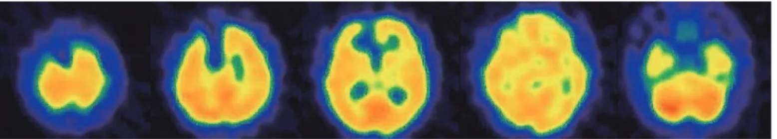
HIV, all within normal limits. Following the surgical pro-cedures for epilepsy and violent behavior, MRI of the brain was performed at ages 41 and 49 years; besides the postsurgical indings in the right frontal lobe and in the posteromedial hypothalami, there were small, non-speciic subcortical lesions in the left frontal lobe (Figure 1). Notably, there was no progressive atrophy or other evidence of neurodegeneration on MRI. Brain perfusion single-photon emission computerized tomog-raphy (SPECT) was also performed, at age 49 years, with no apparent areas of hypoperfusion (Figure 2), except for the area corresponding to the previous surgical resection.
The daughter. his 28-year-old mestizo woman is the only daughter of the patient reported above with a non-consanguineous spouse. During pregnancy, she was exposed to anticonvulsant, sedative, antipsychotic and recreational drugs, as well as maternal seizures. here were no pre- or perinatal events, congenital malforma-tions or neurodevelopmental delay. here was no history of febrile seizures. During childhood, she sufered all sorts of violence committed by her parents, from whom she was legally separated at age 8 years. At this age, she also started exhibiting learning diiculties and aggres-sive behavior.
Between ages 8 and 11 years she had infrequent diurnal, poorly described seizures. Complete seizure
control was achieved after initiation of phenytoin. hroughout adolescence, she gradually developed severe frontal lobe dysfunction, including disinhibition (used to undress in public), puerile behavior (would rather play with children than interact with other teenagers), psychomotor agitation (used to climb furniture and run and jump relentlessly around her house), impaired judgement (engaged in promiscuous relationships with married, homeless, drug-addicted and delinquent men), lack of impulse control, aggressiveness and total lack of hygiene and self-care.
Psychiatric treatment started at age 12. Hormonal therapy with injectable progestogen was required in order to control sexual impulses. She later developed prominent anxiety symptoms, including panic attacks and compulsive behaviors, such as hair pulling (eventu-ally leading to signiicant alopecia) and pushing the lush dozens of times after using the toilet.
As in her mother’s case, there were no other neu-rological symptoms or signs. Apart from obesity and dyslipidemia, there were no other systemic diseases. Multiple scalp EEGs showed relatively synchronous epileptiform discharges in the anterior regions of both hemispheres, suggestive of frontal lobe epilepsy, some with clear left frontal predominance. Brain MRI was unremarkable at age 20 years and again at age 28 (Figure 3), with no signiicant brain atrophy having developed during that interval.
Figure 2. Mother’s recently obtained 99mTc-ECD brain perfusion SPECT axial images. Except for the absence of perfusion over the surgically resected area in the right frontal lobe, brain perfusion is normal.
After many failed attempts with multiple psychotro-pic drugs, satisfactory control of the behavioral symp-toms and some improvement in cognitive function were ultimately achieved with the combination of clo-zapine 100 mg/day, aripiprazole 20 mg/day, paroxetine 20 mg/day, and diazepam 15 mg/day. Despite having been deemed legally incapable at age 23, her ability to live at home is preserved, under the supervision of her grandparents, and she does engage in some self-care and house-keeping activities. In addition, she has been seizure-free on phenytoin 400 mg/day.
Neuropsychological assessment at current age (Table 1), with the same tests used for her mother’s cognitive evaluation, showed average to low average intelligence, as well as deicits in attention and speed of processing, memory, learning, visuospatial abilities and particularly executive functions, with no language dysfunction.
DISCUSSION
We report a family in which a mother and her only daughter presented with progressive and incapaci-tating frontal lobe dysfunction and epilepsy of variable severity. Interestingly, age of onset of the neuropsychi-atric symptoms (6-8 years) and of seizures (8-10 years) were similar in the two patients, as was the course of the behavioral and cognitive worsening, which was relatively rapid throughout adolescence and seemed to plateau during the third decade, with progression at a slower rate. Moreover, neuropsychological proile on formal testing, including severity of the deicits, was quite similar for both mother (before surgery) and daughter, at age 28 years. Finally, refractoriness to several psycho-tropic drugs was another common point. In contrast, gestation and upbringing were very diferent. Whereas the mother was the product of an uneventful pregnancy and raised in a caring environment, the daughter had signiicant drug exposure during gestation, meager prenatal care and sufered severe abuse and neglect during childhood. hus, this pattern of very similar clinical evolution, despite markedly diferent environ-mental upbringing, does suggest a genetic condition.
Executive functions include attentional and inhibi-tory control, working memory, cognitive lexibility, rea-soning, problem solving, and planning.7 Even though
the prefrontal cortex seems to be the main anatomical substrate of these higher-order cognitive processes, the historical linkage of executive functions and the frontal lobes has been reformulated in order to recognize that a one-to-one relationship between structure and func-tion is not possible and other anatomical sites also play a role.7,8
Although psychiatric comorbidities of epilepsy are currently well studied,9,10 the speciic pattern of
behav-ioral and cognitive dysfunction reported here could not be explained solely by any given psychiatric dis-order, such as personality disdis-order, bipolar disorder or schizophrenia. Furthermore, frontal lobe epilepsy, even when refractory to medication, does not usually manifest with such severe degree of neuropsychiatric frontal lobe dysfunction, except when seizures accom-pany frontal lobe destruction, such as those following severe head trauma, tumor resection or massive paren-chymal hemorrhage.11,12 It is known that people with
frontal lobe epilepsy may have behavioral abnormalities, including hyperactivity, conscientiousness, obsession, and addiction,11 but not to the degree seen in these two
patients, which resemble classical, severe frontal lobe syndromes with marked disinhibition and executive dysfunction.1,7,13
Frontal lobe dysfunction can result from several conditions, many of which may have a genetic cause.2
Frontotemporal dementia (FTD) and Alzheimer’s dis-ease may have familial presentations, manifesting at an earlier age in comparison to sporadic cases.14 In a series
of FTD spectrum diseases, Le Ber et al. reported onset of symptoms in a patient with MAPT mutation at age 17 years, which is, however, exceedingly rare.15 Moreover,
these degenerative familial disorders usually do not include epilepsy as a prominent feature. Another group of conditions that should be considered in the diferen-tial diagnosis of our patients include genetically-deter-mined neurodegenerative disorders, such as Hunting-ton’s disease and dentatorubropallidolusyian atrophy, or metabolic diseases, such as Niemann-Pick disease type C and leukodystrophies.2 hese may have childhood onset,
be associated with epilepsy and progress with severe behavioral and cognitive abnormalities. However, these entities are usually associated with a number of other features, especially motor or visual abnormalities, brain MRI indings and systemic signs and symptoms, which were absent in the cases reported here.2,14
Genetic variation has been increasingly recognized as a major etiology of epilepsies.3 Several single genes
whose mutation is clearly associated with epilepsy have already been described and there has been a spate of dis-coveries, particularly in relation to epileptic encephalop-athies.16-18 he latter are entities in which the extremely
of our patients in that the epilepsy is much more severe and development is always delayed from very early in life. In many other instances of probably genetic-related epilepsies, a single genetic mutation cannot be identi-ied; instead, multiple genes and modulation of genetic expression by environmental factors likely play a role.3
hese aspects notwithstanding, two genetically-determined epilepsy syndromes were considered in the diferential diagnosis of our cases. ADNFLE is caused by mutations in the genes CHRNA2, CHRNA4 or CHRNB2, inherited in an autosomal dominant manner, with 70% penetrance, and presents during the irst two decades of life usually with nocturnal focal seizures.19 Familial
partial epilepsy with variable foci is caused by autoso-mal-dominant mutations in the gene DEPDC5 and usu-ally manifests as familial cases of epilepsy in which each individual may have a diferent single focus.16 Both
con-ditions could explain epilepsy in our patients, but would not be expected to account for the early, prominent and incapacitating frontal lobe dysfunction. Moreover, both the mother and the daughter had predominantly diurnal seizures, making the diagnosis of ADNFLE less likely.
To our knowledge, the familial occurrence of severe frontal lobe dysfunction and epilepsy, with onset in the irst decade of life, is extremely atypical and war-rants further investigation. We are well aware that
fur-ther histopathological and genetic workup would be of paramount importance to establish a diagnosis for the cases reported here. Unfortunately, with 20 years having passed since the mother underwent epilepsy surgery, we were not able to recover the pathological samples, thus immunohistochemistry analysis and other molecular techniques currently available could not be performed. We do not know whether mild foci of type I focal cor-tical dysplasia or other relevant pathological indings would be disclosed if these analysis were performed. In addition, a comprehensive genetic panel for epilepsy would be of value, but was not possible to perform due to practical issues. Currently, exome sequencing is being planned and may potentially lead to the identiication of a novel mutation accounting for the development of adolescent-onset severe frontal lobe dysfunction associ-ated with epilepsy.
Author contribution. Giordani Rodrigues dos Passos, Alonso Cuadrado Fernández, William Alves Martins e André Palmini: Involvement in the clinical care of the patients whose case is reported; intellectual contri-bution to the writing of the manuscript. Adriana Machado Vasques: Neuropsychological assessment of the patients whose case is reported; intellectual contri-bution to the writing of the manuscript.
REFERENCES
1. Krudop WA, Pijnenburg YAL. Historical evolution of the frontal lobe syndrome. Psychopathology. 2015;48(4):222-229.
2. Geschwind MD, Yoon G, Goldman J. Adult-Onset Genetic Disorders Involving the Frontal Lobes. In: Miller BL, Cummings JL, editors. The Human Frontal Lobes: Functions and Disorders. New York: Guilford Press; 2007:552-575.
3. Hildebrand MS, Dahl H-HM, Damiano JA, Smith RJH, Scheffer IE, Berkovic SF. Recent advances in the molecular genetics of epilepsy. J Med Genet. 2013;50(5):271-279.
4. Nobili L, Proserpio P, Combi R, et al. Nocturnal frontal lobe epilepsy. Curr Neurol Neurosci Rep. 2014;14(2):424.
5. Lim CX, Ricos MG, Dibbens LM, Heron SE. KCNT1 mutations in seizure disorders: the phenotypic spectrum and functional effects. J Med Genet. 2016;53(4):217-225.
6. Guerrini R, Barkovich AJ, Sztriha L, Dobyns WB. Bilateral frontal polymi-crogyria: a newly recognized brain malformation syndrome. Neurology. 2000;54(4):909-913.
7. Alvarez JA, Emory E. Executive Function and the Frontal Lobes : A Meta-Analytic Review. Neuropsychol Rev. 2006;16(1):17-42.
8. Stuss DT, Alexander MP. Executive functions and the frontal lobes: a conceptual view. Psychol Res. 2000;63(3-4):289-298.
9. Verrotti A, Carrozzino D, Milioni M, Minna M, Fulcheri M. Epilepsy and its main psychiatric comorbidities in adults and children. J Neurol Sci. 2014;343(1-2):23-29.
10. van Ool JS, Snoeijen-Schouwenaars FM, Schelhaas HJ, Tan IY, Alden-kamp AP, Hendriksen JGM. A systematic review of neuropsychiatric comorbidities in patients with both epilepsy and intellectual disability. Epilepsy Behav. 2016;60:130-137.
11. Helmstaedter C. Behavioral Aspects of Frontal Lobe Epilepsy. Epilepsy Behav. 2001;2(5):384-395.
12. Trimble MR. Psychopathology of frontal lobe syndromes. Semin Neurol. 1990;10(3):287-294.
13. Niedermeyer E. Frontal lobe functions and dysfunctions. Clin Electroen-cephalogr. 1998;29(2):79-90.
14. Rossor MN, Fox NC, Mummery CJ, Schott JM, Warren JD. The diag-nosis of young-onset dementia. Lancet Neurol. 2010;9(8):793-806. 15. Le Ber I, Camuzat A, Guillot-Noel L, et al. C9ORF72 repeat expansions
in the frontotemporal dementias spectrum of diseases: a flow-chart for genetic testing. J Alzheimers Dis. 2013;34(2):485-499.
16. Hardies K, Weckhuysen S, De Jonghe P, Suls A. Lessons learned from gene identification studies in Mendelian epilepsy disorders. Eur J Hum Genet. 2016;24(7):961-967.
17. Reid CA, Berkovic SF, Petrou S. Mechanisms of human inherited epilep-sies. Prog Neurobiol. 2009;87(1):41-57.
18. Deng H, Xiu X, Song Z. The Molecular Biology of Genetic-Based Epilep-sies. Mol Neurobiol. 2014;49(1):352-367.