Sedation with Sufentanil and Clonidine in Patients Undergoing
Heart Catheterization
Anita Perpetua Carvalho Rocha, Guilherme Antonio Moreira Barros, Jedson dos Santos Nascimento, Kleber
Pimentel Santos, Lilian Mendes de Vasconcelos, Pedro Augusto Costa Rebouças de Castro
Santa Casa de Misericórdia da Bahia - Hospital Santa Izabel, Salvador, BA, Faculdade de Medicina de Botucatu, Universidade Estadual Paulista - UNESP, São Paulo, SP - Brazil
Mailing address: Anita Perpetua Carvalho Rocha •
Rua Pacífico Pereira, 381/1303 - Garcia - 40100-170 - Salvador, BA - Brazil E-mail: [email protected], [email protected] Manuscript received April 09, 2010; revised manuscript received September 21, 2010; accepted October 04, 2010.
Abstract
Background: Sedation for heart catheterization has been a cause for concern. Benzodiazepines, alpha-2 adrenergic agonists and opioids are used for this purpose. However, each drug has advantages and disadvantages.
Objective: To evaluate the efficacy of sufentanil and clonidine as sedative in patients undergoing heart catheterization, observing their impact on hemodynamic and respiratory parameters, the presence of side effects and satisfaction of the patient and interventional cardiologist with the examination.
Methods: This is a prospective, double-blind, randomized and controlled clinical trial involving 60 patients who received 0.1 µg/kg of sufentanil or 0.5 µg/kg of clonidine before heart catheterization. The score of sedation according to the Ramsay scale, the need for use of midazolam, side effects and hemodynamic and respiratory parameters were recorded, with the data being analyzed at 06 different moments.
Results: The behavior of blood pressure, heart rate and respiratory rate was similar in both groups, but, at moment 2, the patients in the sufentanil group (Group S) had a lower sedation score on the Ramsay scale, and the peripheral oxyhemoglobin saturation was lower than in the clonidine group (Group C) at time 6. Patients in Group S had higher incidence of nausea and vomiting after surgery than patients in Group C. Patient satisfaction was higher in the clonidine group. The interventional cardiologists were satisfied in both groups.
Conclusion: Sufentanil and clonidine were effective as sedative in patients undergoing heart catheterization. (Arq Bras Cardiol 2011;96(3):219-226)
Keywords: Heart catheterization; sufentanil; clonidine.
alpha-2 adrenergic agonists, especially clonidine, have proven to be effective for the control of hemodynamic parameters, while inducing mild sedation, which is desirable in the catheterization room2. However, these medicines should be
used with caution in patients with hypotension, in patients with bradycardia and in users of beta blockers, because they have a synergistic effect with regard to the reduction in blood pressure and heart rate.
On the other hand, opioids represent a class of drug commonly used by anesthesiologists in the operating room, in the catheterization laboratory and in the ward, with the purpose of inducing sedation and controlling acute and chronic pain. The most commonly administered opioids in clinical practice are morphine, fentanyl and meperidine. It is known that other options are available, represented mainly by alfentanil, remifentanil and sufentanil. In contrast to older opioids, such as morphine and meperidine, they have a rapid onset of action and their effects are more closely related to their serum concentration, especially when used in low doses3,4.
The objective of this study is to evaluate the efficacy of sufentanil and clonidine as sedative in patients undergoing cardiac catheterization, observing their impact on
Introduction
The number of diagnostic and therapeutic interventions performed outside the operating room has increased significantly over the past 10 years. This is especially true for procedures performed in the catheterization laboratory, where the most common intervention is heart catheterization, which must be accompanied by monitoring, with a possible need for sedation of the patient1.
hemodynamic and respiratory parameters, the presence of side effects and satisfaction of the patient and interventional cardiologist with the examination.
Methods
The study analyzed sixty patients of both sexes, between 18 and 80 years of age, ASA III, who underwent cardiac catheterization under sedation. The study design was prospective, randomized and double-blind, previously approved by the Ethics Research Committee Professor Dr. Celso Figueirôa.
Upon admission to the catheterization laboratory, the patient was invited to participate in the study and asked to sign the consent form. Two groups were randomly formed with 30 participants each, who received sufentanil (0.1 µg/kg [n = 30]) or clonidine (0.5 µg/kg [n = 30]) intravenously. For the randomization, PEPE (computer programs for epidemiologists), version 4.04, 2001 was used.
Sufentanil or clonidine was administered covertly to each patient, according to the group to which they were allocated. Monitoring was performed during the period in which the patient was hospitalized in the catheterization laboratory, at intervals of 05 minutes, by applying the Ramsay scale, by checking hemodynamic parameters (blood pressure and heart rate), by ascertaining whether the use other sedatives was necessary and by checking for the presence of complications and/or adverse effects.
The inclusion criteria were: patients undergoing cardiac catheterization, in an elective way, based on a positive result for ischemia in a stress test or myocardial scintigraphy. The following people or cases did not take part in or were excluded from the study: individuals under the age of 18 and over the age of 80, patients that are pregnant or breast feeding, indication of insensitivity to the contrasts used in the exam, intolerance or allergy to the drugs studied in the protocol, cognitive impairment , medication dependence, chronic use of opioids or benzodiazepines, morbid obesity, history of sleep apnea, patients with difficult airways and hypotension (characterized by blood pressure below 100 X 60 mmHg).
The drugs of the study were those used for the sedation required for the cardiac catheterization examination. After being admitted to the catheterization laboratory, the subjects were interviewed and selected according to the inclusion and exclusion criteria. Subsequently, such subjects were referred to the procedure room and, after conventional intravenous access, they were monitored with cardioscope in DII lead, with pulse oximetry and the noninvasive/invasive measurement of blood pressure every 05 minutes. The level of sedation was assessed by using the Ramsay scale5 (Table 1).
After the beginning of the administration of 100% oxygen using a nasal catheter (2.0 l/min), sedation was performed with sufentanil (0.1 µg/kg) or clonidine (0.5 µg/kg) according to the randomization. When necessary, midazolam at a dose of 0.02 mg/kg was used for supplementing the sedation. The parameter for administration of this benzodiazepine was to have a score of 01 in the Ramsay scale and/or high blood pressure, characterized by the presence of blood pressure above 180 X 110 mmHg. The moment of the administration
of midazolam was recorded. No pre-anesthetic medication was offered.
After the procedure, the patient was referred to the anesthetic recovery room, where the following characteristics were checked and recorded: the complaint of pain, sedation level, presence of nausea or vomiting, bleeding, altered behavior, tremors, changes in blood pressure, arrhythmia, respiratory depression, allergic reactions, and others that were occasionally detected.
Persistent hypertension, 15 minutes after the use of midazolam, was treated by administration of metoprolol (05 mg/dose). On the other hand, bradycardia, which is characterized by respiratory rate below 50 bpm, was reversed by the application of atropine, at a dose of 20 µg/kg.
Data relating to heart rate, blood pressure, hemoglobin saturation and sedation scores in the Ramsay scale were considered for statistical analysis at the following moments:
• M1 - Control, before the beginning of the examination; • M2 - Five minutes after the injection of drug; • M3 - Beginning of the cardiac catheterization; • M4 - End of the cardiac catheterization;
• M5 - Thirty minutes after the injection of drug; and • M6 - The moment of discharge from the anesthesia
recovery room.
The observation period proposed was 90 minutes, with some variations in it depending on the puncture site and comorbidities of the patients.
The results were analyzed as mean and median ± standard deviation (SD). For comparison of continuous variables between the two groups, the student t test or the Mann-Whitney test was used, whereas for categorical variables, the chi-square statistic was applied with the calculation of X2 and
p6 or Fisher’s exact test when the assumptions of the first one
were not met. The data obtained were considered significant when the p value was less than 0.05.
Results
The study was conducted over a period of two months, from July 1st to August 30th, 2009.
The initial clinical evaluation and demographic characteristics of both groups are shown in Table 2, where it is possible to see that there were no statistically significant differences between the two groups, with regard to age,
Table 1 - Ramsay scale
Ramsay scale
Patient that is anxious and agitated or restless, or both
Co-operative, oriented and calm patient
Patient that is responsive to verbal commands only
Patient exhibiting brisk response to light glabellar tap or to an auditory stimulus
Patient exhibiting a sluggish response to light glabellar tap or auditory stimulus
gender, height, weight, BMI, SBP, DBP, heart rate, respiratory rate, SpO2 and the Ramsay sedation scale.
The comparison between the two groups with respect to the hemodynamic characteristics and Ramsay sedation scale, for each moment in the procedure, demonstrates that there was no difference in the patients’ behavior with regard to SBP, DBP, heart rate and respiratory rate. By looking at the Ramsay sedation scale and SpO2, it is possible to see that there was no statistically significant difference at two moments. At moment 02, the difference was related to the Ramsay sedation scale, indicating that the sedation of patients who used sufentanil was more intense than that of those who used clonidine. At moment 06, there was difference in SpO2, demonstrating that patients who used sufentanil had lower SpO2 value than those that used clonidine. However, the SpO2 value of group S was 97.1 % ± 2.2 against 98.4% ± 1.8 in group C, which is considered a normal value (Table 3).
In a more detailed analysis of the Ramsay scores at each moment of groups S and C, it is possible to notice that both the patients that received sufentanil and those who received clonidine, in most cases, reached a satisfactory level of sedation, and sufentanil has an earlier onset of action than clonidine (Figure 1).
None of the patients in both groups developed respiratory depression or allergy during the procedure.
Seven patients in group S and no patient in group C had nausea and vomiting during the observation period, indicating a 23.3% incidence of nausea and vomiting in the sufentanil group, with p value of 0.01 by the Fisher’s exact test, when compared with the clonidine group.
One patient in group S (3.3%) had urinary retention against none in group C, with p value of 1.0 by Fisher’s exact test.
Table 2 - Comparison of initial clinical evaluation characteristics and demographic characteristics of the groups before the procedure
Group S Group C p
Age (mean ± sd) 60.5 ± 9.2 63.5 ± 8.6 0.20ψ
Gender (%)
Male 40.0% 53.3% 0.30*
Female 60.0% 46.7%
Height (mean ± sd) 1.63 ± 0.07 1.62 ± 0.07 0.55ψ
Weight (mean ± sd) 71.0 ± 10.7 69.8 ± 11.0 0.66ψ
BMI (mean ± sd) 26.6 ± 3.8 26.4±3.8 0.89ψ
SBP (mean ± sd) 150.8 ± 23.4 155.1 ± 22.7 0.47ψ
DBP (mean ± sd) 80.9 ± 8.3 81.1 ± 11.9 0.93ψ
HR (mean ± sd) 66.2 ± 11.6 66.0 ± 10.7 0.95ψ
RR (mean ± sd) 96.0 ± 16.3 99.1 ± 1.2 0.96¥
SpO2 (mean ± sd) 16.5 ± 1.9 17.0 ± 2.3 0.32¥
Ramsay (median/
minimum/maximum) 1/2/2 1/2/2 1.0¥ Sd - standard deviation. *Chi-square. Ψ Student’s t test. ¥ Mann-Whitney test. BMI - body mass index, SBP - systemic blood pressure, DBP - diastolic blood pressure, HR - heart rate, RR - respiratory rate, SpO2 - oxygen saturation, Ramsay - sedation score according to the Ramsay scale.
Table 3 - Comparison between the groups with respect to Ramsay and hemodynamic characteristics for every moment of the procedure
Group S Group C p
Moment 1 (mean ± sd)
SBP 150.8 ± 23.4 155.1 ± 22.7 0.47ψ
DBP 80.9 ± 8.3 81.1 ± 11.9 0.93ψ
HR 66.2 ± 11.6 66.0 ± 10.7 0.95ψ
RR 16.5 ± 1.9 17.0 ± 2.3 0.96¥
SpO2 96.0 ± 16.3 99.1 ± 1.2 0.32¥ Ramsay (median/
minimum/maximum) 1/2/2 1/2/2 1.0¥ Moment 2 (mean ± sd)
SBP 150.0 ± 22.4 151.3 ± 20.0 0.82ψ
DBP 81.0 ± 9.4 79.2 ± 12.4 0.53ψ
HR 65.4 ± 10.7 65.9 ± 10.1 0.84ψ
RR 16.5 ± 2.3 16.7 ± 2.3 0.65¥
SpO2 98.8 ± 2.6 99.3 ± 1.3 0.94¥
Ramsay (median/
minimum/maximum) 2/1/3 2/1/2 0.02¥£ Moment 3 (mean ± sd)
SBP 148.9 ± 25.1 147.6 ± 21.1 0.82ψ
DBP 80.1 ± 10.8 78.5 ± 11.7 0.58ψ
HR 64.7 ± 12.6 65.9 ± 9.7 0.67ψ
RR 16.5 ± 2.3 16.8 ± 2.2 0.56¥
SpO2 98.1 ± 3.6 99.0 ± 1.4 0.70¥
Ramsay (median/
minimum/maximum) 2/1/3 2/1/2 0.61¥ Moment 4 (mean ± sd)
SBP 131.0 ± 29.7 133.4 ± 15.7 0.69 ψ
DBP 73.1 ± 9.1 72.9 ± 11.2 0.93ψ
HR 66.6 ± 10.1 65.2 ± 9.3 0.56ψ
RR 16.6 ± 2.1 16.4 ± 2.1 0.54¥
SpO2 95.5 ± 16.5 99.1 ± 1.3 0.39¥
Ramsay (median/
minimum/maximum) 2/2/3 2/1/3 0.26¥ Moment 5 (mean ± sd)
SBP 136.9 ± 19.9 134.7 ± 15.0 0.64ψ
DBP 75.2 ± 10.7 74.1 ± 11.1 0.68ψ
HR 66.3 ± 10.9 63.9 ± 10.7 0.40ψ
RR 17.2 ± 2.2 16.4 ± 2.1 0.12¥
SpO2 97.8 ± 2.6 98.7 ± 1.8 0.20¥
Ramsay 2/2/3 2/1/3 0.31¥
Moment 6 (mean ± sd)
SBP 126.6 ± 19.0 130.5 ± 13.9 0.37ψ
DBP 74.4 ± 11.8 75.7 ± 11.3 0.66ψ
HR 64.1 ± 10.9 63.0 ± 8.5 0.67ψ
RR 17.2 ± 2.2 16.9 ± 1.8 0,63¥
SpO2 97.1 ± 2.2 98.4 ± 1.8 0.02ψ£
Ramsay (median/
minimum/maximum) 2/1/3 2/2/3 0.67¥ Sd - standard deviation. *Chi-square. Ψ Student’s t test. ¥ Mann-Whitney test. p ≤ 0,05. SBP - systemic blood pressure, DBP - diastolic blood pressure, HR -
heart rate, RR - respiratory rate, SpO2 - oxygen saturation, Ramsay - sedation
Figure 1 -Ramsay scores for each moment of groups S and C.
With respect to the presence of pain, there was only one patient in group C (3.3%) that had low-intensity pain versus
none in group S, with p-value of 01 by Fisher’s exact test. The interventional cardiologist was satisfied with the technique used in all patients studied, without any difference between the two groups. However, not all patients sedated with sufentanil were satisfied with the procedure. After the application of the Fisher’s exact test, 28 patients in the sufentanil group (93.3%) and 30 patients in the clonidine group (100.0%) were satisfied with the anesthetic procedure.
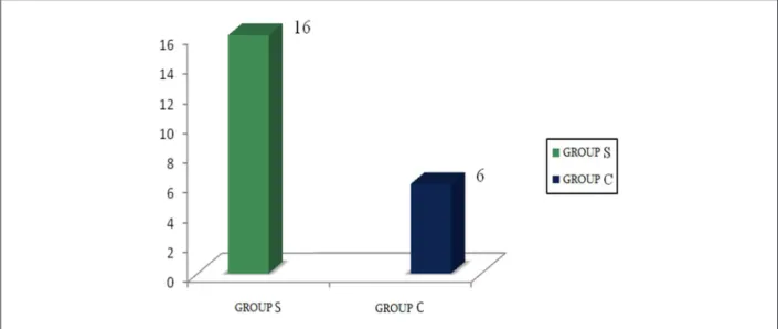
With respect to the value of SpO2, it was possible to notice a reduction in it in 16 patients in group S (53.3%) and 06 patients in group C (20.0%), with p-value of 0.007 by the chi-square test (Figure 2), demonstrating a statistically significant difference in oxygen saturation in the sufentanil group compared to the clonidine group.
Four patients in group S and three patients in group C had to use midazolam for supplementing the sedation, because they had a score of 01 in Ramsay scale, although there was no statistical difference between groups studied. Seven patients in group S used metoclopramide to treat nausea and vomiting and one patient in group C used dipyrone for pain control. The use of atropine was similar in both groups. As to other drugs, it was not necessary to use them.
Discussion
This study evaluated the efficacy of sufentanil versus
clonidine as sedative in patients undergoing cardiac catheterization who had been tested positive for coronary ischemia, comparing their effects on hemodynamic parameters presented during the exam. The choice of these drugs was based on previous studies that demonstrated the benefit of
clonidine as pre-anesthetic medication, and as an adjuvant in patients undergoing cardiac and non-cardiac surgical procedures, reducing the incidence of myocardial ischemia7.
Nascimento et al2 demonstrated the benefit of clonidine
in patients undergoing cardiac catheterization, compared to a control group without sedation. Patients that were sedated with this drug had less variation in blood pressure and heart rate. Although there are no published studies addressing the use of sufentanil in the catheterization laboratory, it is known that this drug has been administered as an adjuvant in anesthetic and analgesic procedures, being also indicated for sedation in adult and pediatric patients8, both inside and
outside the operating room.
In this study, it was possible to notice a similar behavior of the hemodynamic parameters, in both groups, statistically confirmed by the observed values of SBP, DBP and heart rate. Considering the values of SBP, DBP and heart rate measured at M1 and M3, which represent respectively the periods prior to administration of sufentanil and clonidine and the onset of cardiac catheterization, it is possible to notice a similar behavior in both groups. Thus, it can be stated that, for this population studied, sufentanil exhibited characteristics that are similar to those of clonidine with regard to the blocking of hemodynamic changes, usually seen after the beginning of interventional procedures. This suggests that the drugs were effective in blocking the cardiovascular sympathetic response in patients undergoing cardiac catheterization. It is known that the control of hemodynamic parameters reduces the incidence of myocardial ischemia and postoperative mortality in patients who have risk factors for coronary disease9.
The results obtained demonstrate that the efficacy of sufentanil and clonidine is similar with respect to the protection of the cardiovascular system, preventing the presence of hypertension, hypotension and tachycardia, which are considered risk factors for decompensation of preexisting heart disease10,11, which is something that is constantly seen
in the catheterization laboratory.
It is important to remember that sites that have opioid receptors, such as the regulatory centers of the cardiovascular system in the CNS, spinal cord, sympathetic nervous system, the adrenal medulla and vagal nuclei, help the opioids attenuate or eliminate significant hemodynamic responses to harmful stimuli. Thus, these agents used in anesthesia produce negligible cardiac depression, with minimal or no decrease in pre- and post-load, low depression of the great vessels and atrial baroreceptors and no effect on coronary motor functions12. Sufentanil, as a potent opioid, has been
of great value as a drug that provides perioperative analgesia and sedation with minimal deleterious hemodynamic effect13.
On the other hand, clonidine, as an alpha-2 adrenergic agonist, provides hemodynamic stability by reducing dose-dependent blood pressure and heart rate, thereby preventing the occurrence of tachycardia and hypertension during its use14. However, it must be slowly administered in order to
avoid undesirable effects, such as the temporary increase in blood pressure due to stimulation of alpha-2 receptor agonists in the vascular smooth muscle and reflex bradycardia15.
The respiratory parameters that were assessed were respiratory rate and SpO2. The respiratory rate was similar in
both groups, but the patients behaved differently with respect to SpO2, because at M6 the p value was 0.02, demonstrating that, at the time of discharge from the RPA, sufentanil is associated with lower SpO2 value when compared with clonidine. The SpO2 value of Group S at M6 was 97.1% ± 2.2 against 98.4% ± 1.8, which is considered a normal value (Table 3). It should be noted that M6 is the end of the study, with the patient being subjected to less stimulation. This finding is consistent with literature data that demonstrate that respiratory depression is rare in patients undergoing surgical and diagnostic procedures16.
Opioids affect the respiratory function in a dose-dependent manner, causing an increase in the partial pressure of carbon dioxide in arterial blood and the displacement of the CO2 response curve. Opioid agonists act on the respiratory center in the medulla, which could increase the respiratory pause and reduce central sensitivity to increased carbon dioxide17.
The respiratory depression resulting from the use of opioids is dose-dependent, and its incidence increases in proportion to the increase in the drug. In this study, sufentanil was administered at a dose of 0.1 µg/kg/min, which is considered small and unlikely to cause respiratory depression.
The respiratory effects of clonidine have been widely debated, but there is a consensus that alpha-2 adrenergic agonists are associated with minimal respiratory depression18
and this study confirms this. Even though alpha-2 adrenergic agonists are present in the brain, they do not play a central role in the control of breathing19.
The analysis of the values of the Ramsay scale reveals that, at M2, the patients that used sufentanil had a higher level of sedation when compared with those who received clonidine (Table 3). M2 represents 05 minutes after drug administration, which is compatible with the peak effect of sufentanil, which is known to be from three to 05 minutes. However, when we observe the Ramsay scale scores at each moment, in more detail in the two groups, it is possible to notice that most patients reached a satisfactory level of sedation, and sufentanil was associated with an earlier action onset when compared to clonidine, which can be explained by the fact that sufentanil starts its action almost immediately after its application. Clonidine has an onset of action of 05 minutes.
The Ramsay scale was developed 25 years ago. Nevertheless, it continues to be the best way to evaluate, in a subjective manner, the levels of sedation5.
The site for the sedative action of alpha-2 adrenergic agonists is located in the locus coeruleus of the brain stem, and the stimulation of the brain stem leads to inhibition of regulation of sleep and wakefulness20. The main ascending
and descending noradrenergic pathways originate from this important area. The activation of alpha-2 receptors in locus coeruleus leads to the suppression of its activity, resulting in increased activity of inhibitory interneurons, such as the pathway of the g-aminobutyric acid (GABA), which determines the depression of the CNS21.
mild sedation, its results were comparable to fentanyl, avoiding extra doses of sedatives25. It is known that part of the action
of alpha-2 adrenergic receptors derives from the activation of the same potassium channels as the opioid receptors26,
which may explain the behavior similar to that of clonidine in relation to opioids, with regard to sedation. In this study, the sedation obtained with clonidine was similar to that obtained with sufentanil. It should be noted that the dose of clonidine was 0.5 µg/kg, which is lower than that recommended in the literature for deep sedation, which corresponds to values between 02 and 06 µg/kg/dose27.
It is important to note that the sedation induced by clonidine is dose-dependent and that the desired level of sedation for cardiac catheterization is light, which corresponds to level 02 and 03 of the Ramsay scale, which justifies the administration of lower doses of this drug.
Sufentanil, in turn, exerts a sedative effect by acting on opioid receptors located in the central nervous system, with such effect being attributed to the connection between sufentanil and kappa receptors28. The sedation resulting from
the use of fentanyl is dose-dependent. Studies evaluating the actions of opioids on the central nervous system showed that in relatively low doses (0.35 µg/kg), sufentanil leads to a higher incidence of sleep, when compared with fentanyl (3.5 µg/kg).
Patients in Group S had an incidence of nausea and vomiting of 23.3%, versus no patients in the clonidine group. This shows that the incidence of nausea and vomiting found in the sufentanil group was similar to that described in the literature, which ranges from 20 to 30.0%29-31, being
associated with patient discomfort and complications, such as dehydration, increased intracranial and intraocular pressure, aspiration pneumonitis, electrolyte disturbances and even esophageal rupture32,33. The etiology of PONV is
multifactorial, and its presence is justified by the compromising of several neurotransmitters in specific neural pathways, such as dopaminergic, serotonergic, histaminergic and cholinergic pathways. Among the drugs capable of triggering PONV, opioids such as sufentanil gain prominence, with the use of such drugs being considered a major risk factor, together with female sex, abstinence from smoking and previous history of nausea and vomiting34,35. In this work, the presence of such
risk factors was not investigated. However, the groups were considered homogeneous according to other parameters.
It should be noted that opioids can cause nausea and vomiting by stimulating the afferent serotoninergic pathways related to the vagus nerve. These pathways are connected to the chemoreceptor trigger zone, located at the base of the 4th ventricle in the brain and act as partial agonists of dopamine receptors located in the same chemoreceptor zone36.
One patient in Group S had urinary retention, whereas no patient in Group C patient had complaints related to the urinary tract. Despite the low incidence of this event, which was of 3.3%, it is important to comment on such data, because it is known that this low incidence is related to delayed hospital discharge after the performance of outpatient procedures.
Urinary retention is common after anesthesia and surgery, with reports of an incidence of 5 to 70.0% of this adverse event in the period immediately after the surgery. This value is
higher than that found in this study, which may be explained by the small dose of opioid administered. It is believed that the presence of urinary retention is a dose-dependent effect, because studies conducted with patients undergoing cholecystectomy and appendectomy show that the incidence of urinary retention after the operation is proportional to the amount of opioids used37. It is important to remember that
agonists and alpha-2 adrenergic antagonists alter the bladder function, by acting on alpha-2 receptors present in smooth muscles of the lower and upper urinary tract38.
Gentili et al39 studied the effects of clonidine on bladder
function and, like in this study, they discovered that, compared to opioid, clonidine is associated with lower incidence of postoperative urinary retention39. Possible mechanisms
involved in the effects of clonidine are: reduction in sympathetic flow in the spinal cord and supraspinal inhibitory effect, reducing the internal urethral sphincter tone.
Pain was not observed in any patient in Group S, but its incidence in group C was of 3.3%. It is known that the cardiac catheterization procedure is not usually associated with great pain stimulus, and when such stimulus is present, it is weak.
In this study, the primary goal of the administration of sufentanil was not pain control, but its analgesic properties are well established. The analgesic mechanism of sufentanil is similar to any other opioid, occurring after its binding to specific receptors located at central and peripheral levels40.
Despite being an alpha-2 agonist drug, clonidine has analgesic properties that are associated with structures found in the spinal cord and supraspinal sites. Ambrose et al24
described the use of clonidine in the ICU as a substitute for morphine in patients who have become tolerant to opioids or whose sedation was difficult, reporting a good response, with minimal adverse effects24.
When the patients’ satisfaction was evaluated, it was possible to observe that 100% of patients in Group C and 93.3% of patients in group S reported being satisfied with the anesthetic procedure. No patient was asked why he or she was dissatisfied. However, patients in the sufentanil group had a higher incidence of nausea and vomiting, when compared with patients in the clonidine group. Studies have shown that nausea and vomiting are a frequent cause of dissatisfaction and delay in hospital discharge for patients undergoing outpatient procedures35.
All interventional cardiologists were satisfied with the anesthetic procedure performed. This can be explained by the fact that the level of sedation was adequate and there were minimal adverse effects.
In conclusion, sufentanil and clonidine in the doses used were effective as a sedative in patients undergoing cardiac catheterization, with sufentanil being associated with higher incidence of nausea and vomiting and lower patient dissatisfaction with the procedure performed.
Potential Conflict of Interest
References
1. American Society of Anesthesiologists Task Force on Sedation and Analgesia by Non-Anesthesiologists. Practice guidelines for sedation and analgesia by non-anesthesiologists. Anesthesiology. 2002; 96 (4): 1004-17.
2. Nascimento JS, Módolo NS, Carvalho HG, Dórea EM, Santos KP. Clonidina na cineangiocoronariografia: efeitos sedativos sobre a pressão artéria e a frequência cardíaca. Arq Bras Cardiol. 2006; 87 (5): 603-8.
3. Scott JC, Ponganis K, Stanski DR. EEG quantitation of narcotic effect: the comparative phamacodynamics of fentanyl and alfentanil. Anesthesiology. 1985; 62 (3): 234-41.
4. Scott JC, Cooke JE, Stanski DR. Electroencephalographic quantitation of opioid effect: comparative pharmacodynamics of fentanyl and sufentanil. Anesthesiology. 1991; 74 (1): 34-42.
5. Schulte-Tamburen AM, Scheier J, Briegel J, Schwender D, Peter K. Comparison of five sedation scoring systems by means of auditory evoked potentials. Intensive Care Med. 1999; 25 (4): 377-82.
6. Curi, PR. Metodologia e análise da pesquisa em ciências biologicas. 2ª ed. Botucatu: Tipomic; 1998.
7. Dorman BH, Zucker JR, Verrier ED, Gartman DM, Slachman FN. Clonidine improves perioperative myocardial ischemia, reduces anesthetic requirement, and alters hemodynamic parameters in patients undergoing coronary artery bypass surgery. J Cardiothorac Vasc Anesth. 1993; 7 (4): 386-95.
8. Silva YP, Gomez RS, Máximo TA, Silva ACS. Sedação e analgesia em neonatologia. Rev Bras Anestesiol. 2007; 57 (5): 575-87.
9. Wallace A, Layug B, Tateo I, Li J, Hollenberg M, Browner W, et al. Prophylatic atenolol reduces postoperative myocardial ischemia. McSPI Research Group. Anesthesiology. 1998; 88 (1): 7-17.
10. Mantz J. Alpha2-adrenoceptor agonists: analgesia, sedation, anxiolysis, haemodinamics, respiratory function and weaning. Best Pract Res Clin Anesthesiol.. 2000; 14 (2): 433-48.
11. Wallace AW, Galindez D, Salahieh A, Layug EL, Lazo EA, Haratonik KA, et al. Effect of clonidine on cardiovascular morbidity and mortality after noncardiac surgery. Anesthesiology. 2004; 101 (2): 284-93.
12. Bailey PL, Egan TD: Fentanyl and congeners. In: White PF. Intravenous anesthesia. Baltimore: Williams & Wilkins; 1997. p. 213-45.
13. Nociti JR, Serzedo PSM, Nunes AMM, Cagnolati CA, Zuccolotto EB, Angelo MAF, et al. Sufentanil em infusão venosa contínua para cirurgias abdominais. Rev Bras Anestesiol. 1995; 45 (4): 235-43.
14. Aantaa R. Assessment of the sedative effects of dexmedetomidine, an alpha2-adrenoceptor agonist, with analysis of saccadic eye movements. Pharmacol Toxicol. 1991; 68 (5): 394-8.
15. Bloor BC, Ward DS, Belleville JP, Maze M. Effects of intravenous dexmedetomidine in humans. II. Hemodynamic changes. Anesthesiology. 1992; 77 (6): 1134-42.
16. Ribeiro S, Schmidt AP, Schmidt SRG. O uso de opióides no tratamento da dor crônica não oncológica: o papel da metadona. Rev Bras Anestesiol. 2002; 52 (5): 644-51.
17. Gozzani JL. Opióides e antagonistas. Rev Bras Anestesiol. 1994; 44 (1): 65-73.
18. Belleville JP, Wards DS, Bloor BC, Maze M. Effects of intravenous dexmedetomidine in humans. I. Sedation, ventilation and metabolic rate. Anesthesiology. 1992; 77 (6): 1125-33.
19. Hall JE, Uhrich TD, Ebert TJ. Sedative, analgesic and cognitive effects os clonidine infusion in humans. Br J Anaesth. 2001; 86 (1): 5-11.
20. Maze M, Tranquilli W. Alpha-2 adrenoreceptor agonists: defining the role in clinical anesthesia. Anesthesiology. 1991; 74 (3): 581-605.
21. Braz LG, Vianna PTG, Braz JRC. Níveis de sedação determinados pela clonidina e midazolam na medicação pré-anestésica: avaliação clínica e eletroencefálica bispectral. Rev Bras Anestesiol. 2002; 52 (1): 9-18.
22. Knight G, Ramelet AS, Duncan A, Lago P, Piva JP, Garcia P, et al. Analgesia e sedação em UTIP. In: Piva JP, Garcia PCR.(eds) Medicina intensiva em pediatria. Rio de Janeiro: Revinter; 2005. p. 733-52.
23. Fujii Y, Saitoh Y, Tanaka H, Toyooka H. Pretreatment with oral clonidine attenuates cardiovascular responses to tracheal extubation in children. Paediatr Anaesth. 2000; 10 (1): 65-7.
24. Ambrose C, Sale S, Howells R, Bevan C, Jenkins I, Weir P, et al: Intravenous clonidine infusion in critically ill children: dose-dependent sedative effects and cardiovascular stability. Br J Anaesth. 2000; 84 (6): 794-6.
25. Reimer EJ, Dunn GS, Montgomery CJ, Sanderson PM, Scheepers LD, Merrick PM. The effectiveness of clonidine as an analgesic in paediatric adenotonsillectomy. Can J Anaesth. 1998; 45 (12): 1162-7.
26. Jacobi J, Fraser GL, Coursin DB, Riker RR, Fontaine D, Wittbrodt ET, et al. Clinical practice guidelines for sustained use of sedatives and analgesics in the critically ill adult. Crit Care Med. 2002; 30 (1): 119-41.
27. Kress JP, Pohlman AS, O´Connor MF, Hall JB. Daily Interruption of sedative infusions in critically ill patients undergoing mechanical ventilation. N Engl J Med. 2000; 342 (20): 1471-7.
28. Trescot AM, Datta S, Lee M, Hansen H. Opioid pharmacology. Pain Physician. 2008; 11 (2 Suppl.): S133-53.
29. Abreu MP. Controle de náuseas e vômitos – antieméticos. In: Posso I, Poterio GMB, Cangiani LM.(eds). Tratado de anestesiologia SAESP.. 6a ed. São Paulo: Atheneu; 2006. p. 1361-72.
30. Lages N, Fonseca C, Neves A, Landeiro N, Abelha FJ. Náuseas e vômitos no pós-operatório: uma revisão do “pequeno-grande”problema. Rev Bras Anestesiol. 2005; 55 (5): 575-85.
31. Carvalho WA, Vianna PTG, Braz JRC. Náuseas e vômito em anestesia: fisiopatologia e tratamento. Rev Bras Anestesiol. 1999; 49 (1): 65-79.
32. Schmidt A, Bagatini A. Náusea e vômito pós-operatório: fisiopatologia, profilaxia e tratamento. Rev Bras Anestesiol. 1997; 47 (4): 326-34.
33. Ku CM, Ong BC. Postoperative nausea and vomiting: a review of current literature. Singapore Med J. 2003; 44 (7): 366-74.
34. Thomas R, Jones NA, Strike P. The value of risk score for predicting postoperative nausea and vomiting when used to compare patient groups in a randomised controlled trial. Anaesthesia. 2002; 57 (11): 1119-28.
35. Sinclair DR, Chung F, Mezei G. Can postoperative nausea and vomiting be predicted? Anesthesiology.1999; 91 (1): 109-18.
36. Malan TP. Opioid pharmacology: new insights and clinical relevance. ASA Refresher Courses in Anesthesiology. 2000; 28: 109-19.
37. Petros JG, Mallen JK, Howe K, Rimm EB, Robillard RJ. Patient-controlled analgesia and postoperative urinary retention after open appendectomy. Surg Gynecol Obstet. 1993; 177 (2): 172-5.
38. Durant PA, Yaksh TL. Drug effects on urinary bladder tone during spinal morphine-induced inhibition of the micturition reflex in unanesthetized rats. Anesthesiology. 1988; 68 (3): 325-34.
39. Gentili M, Bonnet F. Spinal clonidine produces less urinary retention thanspinal morphine. Br J Anaesth. 1996; 76 (6): 872-3.
40. Inturrisi CE. Clinical pharmacology of opioids for pain. Clin J Pain. 2002; 18 (Suppl 4): S3-S13.
Sources of Funding
There were no external funding sources for this study.
Study Association