CLINICAL SCIENCE
Department of Orthopedics, Faculdade de Medicina da Universidade de São Paulo - São Paulo/SP, Brazil.
Email: [email protected]
Received for publication on September 17, 2009 Accepted for publication on December 17, 2009
ANTEROGRADE REMOVAL OF BROKEN FEMORAL
NAILS WITHOUT OPENING THE NONUNION SITE: A
NEW TECHNIQUE
Henrique Antônio Berwanger de Amorim Cabrita, Eduardo Angeli Malavolta, Otávio Vilhena Reis Teixeira, Nei Botter Montenegro, Fernando Aires Duarte, Rames Mattar Jr.
doi: 10.1590/S1807-59322010000300007
Cabrita HABA, Malavolta EA, Teixeira OVR, Montenegro NB, Duarte FA, Mattar Jr. R. Anterograde removal of broken femoral nails without opening the nonunion site: a new technique. Clinics. 2010;65(3):279-83.
OBJECTIVE: We describe a new technique for removing the distal fragments of broken intramedullary femoral nails without disturbing the nonunion site.
METHODS: This technique involves the application of an AOdistractor prior to the removal of the nail fragments, with subsequent removal of the proximal nail fragment in an anterograde fashion and removal of the distal fragment through a medial parapatel-lar approach. Impaction of the fracture site is then performed with a nail that is broader than the remaining fragmented material.
RESULTS: Nails were removed from ive patients using the technique described above without any complications. After a mean follow-up period of 61.8 months, none of these patients showed worsened knee osteoarthritis.
CONCLUSION: The original technique described in this article allows surgeons to remove the distal fragment of fractured femoral intramedullary nails without opening the nonunion focus or using special surgical instruments.
KEYWORDS: Femoral fractures; Fracture ixation; Intramedullary; Fracture healing; Complications; Orthopedics.
INTRODUCTION
Intramedullary nail placement is currently the treatment of choice for femoral shaft fractures, and has several biomechanical advantages compared to internal ixation with plates,1,2 and consolidation occurs in 97-100% of cases.3
The use of closed focus insertion allows surgeons to avoid damage to the surrounding soft tissue (biological approach). Additionally, the surgical technique is simple and has good reproducibility.
However, this method is not exempt from complications, and pseudarthrosis accompanied by nail fracture is a particularly problematic complication associated with this procedure. This rare event occurs due to the cyclic stress
placed on the synthetic material in the intramedullary nail through the nonunion focus.
The removal of the distal fragment of the intramedullary nail poses an important technical challenge. Most papers describe techniques for the retrograde removal of nail fragments that involve the use of hooks, olive wires, or special instruments.4-6 These methods require the use of
speciic materials that are often not available to the general orthopedist. Additionally, because it is dificult to remove the fragments using this approach, orthopedists performing the procedure may need to open the nonunion site or damage the surrounding tissue.
MATERIAL AND METHODS
We included ive patients (three men and two women) who presented to our clinic with femoral intramedullary nail fracture with pseudarthrosis or delayed union of femoral shaft fracture (classified as AO 32), and who initially underwent surgery between 3/17/03 and 10/20/03. The age range of the included patients was 22-37 years (mean, 27.4 years). All patients were submitted to the novel surgical technique described below. The patients were monitored prospectively after surgery for mean follow-up period of 61.8 months (range: 58-65).
Surgical Technique
The patient is placed in the supine position on a radiolucent table to allow the use of an image intensiier. Next, a femoral distractor (we use an AO distractor, but a similar type could also be used) is applied for fragment stabilization. An incision is then made at the prior site of insertion of the femoral nail with the goal of preserving the gluteal musculature. If the nail is locked, both the proximal and the distal locking screws are removed.
The main fragments of the fracture are then reduced and temporarily stabilized with the assistance of the fracture distractor. Next, a guide wire is passed through the proximal fragment of the nail and, after the reduction of the pseudarthrosis focus, through the fractured distal fragment. This step is not performed if the broken fragment is solid. The proximal fragment is then removed with the type of extractor that is suitable for the type and size of pin that was used. If the passage of the guide wire in the distal segment proves to be dificult, the proximal fragment is removed irst to facilitate access to the distal fragment.
A small medial parapatellar incision is then made in the direction in which the tip of the femoral nail will be removed. This direction is chosen with the help of an image intensiier. The local soft tissue is then divided to allow exposure of the femoral intercondylar notch.
Two types of procedures can be performed at this stage. If the guide wire is directed towards the exposed region, it can be used to perforate the cortical bone of the femoral intercondylar notch; however, care should be taken not to injure the cruciate ligaments. On the other hand, if the wire is not properly positioned or if the broken nail is bulky, a number two Steinmann wire is inserted in the femoral intercondylar notch in the direction of the fractured nail.
Intercondylar reaming is then performed using the guide wire or the distally fastened Steimann wire until the opening reaches the diameter of the nail that must be removed.
If the reamed nails are broken, the guide wire is propelled through this opening in the distal femur.
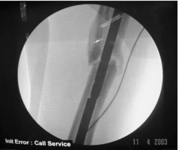
A femoral nail with a diameter that is 2 mm larger than the diameter of the extracted nail is then inserted in an anterograde manner. The anterograde approach was chosen so that the nail could be carefully introduced such that its distal fragment could be propelled through the opening that had been created in the femoral intercondylar notch (Figures 1 and 2).
After removal of the nail fragments, the surgeon has the option of reaming the femoral canal again, but the decision of whether or not to perform this additional step is left to the discretion of the treating surgeon. The new intramedullary nail with the appropriate diameter is then inserted and the pseudarthrosis is repaired. Finally, the distractor is removed and the incisions are sutured to complete the procedure. Figure 1 - Intramedullary nail with a diameter that was 2 mm larger than the fractured nail propelling the distal fragment (luoroscopic view)
RESULTS
All of the included patients’ sites of nonunion healed within 4-9 months (mean, 6.6 months). No patients experienced functional impairment of their knees or hips (case 5 had impaired function of the knee preoperatively). Only one patient presented with femoropatellar crepitus (case 1). However, this patient was asymptomatic. The radiographic evaluation that was performed at the last follow-up appointment (mean, 61.8 months postoperatively) showed that one patient had knee osteoarthritis (case 5), but that patient’s Ahlbäck classiication was the same as it had been preoperatively (Table 1).
DISCUSSION
The removal of the distal fragment of a fractured femoral nail is challenging for the orthopedic surgeon, and countless operative techniques for fractured femoral nail removal have been previously described.2,4,6 Franklin et al.5 described their
experience with the treatment of 60 broken femoral or tibial nails. In their series, 20 distal fragments were extracted without auxiliary surgical methods and 28 nail fragments were removed using long hooks. The hooks that were used have a proile that is similar to the proile of Ender pin extractors.
Brewster et al.6 and Hahn et al.7 also endorsed the
removal of reamed nails with the use of long hooks. However, they mention that the hooks can slip several times at the tip of the nail, become stuck in the distal fragment, and bend (or even break) inside the nail.8 These complications
prolong the patient’s surgery and exposure to the image intensiier, test the surgeon’s patience, and increase the risk of postoperative complications.9
Giannoudis et al.4 described the extraction of fragments
with special tools, such as long graspers and hooks. This technique involves the use of long trephines, hooks, and auxiliary pins. The technique is costly and labor-intensive, but it is a good alternative method, especially for fractures of rigid and unreamed tibial pins.
Table 1 - Patient characteristics and outcomes
Case Age Time
between fracture and surgery
(months)
Time to partial load
bearing (weeks)
Number of previous
femoral surgeries
Time to ra-diographic fracture
con-solidation
(months)
Previous knee range
of motion (degrees)
Current knee range
of motion (degrees)
Ahlbäck OA clas-siication
(preopera-tive)
Ahlbäck OA clas-siication (at the last
follow-up
appoint-ment)
Femoro-patellar crepitus
(asymptom-atic)
Surgical time (hours)
Follow-up time (months)
1 27 16 3 1 9 0-145 0-145 0 0 Yes 1.5 64
2 29 42 Paraplegic 1 4 0-145 0-145 0 0 No 1.0 65
3 37 48 Immediate 3 5 0-160 0-160 0 0 No 0.8 61
4 22 16 2 1 6 0-160 0-160 0 0 No 1.1 58
5 22 60 6 4 9 0-90 0-90 2 2 No 1.0 61
The radiographic images of one of the patients (case 1) are presented in the igures (igures 3, 4, 5 and 6).
Figure 3 and 4 - A fractured femoral nail in an unhealed femoral fracture (Anterior-Posterior and lateral radiographs)
Levy et al.9 described yet a different surgical approach
in which they impact a nail of smaller diameter than the original nail inside the distal fragment of a broken reamed femoral nail to facilitate local impaction and anterograde extraction.
Middleton et al.10 suggested illing the internal space
of a reamed nail with several guide wires to allow the anterograde extraction of the distal fragment.
Maini et al.11 proposed passing an olive guide wire
through the distal fragment of a reamed femoral nail and then illing the nail with long Steinmann wires to facilitate its removal.
Finally, Marwan and Ibrahim8 described a technique in
which they pass a metallic wire through the middle of the fractured nail and through its distal hole. They then fasten this wire to the distal fragment through a small incision at the level of the distal hole.
Unreamed nails are being used increasingly often these days (especially in polytrauma patients) due to the lower associated rate of fat embolism. The methods described above cannot be used in patients with fractured unreamed nails because they involve the passing of wires or hooks through the hollow diameter of the nail. Likewise, the anterograde removal of fractured Marchetti-type nails without disturbance of the nonunion focus is impossible.
Hellemondt and Haeff12 described a technique in which
they extract the solid distal fragment of a tibial nail via the passage of a reamed nail with a larger diameter than that of the broken fragment and the assistance of pins and long hooks. This method cannot be used when the fractured fragment is of the same diameter as the medullary canal.
Franklin et al.5 also described the use of a long
grasper-type instrument for the removal of distal fragments of broken solid nails, but stated that this approach is associated with a high degree of technical dificulty during the surgery.
Kretteck et al.13 described a technique in which they
remove the distal fragment by creating an opening directly adjacent to the ixation hole of the proximal screw of the distal segment of the nail. They then place a Hohmann-type lever into this opening to impel the fragment in the direction of the fracture focus.
Fractured nail removal via the opening of the nonunion focus and resection of the bone lute followed by traction with a hook was described by Khan.14 Gregory15 described a
different approach, in which the distal fragment is extracted by means of a new incision through the pyriform fossa. An auxiliary opening is then created in the femoral canal to allow clamping of the distal fragment. These types of approaches are aggressive, but they become the best option when special instruments are not available.
The method presented in this study does not depend on
special surgical instruments and can be performed in any operating room. Moreover, it does not protract the operative time or require excessive use of the image intensiier.
The fracture focus is not disturbed in this technique, which allows us to avoid aggressive dissection, excessive bleeding, and the exposure of the surrounding soft tissue. Additionally, by not disturbing the local soft tissue, the infection rate should be decreased. In a study that compared the treatment of femoral pseudarthrosis using a technique that involved a pseudarthrosis approach with an approach that did not, Wu et al.16 observed shorter operative times
(1.5+/-0.4 hrs vs. 2.4+/-0.4 hrs) and shorter time to radiographic fracture consolidation (4.4+/-0.9 months vs. 5.7+/-1.5 months) in patients who underwent surgery that did not involve the pseudarthrosis focus approach. In the ive cases presented in this paper, the mean surgical time was 1.08 hours (range, 0.8 to 1.5).
By accessing the proximal fragment of the nail through the previous incision site, we follow the principles of minimally invasive osteosynthesis that serve as the current guidelines for the treatment of fractures.
The insertion of the pin that is used to impel the distal fragment should be performed carefully to avoid fractures at the level of the pseudarthrosis focus as well as fractures of the distal femur.
The method we describe is cost-effective because it does not require auxiliary instruments to remove the distal fragment of the nail; the pin that is used is both the instrument of removal of the fractured nail and the method of deinitive treatment.
The disadvantage of our technique is that it requires an additional incision at the knee. Ostrum et al.17 prospectively
randomized 100 patients with femoral fractures to 2 treatment groups: nail insertion through an anterograde approach vs. nail insertion through a retrograde approach. Differences regarding knee symptoms were not observed between the two groups, which reinforces the concept that when local dissection is executed well, the passage of a nail through the knee does not lead to adverse effects in the femoropatellar region. In a prospective series of 284 patients with femoral fractures, Ricci et al.18 found that 36%
of patients complained of knee pain after retrograde ixation as compared to 9% of patients who underwent anterograde ixation.
Milia et al.19 described a technique for the removal of
a fractured proximal femoral nail from a subtrochanteric fracture that involves the use of a medial arthrotomy of the knee and retrograde impulsion of the distal fragment. The only patient who was treated in this manner was without knee pain a year after surgery.
in this study, with a minimally invasive approach in the intercondylar region, did not lead to pain or functional impairment in our patients. The patients all recovered without complaints of knee pain and had good range of knee motion by their last follow-up appointment. Only one patient experienced femoropatellar crepitus, which was asymptomatic. By the last follow-up appointment, all patients had knee range of motion measurements that were unchanged from their preoperative measurements. None of the patients showed progression of their osteoarthritis after a mean follow-up period of 61.8 months.
All patients experienced fracture consolidation, which proves that through new ixation with larger nails (i.e., nails that have a greater diameter than the nails that were initially
used) and the resultant increase in stability of the fracture focus, the use of a bone graft is made unnecessary.16
We believe that this method, which does not involve opening of the nonunion focus, is reproducible and can be easily implemented by other orthopedists.
CONCLUSION
The original technique described in this article allows surgeons to remove the fractured distal fragment of intramedullary femoral nails without disturbing the nonunion focus. It can be used for reamed and unreamed nails and does not require the use of special surgical instruments, making it accessible to the general orthopedist.
REFERENCES
1. Bucholtz RW, Brumback RJ. Fractures of the shaft of the femur. In: Rockwood J, Green DP, Bucholtz RW, Heckman JD, editors. Fractures in adults. Philadelphia: Lippincot-Raven; 1996. p.1918-27.
2. Winquist RA, Hansen ST, Clawson DK. Closed intra-medullary nailing of femoral fractures. J Bone Joint Surg Am. 1984;66:529-39. 3. Wolinsky PR, McCarty E, Shyr Y, Johnson K. Reamed intramedullary
nailing of the femur: 551 cases. J Trauma. 1999;46:392-9.
4. Giannoudis PV, Matthews SJ, Smith RM. Removal of the retained fragment of broken solid nails by the intra-medullary route. Injury. 2001;32:407-10.
5. Franklin JL, Winquist RA, Benirschke SK, Hansen ST Jr. Broken intramedullary nails. J Bone Joint Surg Am. 1988;70:1463-71. 6. Brewster NT, Ashcroft GP, Scotland TR. Extraction of broken
intramedullary nails--an improvement in technique. Injury. 1995;26:286. 7. Hahn D, Bradbury N, Hartley R, Radford PJ. Intramedullary nail
breakage in distal fractures of the tibia. Injury. 1996;27:323-7. 8. Marwan M, Ibrahim M. Simple method for retrieval of distal segment
of the broken interlocking intramedullary nail. Injury. 1999;30:333-5. 9. Levy O, Amit Y, Velkes S, Horoszowski H. A simple method for removal of a fractured intramedullary nail. J Bone Joint Surg Br. 1994;76:502-3. 10. Middleton RG, McNab IS, Hashemi-Nejad A, Noordeen MH. Multiple guide wire technique for removal of the short distal fragment of a fractured intramedullary nail. Injury. 1995;26:531-2.
11. Maini L, Upadhyay A, Aggarwal A, Dhaon BK. A new method of removing a fractured interlocked nail. Injury. 2002;33:261-2. 12. Hellemondt FJ, Haeff MJ. Removal of a broken solid intramedullary
interlocking nail. A technical note. Acta Orthop Scand. 1996;67:512. 13. Krettek C, Schandelmaier P, Tscherne H. Removal of a broken solid
femoral nail: a simple push-out technique. A case report. J Bone Joint Surg Am.1997;79:247-51.
14. Khan FA. Retrieval of a broken intramedullary femoral nail. Injury. 1992;23:129-30.
15. Gregory PR Jr. Removal of a broken solid-core intramedullary femoral nail using both antegrade and retrograde starting points. Orthopedics. 1997;20:1087-9.
16. Wu CC, Shih CH, Chen WJ, Tai CL. Treatment of ununited femoral shaft fractures associated with locked nail breakage: comparison between closed and open revision techniques. J Orthop Trauma. 1999;13:494-500. 17. Ostrum RF, Agarwal A, Lakatos R, Poka A. Prospective comparison of retrograde and antegrade femoral intramedullary nailing . J Orthop Trauma. 2000;14:486-501.
18. Ricci WM, Bellabarba C, Evanoff B, Herscovici D, DiPasquale T, Sanders R. Retrograde versus antegrade nailing of femoral shaft fractures. J Orthop Trauma. 2001;15:161-9.