Digital models: How can dental arch form
be verified chairside?
Alana Tavares1, Emanuel Braga2, Telma Martins de Araújo2
Introduction: Plaster dental casts are routinely used during clinical practice to access maxillary dental arch form and assist on fabrication of individualized orthodontic archwires. Recently introduced, digital model technology may offer a limitation for the obtainment of a dental physical record. In this context, a tool for dental arch form assessment for chairside use is necessary when employing digital models. In this regard, paper print of the dental arch seems thus to be useful. Methods: In the present study, 37 lower arch models were used. Intercanine and intermolar widths and dental arch length measurements were performed and compared using plaster dental casts, digital models and paper print im-age of the models. Ortho Insight 3D scanner was employed for model digitalization. Results: No statistically significant differences were noted regarding the measurements performed on the plaster or digital models (p > 0.05). Paper print images, however, showed subestimated values for intercanine and intermolar widths and overestimated values for dental arch length. Despite being statistically significant (p < 0.001), the differences were considered clinically negligible. Con-clusion: The present study suggests that paper print images obtained from digital models are clinically accurate and can be used as a tool for dental arch form assessment for fabrication of individualized orthodontic archwires.
Keywords: Orthodontics. Dental arch. 3D imaging. Computer generated.
1 Universidade Federal da Bahia, Mestrado em Odontologia e Saúde
(Salvador/BA, Brasil).
2 Universidade Federal da Bahia, Departamento de Ortodontia
(Salvador/BA, Brasil).
» The authors report no commercial, proprietary or financial interest in the products or companies described in this article.
DOI: https://doi.org/10.1590/2177-6709.22.6.068-073.oar
How to cite: Tavares A, Braga E, Araújo TM. Digital models: How can dental arch form be verified chairside? Dental Press J Orthod. 2017 Nov-Dec;22(6):68-73. DOI: https://doi.org/10.1590/2177-6709.22.6.068-073.oar
Submitted: May 05, 2017 - Revised and accepted: August 22, 2017
Contact address: Alana Tavares Ribeiro Meneses
Rua Priscila Dutra, 1229, casa 14, Vilas do Atlântico - Lauro de Freitas/BA CEP: 42.700-000 – E-mail: [email protected]
Introdução: os modelos de gesso são usados rotineiramente, durante a prática clínica, para avaliação da forma da arcada inferior e para auxiliar na confecção de arcos ortodônticos individualizados. A tecnologia dos modelos digitais, introduzida recente-mente, pode oferecer uma limitação na obtenção de um registro físico da arcada dentária. Assim, quando se utilizam modelos digitais, faz-se necessária uma ferramenta clínica para obtenção da forma da arcada. Com essa finalidade, poderia-se imprimir, em papel, uma imagem da arcada dentária obtida a partir do modelo de gesso. Métodos: nesse estudo, 37 modelos da arcada inferior foram utilizados, nos quais foram realizadas medições das distâncias intercaninos, intermolares e comprimento da arcada; sendo, então, comparadas entre modelos de gesso, modelos digitalizados com um scanner Ortho Insight 3D e imagens impressas em folha de papel A4. Resultados: não foram encontradas diferenças estatisticamente significativas nas medidas realizadas nos modelos de gesso e modelos digitais (p > 0,05). As imagens impressas, contudo, mostraram valores subestimados para as distâncias intercaninos e intermolares, e superestimados para o comprimento da arcada. Apesar de serem estatisticamente significativas (p < 0,001), as diferenças foram consideradas clinicamente insignificantes. Conclusão: o presente estudo sugere que as imagens obtidas por meio dos modelos digitais e impressas em papel são clinicamente acuradas e podem ser utilizadas como uma ferramenta auxiliar na confecção dos arcos ortodônticos individualizados.
INTRODUCTION
Plaster models are traditionally used as an essential part
of the orthodontic documentation process.1,2 Combined
to photographs, radiographs and clinical examination, plaster models provide important information for dental
and skeletal malocclusions diagnosis and treatment.3
Plaster models are very convenient but indeed pres-ent disadvantages, such as need for signiicant physical space for storage, possible breakages or damages, mi-croorganisms colonization in the long-term, possibili-ty of loss, and diiculpossibili-ty to exchange information with other professionals.
Reducing physical iles volume in dental oices is widely needed. In this context, digital records of patients have been increasingly incorporated in or-thodontic oices. Digital models have recently been introduced in clinical orthodontics, having potential to replace plaster models and eliminate storage space
issues.4 On the other hand, digital models also have
some limitations, such as inability to be manually han-dled, need for sotware technical support and possi-ble information loss. However, it is believed that such problems are less important compared to what digital
technology may ofer.1,5
In orthodontics, dental arch form maintenance is important, being directly related to function,
aesthet-ics and stability.6-8 Therefore, arch shape, especially the
maxillary arch, should not be altered throughout the treatment, in order to ensure outcome stability.
It is common in clinical practice to use plaster models to assist in the preparation of individualized orthodontic archwires. In this sense, replacing plaster models by digital models implies the loss of a physical record to guide the orthodontist. Thereby, the present study aimed to test the idelity of printed images ob-tained from the digitized model.
MATERIAL AND METHODS
The study sample consisted of 37 lower dental arch plaster models depicting initial malocclusion of patients who undergone treatment in the Prof. José Édimo J. Soares Martins Orthodontics and Den-tofacial Orthopedics Center, Federal Universi-ty of Bahia (FOUFBA). The study was approved by FOUFBA ethics committee with the protocol number 35868414.5.0000.50.24. All participants signed an in-formed consent.
Patients were randomly selected and met the follow-ing inclusion criteria: complete permanent dentition up to irst molars; no prosthetic restoration; and plaster models in perfect preparation and conservation state, without pos-itive or negative bubbles or dental crown defects.
Evaluations were made in plaster models, digital models and printed images generated from digitized models. In order to evaluate plaster models, a Cen-Tech 4” (Harbor Freight Tools, Calabasas, CA, USA) digital caliper with 0.01mm accuracy and a speciic plate made in CorelDRAW X5 containing two bold lines, one vertical and one horizontal, were used. Sub-sequently, a transparent adhesive was printed and ixed on a three millimeters thick glass plate, in order to keep the grid lat and facilitate assessments.
Evaluation was performed considering the following measures:
» Arch length – measured in millimeters on a verti-cal line between lower central incisors to a horizontal line connecting the distal surfaces of teeth #36 and #46. A graduated plate specially designed for the study was used. The vertical line perpendicular to the horizontal line was positioned in a point between central incisors. Arch length was evaluated on the plate with a digital caliper (Fig 1).
» Intercanine width – distance from tooth #33 cusp tip to tooth #43 cusp tip, measured in millimeters with a digital caliper (Fig 2).
» Intermolar width – distance from tooth #36 me-siobuccal cusp tip to tooth #46 meme-siobuccal cusp tip, measured in millimeters with a digital caliper (Fig 3).
In order to evaluate digital models, lower arch virtu-al images were created from the plaster model through Ortho Insight 3D scanner, v. 5.0 (Motion View Sot-ware, LLC, Chattanooga, Tennessee, USA). Subse-quently, arch length, and intercanine and intermolar widths measures were automatically generated by the program, following the same reference points used for the plaster model (Fig 4).
Figure 1 - Plaster model arch length evaluation using the graduated plate and digital caliper.
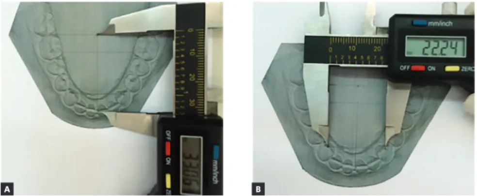
Figure 5 - Printed image evaluation, as follows: A) arch length, B) intercanine and C) intermolar widths.
Figure 2 - Plaster model intercanine width evalu-ation.
Figure 3 - Plaster model intermolar width evalu-ation.
arch length, intercanine and intermolar widths were measured with a digital caliper using the same reference points used for plaster and digital models (Fig 5).
Prior to measurements, in order to determine re-searcher calibration, i ve plaster models were random-ly selected. Plaster model, digital model and printed image measurements were performed at two dif erent times, two weeks apart, under the same conditions by the same researcher, which was properly trained.
Measurements were subjected to statistical test to de-termine method error. For all variables, error was cal-culated according to Dahlberg’s formula, in order to verify intra-rater agreement. Measurement reproduc-ibility analysis was conducted by intraclass correla-tion test. Both tests were set to 95% coni dence level. The Bland-Altman test was also performed for mea-surement reproducibility analysis and the results were considered not clinically important.
A B
Figure 4 - Digital model evaluation, as follows: A) arch length, B) intercanine and C) intermolar widths.
A B C
Descriptive analysis was used to express the results as mean and standard deviation. To compare the den-tal arch measurements in the diferent methods, Vari-ance Analysis for Repeated Measurements (ANOVA) was employed. Data distribution was evaluated with Shapiro-Wilk test. Signiicance level was set at 5%
(α= 0,05). Data was tabulated and veriied using IBM
SPSS Statistics for Windows (IBM SPSS. 21.0, 2012, Armonk, NY: IBM Corp.).
RESULTS
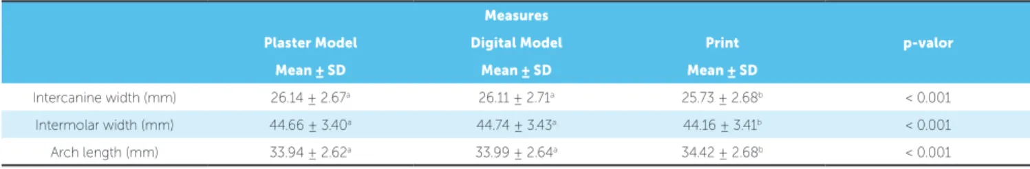
Table 1 shows the results for intercanine and inter-molar widths and arch length, measured in the plaster models, digital models or paper prints. No statistically signiicant diferences were noted regarding the mea-surements performed on the plaster or digital models
(p > 0.05). Paper print images, however, showed
subesti-mated values for intercanine and intermolar widths and overestimated values for dental arch length; these
difer-ences were considered statistically signiicant (p < 0.001)
for the tested parameters.
DISCUSSION
Literature reports that arch length, perimeter and incisors position change over time due to
physiologi-cal reasons, regardless of orthodontic treatment.9 It is a
consensus for most authors that arch shape and length should be preserved during orthodontic therapy, in
or-der to achieve greater post-treatment stability.6,7,10
Plas-ter models have been the tool used to help orthodontists to reproduce individual dental arch shape of each pa-tient during archwire bending. However, plaster models presents some disadvantages, such as need for signii-cant physical space for storage, possible breakages and
damage, possibility of loss and diiculty to exchange
information with other professionals.1,11 In this context,
three-dimensoinal (3D) scanned models have been pro-posed as a means to overcome plaster models limitations and facilitate orthodontics diagnose and planning.
As advantages, virtual models do not require physi-cal space for storage, allow faster information exchange
among professionals,12 and provide working options
such as the virtual setup preparation.13 On the other
hand, virtual models also have some limitations, such as
the inability to be manually handled.1 Thus, it is
neces-sary a mechanism to assist the orthodontist on accessing the arch shape when dealing with digital models.
The present study compared traditional plaster mod-els, digitized models and paper prints obtained through the virtual models, aiming at providing a tool to help the orthodontist on accessing the dental arch shape in the daily clinical practice. No similar studies were found in the literature.
This study found no statistically significant dif-ference (p > 0.05) comparing arch length, intercanine and intermolar width measurements between plaster and digital models. Different results were found by
Santoro et al,2 but it was highlighted by the authors
that the differences were within a clinical acceptable range and, thus acceptable for orthodontic use.
Keat-ing et al11 found no statistically significant differences
between the two methods. Oliveira et al14 have also
observed not statistically significant differences be-tween methods, showing measurement
reproducibil-ity and reliabilreproducibil-ity using digital models. Kim et al15
found excellent agreement between plaster and dig-italized models, considering digdig-italized models reli-able to replace traditional ones.
Measures
p-valor
Plaster Model Digital Model Print
Mean ± SD Mean ± SD Mean ± SD
Intercanine width (mm) 26.14 ± 2.67a 26.11 ± 2.71a 25.73 ± 2.68b < 0.001
Intermolar width (mm) 44.66 ± 3.40a 44.74 ± 3.43a 44.16 ± 3.41b < 0.001
Arch length (mm) 33.94 ± 2.62a 33.99 ± 2.64a 34.42 ± 2.68b < 0.001
Table 1 - Comparison between the different methods for evaluating intercanine and intermolar widths and arch length.
a,b Horizontal values (line) with distinct letters indicate statistical difference (p < 0.05, comparisons between pairs with Bonferroni adjustment). Values are expressed
Paper print images, however, showed subestimated values for intercanine and intermolar widths and over-estimated values for dental arch length. The
diferenc-es were found to be statistically signiicant (p < 0.001).
However, the comparison between the digital model and the paper print obtained from it, showed that for intercanine and intermolar widths the mean difer-ences were 0.38mm and 0.58mm, respectively. Re-garding arch length, the mean diference observed was
-0.52mm. Previous published research,2 so as the
au-thors of the present study, considered such diferenc-es as clinically negligible. It is thus suggdiferenc-ested that the presented method is accurate for clinical use without bringing any potential distortions for the fabrication of orthodontic archwires or arch shape observation.
It is important to note that the image was obtained according to the mentioned methodology and, there-fore, must be reproduced. Diferent scanners may show diferent results, thus, proper studies for diferent man-ufacturers are recommended.
Outcome stability, especially in relation to lower teeth irregularities, constitutes a key factor in ortho-dontic treatment. Several factors may contribute for increased stability and, among them, intercanine and
intermolar widths maintenance are highlighted.16
According to accessed literature, arch shape change during orthodontic therapy potentiates relapse
occur-rence, making clear that, when possible, patient’s initial
arch form is the best guide for future stability.7,10
The literature also shows evidence that intercanine and intermolar width decrease ater treatment,
especial-ly if expansion was performed17. Glenn et al18 showed
intercanine width and arch length decrease in the
post-retention period. Park et al19 have also found intercanine
and intermolar width decrease ater retainer removal.
Moreover, Myser et al20 have also conirmed that there
is intercanine width decrease ater treatment, showing the importance of preserving the aforementioned dis-tances and the arch shape.
Many researchers have sought methods for lower arch representation. A single and universal way may not represent the various features found in diferent indi-viduals. Common arch shapes are simple to work with, yet challenging malocclusions may bring unusual arch
forms.21 Therefore, in order to determine the arch shape
of each patient, a reliable record is required, to preserve this shape during orthodontic therapy, thus contribut-ing to treatment stability.
CONCLUSIONS
1. Jofe L. OrthoCAD: digital model for a digital era. J Orthod. 2004 Dec;31(4):344-7.
2. Santoro M, Galkin S, Teredesai M, Nicolay OF, Cangialosi TJ. Comparison of measurements made on digital and plaster models. Am J Orthod Dentofacial Orthop. 2003 July;124(1):101-5.
3. Han KU, Vig KWL, Welntraub JA, Vig PS, Kowalski CJ. Consistency of orthodontic treatment decisions relative to diagnostic records. Am J Orthod Dentofacial Orthop. 1991 Sept;100(3):212-9.
4. Quimby ML, Vig KWL, Rashid RG, Firestone AR. The accuracy and reliability of measurements made on computer-based digital models. Angle Orthod. 2004 June;74(3):298-303.
5. Mayers M, Firestone AR, Rashid R, Vig KW. Comparison of peer assessment rating (PAR)index scores of plaster and computer-based digital models. Am J Orthod Dentofacial Orthop. 2005 Oct;128(4):431-4.
6. Riedel AR. A review of the retention problem. Angle Orthod. 1960 Oct;30:179-99.
7. de la Cruz A, Sampson P, Little RM, Artun J, Shapiro PA. Long-term changes in arch form after orthodontic treatment and retention. Am J Orthod Dentofacial Orthop. 1995 May;107(5):518-30.
8. Ronay V, Miner M, Will LA, Arai K. Mandibular arch form: the relationship between dental and basal anatomy. Am J Orthod Dentofacial Orthop. 2008 Sept;134(3):430-8.
9. Pancherz H, Bjerklin K, Lindskog-Stokland B, Hansen K. Thirty-two-year follow-up study of Herbst therapy: a biometric dental cast analysis. Am J Orthod Dentofacial Orthop. 2014 Jan;145(1):15-27.
10. Felton JM, Sinclair PM, Jones DL, Alexander RG. A computerized analysis of the shape and stability of mandibular arch form. Am J Orthod Dentofacial Orthop. 1987 Dec;92(6):478-83.
11. Keating AP, Knox J, Bibb R, Zhurov AI. A comparison of plaster, digital and reconstructed study model accuracy. J Orthod. 2008 Sept;35(3):191-201; discussion 175.
REFERENCES
12. Dalstra M, Melsen B. From alginate impressions to digital virtual models: accuracy and reproducibility. J Orthod. 2009 Mar;36(1):36-41; discussion 14.
13. Fleming PS, Marinho V, Johal A. Orthodontic measurements on digital study models compared with plaster models: a systematic review. Orthod Craniofac Res. 2011 Feb;14(1):1-16.
14. Oliveira DD, Ruellas ACO, Drummond MEL, Pantuzo MCG, Lanna AMQ. Coniabilidade do uso de modelos digitais tridimensionais como exame auxiliar ao diagnóstico ortodôntico: um estudo piloto. Rev Dental Press Ortod Ortop Facial. 2007;12(1):84-93.
15. Kim J, Heo G, Lavravère MO. Accuracy of laser-scanned models compared to plaster models and cone-beam computed tomography. Angle Orthod. 2014 May;84(3):443-50
16. Ramalho DCV, Motta AFJ, Motta ATS, Mucha JN. A manutenção da forma do arco inferior – diagrama individualizado da forma de arco Mucha (DIFAM–UFF). Orthod Sci Pract. 2013;6(23):405-9.
17. Uhde MD, Sadowsky C, Begole EA. Long-term stability of dental relationships after orthodontic treatment. Angle Orthod. 1983 July;53(3):240-52.
18. Glenn G, Sinclair PM, Alexander RG. Nonextraction orthodontic therapy: Posttreatment dental and skeletal stability. Am J Orthod Dentofacial Orthop. 1987 Oct;92(4):321-8.
19. Park H, Boley JC, Alexander RA, Buschang PH. Age-related long-term posttreatment occlusal and arch changes. Angle Orthod. 2010 Mar;80(2):247-53.
20. Myser SA, Campbell PM, Boley J, Buschang PH. Long-term stability: postretention changes of the mandibular anterior teeth. Am J Orthod Dentofacial Orthop. 2013 Sept;144(3):420-9