Periodontal and dental effects of surgically
assisted rapid maxillary expansion, assessed by using
digital study models
Danilo Furquim Siqueira1, Mauricio de Almeida Cardoso2, Leopoldino Capelozza Filho2,
Dov Charles Goldenberg3, Mariana dos Santos Fernandes4
How to cite this article: Siqueira DF, Cardoso MA, Capelozza Filho L, Gold-enberg DC, Fernandes MS. Periodontal and dental effects of surgically assisted rapid maxillary expansion, assessed by using digital study models. Dental Press J Orthod. 2015 May-June;20(3):58-63.
DOI: http://dx.doi.org/10.1590/2176-9451.20.3.058-063.oar
Submitted: March 20, 2014 - Revised and accepted: November 25, 2014
» The authors report no commercial, proprietary or financial interest in the prod-ucts or companies described in this article.
Contact address: Danilo Furquim Siqueira
Rua Moyses Leme da Silva, 8-38, Jd América, Bauru, SP, Brazil E-mail: [email protected]
1 Coordinator of the Postgraduate course in Orthodontics, Sociedade Paulista de
Ortodontia, Botucatu, São Paulo, Brazil.
2 Professor of Orthodontics, Universidade Sagrado Coração (USC), Bauru, São
Paulo, Brazil.
3 Full professor, Universidade de São Paulo (USP), School of Medicine,
Department of Surgery, São Paulo, São Paulo, Brazil.
4 MSc in Orthodontics, Universidade Metodista de São Paulo (UMESP), São
Bernardo do Campo, São Paulo, Brazil.
Objective: The present study assessed the maxillary dental arch changes produced by surgically assisted rapid
maxil-lary expansion (SARME). Methods: Dental casts from 18 patients (mean age of 23.3 years) were obtained at treatment
onset (T1), three months after SARME (T2) and 6 months after expansion (T3). The casts were scanned in a 3D scanner
(D-250, 3Shape, Copenhagen, Denmark). Maxillary dental arch width, dental crown tipping and height were measured
and assessed by ANOVA and Tukey’s test. Results: Increased transversal widths from T1 and T2 and the maintenance of
these values from T2 and T3 were observed. Buccal teeth tipping also showed statistically significant differences, with an
increase in all teeth from T1 to T2 and a decrease from T2 to T3. No statistically significant difference was found for dental
crown height, except for left first and second molars, although clinically irrelevant. Conclusion: SARME proved to be
an effective and stable procedure, with minimum periodontal hazards.
Keywords:Orthodontics. Periodontics. Palatal expansion technique. Dental casts.
DOI: http://dx.doi.org/10.1590/2176-9451.20.3.058-063.oar
Objetivos: o presente estudo teve o objetivo de avaliar as alterações dentárias e periodontais decorrentes da Expansão Rápida
da Maxila Assistida Cirurgicamente (ERMAC). Métodos: foram obtidos os modelos de gesso de 18 pacientes (média de idade
de 23,3 anos), ao início (T1), 3 meses após a ERMAC (T2) e 6 meses após a expansão (T3). Os modelos foram digitalizados
(Scanner 3D 3Shape D-250) e mensuraram-se as distâncias transversais, bem como a inclinação e a altura da coroa clínica dos
dentes posteriores. Para análise dos resultados, aplicou-se a análise de Variância e o teste de Tukey. Resultados: nas distâncias
transversais, observou-se um aumento de T1 para T2 e uma manutenção de T2 para T3. As inclinações dentárias demonstraram
diferenças estatisticamente significativas em alguns dentes; porém, numericamente tenderam a um aumento de T1 para T2 e a
uma diminuição de T2 para T3. Não se observou diferença estatisticamente significativa na altura da coroa clínica, exceto nos
primeiros e segundos molares do lado esquerdo, porém, clinicamente irrelevante. Conclusões: a ERMAC demonstrou ser um
procedimento efetivo e estável, com mínima repercussão periodontal.
INTRODUCTION
Proper maxillary transverse dimension is a key com-ponent of optimal, stable occlusion. Rapid maxillary expansion (RME) is a procedure commonly employed by orthodontists treating transverse issues.1-5 Despite
being successful in children and adolescents, this pro-cedure fails when performed in patients in the inal growth phase and in adults.1,2,6,7,8
Ater growth ends, the amount of force required to split the midpalatal suture is relatively high due to in-creases both in the complexity of this suture and in the rigidity of adjacent facial structures. Thus, enlarging the maxillary complex by nonsurgical expansion in adults can cause side efects, such as higher relapse rates, tipping of support teeth, severe pain and gingival recession,1,2,6,9
since the forces delivered during expansion may produce buccal tipping of teeth, thereby generating areas of com-pression in the periodontal ligament of support teeth.10,11
In these cases, midpalatal suture splitting must be com-bined with a surgical procedure known as surgically as-sisted rapid maxillary expansion (SARME) which breaks down sutural resistance and enables maxillary expansion without the aforementioned side efects.1,3,4,6,9,12,13
The beneits of treating transverse maxillary deicien-cy include improvements in dental and skeletal stability, decreased need for extractions to perform alignment and leveling, increased teeth visibility at smiling, and, occa-sionally, improvements in nasal breathing.5,12,14,15
There are numerous ways to assess changes resulting from SARME, but in the last two decades, thanks to remarkable technological advances in Dentistry, cut-ting edge analysis tools have emerged. In Orthodontics, these advances have primarily occurred in diagnostic elements, such as the use of photography and digital ra-diography. The use of digital dental casts was introduced by the orthodontic industry as a component of the new, now fully digitized and highly accurate orthodontic re-cords.7,16-23 Thus, this study aims at analyzing, with the
aid of digital models, the major changes produced in the transverse dimension and tipping of maxillary teeth, as well as the potential impact of this procedure on adult patients undergoing SARME.
MATERIAL AND METHODS
This project was submitted to Universidade Meto-dista de São Paulo Institutional Review Board, and ap-proved under protocol number 142.170/07.
This is a retrospective study of which sample com-prised 54 maxillary dental casts obtained from 18 adult patients with maxillary atresia, 6 men and 12 women, with a mean age of 23.3 years (minimum of 18 and maximum of 35 years old) from the Postgraduate Clinic of Universidade Metodista de São Paulo. All subjects underwent SARME.
To perform the expansion procedure, a 13-mm Hyrax expansion screw was used.24 Moreover, a conservative
sur-gical technique consisting of LeFort I osteotomy was em-ployed to approach the midpalatal suture without involving the pterygopalatine suture.25 All surgeries were conducted
by the same surgeon.
The expansion screw was irst activated on the third day ater surgery, and patients were instructed to make two daily activations, one in the morning (1/4 turn) and one at night (1/4 turn), until the screw was fully opened, or until it reached the desired overcorrection (palatal cusp of the maxillary irst molar edge-to-edge with the buccal cusp of the mandibular irst molar).
The appliance (Hyrax) remained in the oral cavity for three months, functioning as a retainer. Ater this period, the expander was removed and an acrylic plate (with retention clips between premolars) was inserted and remained in place for three months until a ixed orthodontic appliance was placed.
For variables assessment, dental casts were scanned with a 3D scanner (D-250, 3Shape, Copenhagen, Denmark). Only the maxillary models during phases T1 (initial), T2 (three months post-expansion) and T3 (six months post-expansion) were used.
Linear measurements were taken by means of Geo-magic Studio 5TM (Research Triangle Park, USA), a
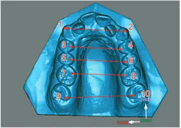
sotware that allows viewing and manipulating digital representations on a computer screen. Transverse changes resulting from SARME were assessed by means of intercanine, interpremolar and intermolar widths (Fig 1), using the points described by Currier26 and
Berger et al27 as reference.
The height of the clinical crown of canines, premo-lars and mopremo-lars was measured based on the distance be-tween the buccal cusp and the most apical point of the gingival margin,5,9 as shown in Figure 2.
Angular measurements were taken with the aid of OrthoDesignerTM sotware (3Shape, Copenhagen,
Figure 3 - Defining lines a, b and c Figure 4 - Defining clipping plane.
Figure 1 - Points and transverse widths in the digital models: 1) Cusp tip of right canine; 2) Cusp tip of left canine; 3) Palatal cusp tip of right maxillary first premolar; 4) Palatal cusp tip of left maxillary first premolar; 5) Palatal cusp tip of right maxillary second premolar; 6) Palatal cusp tip of left maxil-lary second premolar; 7) Mesio-palatal cusp tip of right maxillary first molar;
8) Mesio-palatal cusp tip of left maxillary first molar; 9) Mesio-palatal cusp tip of right maxillary second molar; 10) Mesio-palatal cusp tip of left maxil-lary second molar.
Figure 2 - Height of clinical crowns.
Figure 5 - Enabling clipping plane tool, shown in red.
Intercanine, interpremolar and intermolar tipping was calculated using the following references5: Line a=
dis-tance between the let and right midpoints of the deep-est region of buccal and palatal surfaces in the gingival margin; Line b= distance between the geometric mid-point on the right side of the center of buccal and pala-tal cusps, and the midpoint of the deepest region in the gingival margin; Line c= distance from the let side of the geometric midpoint at the center of buccal and pala-tal cusps, and the midpoints of the deepest buccal and palatal portions of the gingival margin. With these ref-erence lines, the internal angles formed by lines a-b and a-c were calculated with the aid of the sotware. Ater this deinition, the bilateral angulation of posterior teeth was calculated (Fig 3).
To this end, it was necessary to create a clipping plane in the models (Fig 4) to allow teeth to be viewed mesially. The reference plane met the aforementioned criteria.
In selecting the clipping plane, the tool “enable clip-ping plane” was used. This allowed the mesial view-ing of the models, as it excluded their anterior portion (Fig 5). The changes in each parameter occurring dur-ing treatment were calculated in the models at the times described before.
Statistical analysis
To determine the error of the method, 30% of the sample was randomly selected and measured ater at least one week, using the same material and applying the same aforementioned criteria. Paired t-test was used to determine intraexaminer systematic error. Random error was calculated by Dahlberg’s formula.28
In order to compare the three assessment periods, analysis of variance (ANOVA) was used with a criterion
1
23 4
5 6
7 8
9 10
Y-Axis Z-Axis
X-Axis
a
b 45.12 c
for repeated measurements. When ANOVA revealed statistically signiicant diference, Tukey’s test for mul-tiple comparisons was applied. A level of signiicance of 5% (p < 0.05) was adopted for all tests.
RESULTS
From the foregoing, one can argue that the results found in this study are reliable, since, ater further mea-surements were carried out in the dental casts of ive randomly selected patients, no intraexaminer errors that might compromise this research were identiied. Measurements of tooth tipping are more error-prone due to inconsistencies in (a) the location of points, (b) trimming of casts, and (c) construction of lines.
Table 1 depicts means and standard deviation values of transverse widths in the upper dental arch, expressed in millimeters, at the three evaluation periods, and re-sults from ANOVA and Tukey’s test. It shows an in-crease in transverse width with means of 9.26 mm for irst molars, 5.4 mm for second molars, 9.8 mm for irst premolars, 9.49 mm for second premolars, and 5.87 mm for canines from T1 to T2. These values remained un-changed from T2 to T3.
Table 2 presents the mean size of crowns in the max-illary arch, expressed in millimeters, at T1, T2 and T3, and the results of ANOVA and Tukey’s test showing diferences only in let irst and second molars.
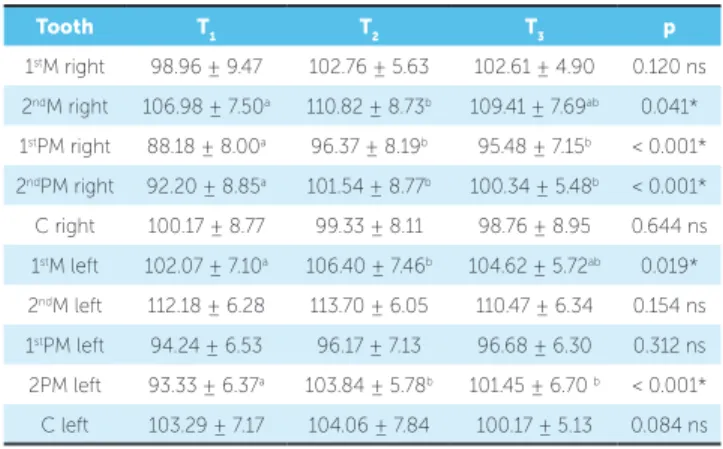
Table 3 shows means and standard deviation values of maxillary teeth tipping, expressed in degrees, at T1, T2 and T3, and the results of ANOVA and Tukey’s test. All values increased, thereby pointing to buccal tipping, although signiicant only in some teeth.
DISCUSSION
The literature presents diferent methods to assess changes induced by SARME in dental casts, namely: as-sessment with a bow compass,27 digital calipter4 and
laser-scanned models. Laser scanning is common in industrial engineering and medicine as a noninvasive alternative to generate 3D images. The measurement method using a 3D scanner has been studied and proved reliable and convenient.7,18,21 It has also been proven that analyses in
digital models can be performed in both clinical practice and research, with extremely accurate outcomes.16,17,19,20
Digital models have the added advantage of allowing images to be sliced, providing superior viewing of points not visible in dental casts. Furthermore, they can
Table 1 - Means and standard deviation values of transverse widths in the up-per dental arch, in mm, at the three assessment up-periods, and results of ANOVA and Tukey’s test.
*Statistically significant difference found by ANOVA (p < 0.05).
Periods of time with the same letter are not statistically different (Tukey’s test).
Tooth T1 T2 T3 p
1stM 35.94 ± 4.43a 45.20 ± 3.96b 45.26 ± 4.41b < 0.001*
2ndM 43.45 ± 4.58a 48.85 ± 4.53b 48.82 ± 4.72b < 0.001*
1stPM 26.02 ± 2.56a 35.82 ± 2.97b 35.47 ± 2.69b < 0.001*
2ndPM 31.28 ± 3.24a 40.77 ± 3.05b 40.73 ± 3.20b < 0.001*
C 30.56 ± 2.39a 36.43 ± 2.41b 36.14 ± 2.57b < 0.001*
Table 2 - Means and standard deviation values of crown heights in the upper dental arch, in mm, at the three assessment periods, and results of ANOVA and Tukey’s test.
ns = No statistically significant difference.
*Statistically significant difference found by ANOVA (p < 0.05).
Periods of time with the same letter are not statistically different (Tukey’s test).
Tooth T1 T2 T3 p
1stM right 7.25 ± 1.08 7.25 ± 0.84 7.23 ± 0.81 0.996 ns
2ndM right 6.88 ± 1.00 7.19 ± 0.78 7.05 ± 0.79 0.182 ns
1stPM right 7.66 ± 0.89 7.92 ± 0.94 8.04 ± 0.88 0.062 ns
2ndPM right 6.88 ± 1.11 6.88 ± 1.15 6.98 ± 0.94 0.754 ns
C right 9.29 ± 1.15 9.47 ± 1.14 9.40 ± 0.98 0.492 ns
1stM left 6.96 ± 0.74a 7.17 ± 1.03ab 7.39 ± 1.11b 0.035*
2ndM left 6.45 ± 0.78a 6.89 ± 0.89b 6.87 ± 0.91b 0.006*
1stPM left 7.92 ± 1.03 8.00 ± 0.79 8.03 ± 0.70 0.748 ns
2ndPM left 6.81 ± 0.99 6.84 ± 0.93 6.84 ± 0.97 0.930 ns
C left 9.21 ± 1.00 9.35 ± 1.15 9.38 ± 0.99 0.597 ns
Table 3 - Means and standard deviation values of tipping of teeth in the upper dental arch, in mm, at the three assessment periods, and results of ANOVA and Tukey’s test.
ns – No statistically significant difference.
*Statistically significant difference found by ANOVA (p < 0.05)
Periods of time with the same letter are not statistically different (Tukey’s test).
Tooth T1 T2 T3 p
1stM right 98.96 ± 9.47 102.76 ± 5.63 102.61 ± 4.90 0.120 ns
2ndM right 106.98 ± 7.50a 110.82 ± 8.73b 109.41 ± 7.69ab 0.041*
1stPM right 88.18 ± 8.00a 96.37 ± 8.19b 95.48 ± 7.15b < 0.001*
2ndPM right 92.20 ± 8.85a 101.54 ± 8.77b 100.34 ± 5.48b < 0.001*
C right 100.17 ± 8.77 99.33 ± 8.11 98.76 ± 8.95 0.644 ns
1stM left 102.07 ± 7.10a 106.40 ± 7.46b 104.62 ± 5.72ab 0.019*
2ndM left 112.18 ± 6.28 113.70 ± 6.05 110.47 ± 6.34 0.154 ns
1stPM left 94.24 ± 6.53 96.17 ± 7.13 96.68 ± 6.30 0.312 ns
2PM left 93.33 ± 6.37a 103.84 ± 5.78b 101.45 ± 6.70 b < 0.001*
be superimposed, which facilitates viewing of the me-chanics used in a given treatment.21
The time spent while taking measurements in the digital models was relatively shorter, given the user-friendliness of the programs and the measuring resources available, which yield very accurate measurements.23
Treatment including SARME proved successful for adult patients requiring maxillary expansion, a inding reported by several authors.2,4,6,12,13,25
The present study disclosed an increase in trans-verse width in all teeth from T1 to T2, with measure-ments remaining unchanged from T2 to T3 (Table 1). Thus, it is reasonable to assert that SARME demon-strated efectiveness and stability during the assessment period (6 months).
The slight increase found in intercanine width can be attributed to the fact that patients with indication for SARME oten have canines in infralabioversion. As an-terior space is gained, these teeth tend to align, conse-quently taking on a more lingual position and not show-ing so much increase in width.1,4,13,27
In comparison to irst molars, there was less increase in transverse width in second molars (5.4 mm and 9.4 mm, respectively). This diference can be probably linked to release of the pterygopalatine process due to the surgical technique employed, and also to the fact that this tooth was not included in the appliance.25
In adults, both surgical and nonsurgical procedures can correct maxillary transverse deiciency and achieve stability,4,5,8,9 but comparison showed greater transverse
increase in surgical cases.
SARME did not interfere in gingival attachment at the three assessment periods, except for irst and second molars on the let side. Bassareli, Dalstra and Melsen5
as well as Handelman et al8 reported that nonsurgical
maxillary expansion is efective in adults. However, these studies demonstrated greater dentoalveolar com-pensation due to increased tipping. Furthermore, they found no connection between the development of gin-gival recession and the amount of transverse expansion in adults, since there was no change in clinical crown height. In comparing the two types of treatment, i.e., SARME versus nonsurgical expansion, Carmen et al9
found that these treatment modalities result in increased transverse dimension and show no statistically signii-cant diferences in the development of gingival reces-sion. Nevertheless, SARME proved more efective and
less harmful to the periodontium, thereby corroborat-ing Northway and Meade,4 who argued that crown
length displayed greater increase in nonsurgical patients. The literature has shown that bone dehiscence can be produced in the alveolar bone when teeth are tipped bucally, but orthodontic movement would not neces-sarily be accompanied by loss of connective tissue.10,11
It has been acknowledged that teeth positioned or moved bucally, bone dehiscence and the presence of thin and brittle keratinized mucosa are the main predis-posing factors of gingival recession.15,29 Gingival
reces-sion, however, is only triggered by mechanical trauma caused by brushing, or inlammation induced by the presence of plaque.15 Therefore, the quality of the
kera-tinized mucosa and tooth brushing in particular should be closely monitored in patients undergoing SARME.
The surgical procedure resulted in dentoalveo-lar tipping, with statistical significance (Table 3), in the second molar, first and second premolars on the right side, and first molar and second premolars on the left side from T1 to T2. From T2 to T3, ping remained unchanged. In this study, crown tip-ping was calculated by means of the angle formed by the long axis of the tooth with a line that connects the buccal and lingual surfaces of the gingival most points. Thus, calculating tipping was less dependent on crown morphology,5 since other methods are
in-fluenced by changes in cusp height.1,4
This diference in the amount of tipping may be related to the way in which expansive force is delivered. Second premolars experienced expansion forces through contact between the lingual connection wire and its homonymous surface. With simple force applied to the crown, away from the center of resistance, a moment of force was cre-ated in the buccal direction, ultimately yielding some tip-ping component. Furthermore, anchorage teeth received expansion forces by means of bands rigidly ixed to the appliance. As the screw was activated, the bands, which were wide in the cervico-occlusal direction, resisted the tendency to tip by moving the anchorage teeth predomi-nantly through a bodily movement in buccal direction.15
This clearly shows that overcorrection was necessary due to relapse induced by the efects of tipping.3,4,8
CONCLUSION
1. Adkins MD, Nanda RS, Currier GF. Arch perimeter changes on rapid palatal expansion. Am J Orthod Dentofacial Orthop. 1990;97(3):194-9. 2. Atac ATA, Karasu HA, Aytac D. Surgically assisted rapid maxillary expansion
compared with orthopedic rapid maxillary expansion. Angle Orthod. 2006;76(3):353-9.
3. Chung C, Goldman AM. Dental tipping and rotation immediately after surgically assisted rapid palatal expansion. Eur J Orthod. 2003;25(4):353-8.
4. Northway WM, Meade JB Jr. Surgically assisted rapid expansion: a comparison of technique, response, and stability. Angle Orthod. 1997;67(4):309-20. 5. Bassarelli T, Dalstra M, Melsen B. Changes in clinical crown height as a result of
transverse expansion of the maxilla in adults. Eur J Orthod. 2005;27(2):121-8. 6. Bays R, Greco JM. Surgically assisted rapid palatal expansion: an outpatient
technique with long-term stability. J Oral Maxillofac Surg. 1992;50(2):110-5. 7. Bell A, Ayoub F, Siebert P. Assessment of the accuracy of a three-dimensional
imaging system for archiving dental study models. J Orthod. 2003;30(3):219-23. 8. Handelman CS, Wang L, BeGole EA, Haas AJ. Nonsurgical rapid maxillary
expansion in adults: report on 47 cases using the Haas expander. Angle Orthod. 2000;70(2):129-44.
9. Carmem M, Marcella P, Giuseppe C, Roberto A. Periodontal evaluation in patients undergoing maxillary expansion. J Craniofac Surg. 2000;11(5):491-4. 10. Garib DG, Henriques JFC, Janson G, Freitas MR, Fernandes AY. Periodontal
efects of rapid maxillary expansion with tooth-tissue-borne and tooth-borne expanders: a computed tomography evaluation. Am J Orthod Dentofacial Orthop. 2006;129(6):749-58.
11. Steiner GG, Pearson JK, Ainamo J. Changes of the marginal periodontium as a result of labial tooth movement in monkeys. J Periodontol. 1981;52(6):314-20. 12. Betts NJ, Vanarsdall RL, Barber HD, Higgns-Barber K, Fonseca RJ. Diagnosis and
treatment of transverse maxillary deiciency. Int J Adult Orthodon Orthognath Surg. 1995;10(2):75-96.
13. Bylof FK, Mossaz CF. Skeletal and dental changes following surgically assisted rapid palatal expansion. Eur J Orthod. 2004;26(4):403-9.
14. Haas A. Long-term post treatment evaluation of rapid palatal expansion. Angle Orthod. 1980;50(3):189-217.
15. Garib DG, Henriques JFC, Janson G, Freitas MR, Coelho RA. Rapid maxillary expansion — tooth-tissue-borne versus tooth-borne expanders: a computed tomography evaluation of dentoskeletal efects. Angle Orthod. 2005;75(4):548-57.
16. DeLong R, Heinzen M, Hodges JS, Ko CC, Douglas WH. Accuracy of a system for creating 3D computer models of dental arches. J Dent Res. 2003;82(6):438-42.
REFERENCES
17. Hayasaki H, Martins RP, Gandini LG Jr, Saitoh I, Nonaka K. A new way of analyzing occlusion 3 dimensionally. Am J Orthod Dentofacial Orthop. 2005;128(1):128-32.
18. Kusnoto B, Evans CA. Reliability of a 3D surface laser scanner for orthodontic applications. Am J Orthod Dentofacial Orthop. 2002;122(4):342-8. 19. Motohashi N, Kuroda T. A 3D computer-aided design system applied to
diagnosis and treatment planning in orthodontics and orthognathic surgery. Eur J Orthod. 1999;21(3):263-74.
20. Okumura H, Chen L, Tsutsumi S, Oka M. Three-dimensional virtual imaging of facial skeleton and dental morphologic condition for treatment planning in orthognathic surgery. Am J Orthod Dentofacial Orthop. 1999;116(2):126-31. 21. Oliveira NL, Silveira ACd, Kusnoto B, Viana G. Three-dimensional assessment
of morphologic changes of the maxilla: A comparison of 2 kinds of palatal expanders. Am J Orthod Dentofacial Orthop. 2004;126(3):354-62. 22. Stevens DR, Flores-Mir C, Nebbe B, Raboud DW, Heo G, Major PW. Validity,
reliability, and reproducibility of plaster vs digital study models: comparison of peer assessment rating and Bolton analysis and their constituent measurements. Am J Orthod Dentofacial Orthop. 2006;129(6):794-803.
23. Sousa MVS, Vasconcelos EC, Janson G, Garib D, Pinzan A. Accuracy and reproducibility of 3-dimensional digital model measurements. Am J Orthod Dentofacial Orthop 2012;142(2):269-73.
24. Biederman W. A hygienic appliance for rapid expansion. JPO, J Pract Orthod. 1968;2(2):67-70.
25. Goldenberg DC, Alonso N, Goldenberg FC, Gebrin E, Amaral TS, Scanavini MA, et al. Using computed tomography to evaluate maxillary changes after surgically assisted rapid palatal expansion. J Craniofacial Surg 2007;18(2):302-11.
26. Currier JHA. A computerized geometric analysis of human dental arch form. Am J Orthod. 1969;56(2):164-79.
27. Berger JL, Pangrazio-Kulbersh V, Borgula T, Kaczynski R. Stability of orthopedic and surgically assisted rapid palatal expansion over time. Am J Orthod Dentofacial Orthop. 1998;114(6):638-45.
28. Houston WJB. The analysis of errors in orthodontic measurements. Am J Orthod. 1983;83(5):382-90.