Discoloration and force
degradation of orthodontic elastomeric ligatures
Samaneh Nakhaei1, Raha Habib Agahi2, Amin Aminian3, Masoud Rezaeizadeh4
Objective: The aim of the present study was to evaluate color changes and force degradation of orthodontic elastomeric ligatures in different stretching patterns during a 8-weeks period. Methods: Two elastomers with the minimum and two with the maximum color changing, and gray elastomers of two brands (American Orthodontics and Ortho Technology) were selected according to an opinion poll with clinicians and color changes after 4 weeks of intraoral use were evaluated. These elastomers were mounted on special jigs fabricated using a CAD-CAM technique, underwent different stretching patterns and the force was measured in 0, 24 hours, 2, 4 and 8 weeks. During in vivo part of the study, force levels of elastomers were measured after 4 weeks on a material testing machine. Data were analyzed with four-way ANOVA and Tukey post hoc tests. Results: All the elastomers showed color changing but the degree of color stability was significantly different. The mean force degradation was higher in 1-mm stretch groups. After 8 weeks, the average residual force of elastomers was 1.45 ± 0.18 N and the maximum force decay was seen in the elastomers that exhibited the maximum initial force. Conclusion: There is significant relationship between the stretching pattern and the amount of residual force of elastomers. Elastomers with higher initial forces exhibited higher percentages of force loss after 8 weeks. It seems that there is a relationship between initial color and color changing of elastomers.
Keywords: Color. Elastomers. Orthodontics. Force degradation.
1 Assistant Professor, Department of Orthodontics, Birjand Dental School,
Birjand University of Medical Science, Birjand, Iran.
2 Oral and Dental Disease Research Center, Kerman Dental School, Kerman
University of Medical Science, Kerman, Iran.
3 Assistant Professor, Oral and Dental Diseases Research center, Department of
Orthodontics, Kerman Dental School, Kerman University of Medical Science, Kerman, Iran.
4 Assistant Professor, Graduate University of Advanced Technology, Mechanical
Engineering Department , Kerman, Iran.
DOI: http://dx.doi.org/10.1590/2177-6709.22.2.045-054.oar
How to cite this article: Nakhaei S, Habib Agahi R, Aminian A, Rezaeiza-deh M. Discoloration and force degradation of orthodontic elastomeric ligatures. Dental Press J Orthod. 2017 Mar-Apr;22(2):45-54.
DOI: http://dx.doi.org/10.1590/2177-6709.22.2.045-054.oar
» The authors report no commercial, proprietary or financial interest in the products or companies described in this article.
Submitted: February 18, 2016 - Revised and accepted: August 27, 2016 Contact address: Raha Habib Agahi
E-mail: [email protected]
Objetivo: o objetivo do presente estudo foi avaliar as alterações de cor e a degradação da força de ligaduras elásticas em diferentes padrões de estiramento, durante um período de 8 semanas. Métodos: duas ligaduras elásticas com alteração mínima de cor, duas com alteração máxima de cor e ligaduras elásticas na cor cinza de duas marcas comerciais (American Orthodontics e Ortho Technology) foram selecionadas de acordo com uma pesquisa de opinião com clínicos, e mediante a avaliação das alterações de cor após quatro semanas em uso intrabucal. Essas ligaduras elásticas foram montadas em jigs fabricados por meio da técnica CAD-CAM e foram expostas a diferentes padrões de estiramento, e a força foi medida nos tempos de 0h, 24h; 2, 4 e 8 semanas. Durante a parte in vivo
do estudo, os níveis de força das ligaduras elásticas foram medidos após 4 semanas, em uma máquina de teste de materiais. Os dados foram analisados com os testes ANOVA de quatro vias e post-hoc de Turkey. Resultados: todas as ligaduras elásticas mostraram alterações de cor, mas houve diferenças significativas quanto ao grau de estabilidade da cor. A média de degradação da força foi mais alta nos grupos com estiramento a 1 mm. Após 8 semanas, a média da força residual nas ligaduras elásticas foi de 1,45 ± 0,18 N e a maior degradação na força foi observada nas ligaduras elásticas que apresentaram a maior força inicial. Conclusão: há uma relação significativa entre o padrão de estiramento e a quantidade de força residual nas ligaduras elásticas. As ligaduras elásticas com as maiores forças iniciais exibiram as mais altas porcentagens de degradação da força após 8 semanas. É, também, possível que haja uma relação entre a cor inicial da ligadura elástica e sua alteração de cor.
INTRODUCTION
Clinicians may use pins, stainless steel ligature; self ligating clips and circular elastomers to ligate
orthodon-tic archwires to the brackets.1,2 Elastomeric ligatures are
most commonly used by clinicians due to their several advantages including low cost, easy application, reduced
chair time, patient comfort and satisfaction.3
These ligatures are manufactured by many com-panies in a variety of different colors that meet the growing global demand for esthetic orthodontic ap-pliances. Also, the possibility of choosing the liga-tures color facilitate young people adhesion to
treat-ment.4 Although adding pigments to ligatures seems
to represent a great advantage, there is two concerns regarding colored elastomers. The first one is that clinicians and patients may choose ligatures of pleas-ing color at placement time, but this chosen color is susceptible to color degradation over time, which is
a critical concern. Ardeshna et al5 reported that food
diet may affect elastomers color. The second question is whether force delivery is affected by adding
pig-ments to elastomers. Oliveira et al6 reported that
elas-tomers of different colors have different initial force and residual force over time.
The important clinical issue about elastomers is force delivery and force degradation of these materi-als over time. The force exerted by elastomers depends on the initial force and force decay rate, many studies
reported 50-70% force loss in the irst 24 hours.3,4,6
Under clinical conditions, diferently from usual laboratory conditions in force decay studies, the wire is not placed uniformly within the bracket slot and is not passive. Normally, during the early stages of treat-ment, high-stress areas that are under tension are pro-duced in the ligature, depending on the extent of tooth misalignment and the diferences in the buccolingual position of adjacent teeth. In studies carried out so far on elastomers, less attention has been paid to the ex-act simulation of the clinical situation and to the efect of stretching on the elastic properties of elastomers. In this study an attempt to evaluate the relationship between stretching pattern and the elastic properties of elastomeric modules as close to the clinical situation as possible was made by designing a speciic instrument using CAD-CAM technology.
Knowledge about changes in the physical (like color changes) and mechanical properties (like force delivery)
of elastomers is of great interest for their clinical
applica-tion.7 Since elastomers might remain in the oral cavity
for an average of 30 days, it is very important to preserve their properties, including force consistency and color
stability, and their relationship during this period.5,8,9
In this study, an attempt was made to evaluate color changes and force degradation of orthodontic elasto-meric ligatures under diferent stretching conditions over a period of 8 weeks, and test the hypothesis that elastomeric ligatures of diferent color under diferent stretching pattern have diferent force decay rates.
MATERIAL AND METHODS
To determine the elastomers which exhibit the maxi-mum and minimaxi-mum color changes from one orthodontic treatment session to another, diferent colors of the most commonly used elastomeric ligatures (Unistick, Ameri-can Orthodontics, USA; Power Sticks, Ortho Technol-ogy, USA) were selected and delivered to ten orthodon-tists, along with questionnaires. All the orthodontists had at least three years of experience working with those commercial brands. Based on the results of an opinion poll with these clinicians, two colored elastomers with maximum color changes and two colored elastomers with minimum color changes were selected from each brand, adding up to eight colored elastomers (Table 1). In the process of selecting the elastomers with the smallest and largest color changes, the next step was an intraoral stage: forty ixed orthodontic patients with good oral hy-giene and no speciic type of diet were assigned to two groups (n = 20), by simple randomization. Four selected colored elastomers of each brand were placed in the oral cavity of 20 patients from each group. Selected ligatures from each brand were placed in a crisscross pattern on teeth #4 and #5 in each quadrant to preserve esthetic ap-pearance and to avoid the bias resulting from the patient’s eating on only one side. Ater thirty days, the ligatures were removed from the oral environment, separated by quadrant and the brands were identiied by numbers.
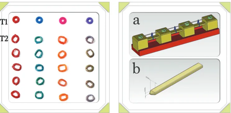
A total of 168 photographs [8 at T1 (not used elastomers),
and 160 at T2 (elastomers ater 30 days use in oral
Table 1 - Percentage of color changing in elastomeric ligatures based on clinician’s opinion* of digital photograph.
*Color changing score: 1 = unpigmented ligatures; 2 = pigmented ligatures; 3 = heavily pigmented ligatures.
self-timer mode set to trigger ater 15 seconds. The im-ages were stored in JPEG format (Fig 1). In order to carry out a visual analysis of the elastomeric ligatures, a Power Point presentation was created. The images were assessed independently by a panel of two orthodontists who rated the degree of discoloration of the elastomeric ligatures using a numerical scale ranging from 1 to 3, in which 1 was assigned to unpigmented ligatures, 2 to moderately pigmented ligatures, and 3 to heavily pigmented ligatures (Table 1). Finally, from each brand, one elastomer with the most color change and one elastomer with the least color change under clinical conditions were selected. The gray color elastomers from each brand served as control group.
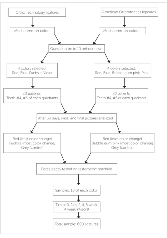
Two types of storage jigs for ‘1-mm point stretch’ and ‘uniform stretch’ groups were designed and fabricated by using a CAD-CAM technique (Fig 2) in which the de-signing process (CAD) was carried out with the sotware SolidWorks 2011 (3D design, SolidWorks Corp., USA) and the manufacturing phase (CAM) was carried out with a CNC machine (VMC Machine, model 850, Machine Sazi Tabriz corp., Iran) and Power MILL sot-ware (Delcam corp., UK). The ‘uniform stretch’ jig was prepared from a rectangular aluminum bar mea-suring 2.9 × 1.1 mm, equal to the width of a premolar bracket (Gemini Metal bracket, 0.022-in slot, 3M Uni-tek). The ‘1-mm point stretch’ jigs were designed to
simulate the elastomers stretch condition during the irst stage of ixed orthodontic treatment with malpositioned teeth. To create stretch and stress points in elastomers, 0.016-in SS wires were used (Fig 3). These holding jigs were used to store the elastomers during the study pe-riod in containers with artiicial saliva in an incubator at 37 ± 1°C under a traction force. All the elastomer groups were tested for force on a materials testing machine (model 10 KN, M350-10CT, Testometric Company, UK). The ligatures were stretched at a rate of 5 mm per
minute until rupture according to Kovatch et al10 study.
As each elastomer was stretched, force (N) was constant-ly measured and recorded. Force–extension curve was plotted by WinTest Analysis Materials Testing sotware (Testometric Company, UK). The ixture of materials testing machine was designed by the CAD-CAM tech-nique and was manufactured from two equal-sized alu-minum cubes, with two semi-circular rods, measuring 1.1 mm in radius (Fig 4). At the beginning of the test the two halves of the ixture were placed next to each other with no space between them, hence producing a 2.2-mm diameter circle. To calculate distance between the two parts of ixture (Fig 4) at which the force levels of elastomeric modules were measured (‘X’ in Fig 4), the inner circumferences of the elastomer on the ixture of the testing machine were adjusted to match the inner
cir-American Orthodontics Ortho Technology
Color Color changing score*
Percentage of
color changing Color
Color changing score*
Percentage of color changing
Red
1 63%
Red
1 58%
2 27% 2 29%
3 10% 3 13%
1 55% 1 51%
Blue
2 28%
Cobalt blue
2 34%
3 17% 3 15%
1 24% 1 21%
Pink
2 30%
Violet
2 27%
3 46% 3 52%
1 15% 1 10%
Bubble gum pink 2 28% Fuchsia 2 27%
cumferences of the specimens placed on the storage jigs of the relevant groups, and all the measurements were made at 0.1-mm accuracy, using SolidWorks sotware. The ‘X’ distance was calculated for all groups using the following formula:
X = y -2z 2 Whereas:
y = the circumference of the elastomer on the storage jig;
z = the circumference of the half-circle of the fixture.
X was calculated at 1.5 mm in the ‘uniform stretch’ group and at 2 mm in the ‘1-mm point stretch’ group. A total of 10 elastomers from each color were tested for the amount of initial force and residual force after 24 hours, 2, 4 and 8 weeks in the two groups with uniform and 1-mm point stretch patterns.
In order to confirm the accuracy of the results of the in vitro study with the results of the intraoral study, 10 elastomeric ligatures from each three se-lected colors (least color change, most color change, control) from both companies were placed on the premolar teeth of 10 patients using ixed appliances. They all used 3M-Unitek bracket system and were in the inishing stage with 0.016-in SS archwires. In order to make the in vitro and the oral cavity conditions as similar as possible, the elastomers were placed in both conditions with a ligature gun (Straight Shooter, TP Orthodontics, USA). The elastomers were retrieved from the oral cavity at a 4-week interval, rinsed with copious distilled water and placed in artiicial saliva. The forces were measured in less than 30 minutes on a materials testing machine. Sixty samples were evalu-ated in the intraoral stage and a total of 600 samples were evaluated in the present study (Fig 5).
Figure 1 - Photographic evaluation of color changing of elastomers after in-traoral use (T1: not used elastomers, T2: inin-traoral used elastomers).
Figure 3 - The 1-mm point (A) and the uniform stretch (B) jigs. Figure 4 - Fixture fabricated for testometric machine.
A
B
A study of ethics in medical ethics committee re-viewed and approved the research. The aim of the study was explained to the patients, which volun-tarily participated in the study. Informed consent to participate in the study was obtained from pa-tients before their involvement and they were given a choice to leave the study whenever they want. Pa-tient’s data were all kept confidential, and they did not had to pay any additional costs to take part in the study. Photographs were taken including only premolar of the patients, pictures of the face were not taken, to follow the ethical guidelines.
The SPSS sotware v. 18 (SPSS Inc. Chicago, IL, USA) was used for statistical analysis of data (at
signii-cance level of p < 0.05). Descriptive statistics were used to
report the clinician’s opinion regarding the most com-monly used colored elastomers and photographic evalu-ation of elastomers with minimum and maximum color
changes in the mouth. Results of force measurements were statistically analyzed with four-way ANOVA using
brands, stretching pattern (uniform versus 1-mm), color
and time as variables, followed by Tukey post-hoc tests.
RESULTS
The results of four-way ANOVA showed that all four variables (commercial brand, color, time and stretching pattern) had signiicant efects on the force
levels of the elastomers (p < 0.001) (Table 2).
The mean residual forces and mean percentages of force degradation for elastomeric ligatures of each brand are sum-marized in Table 3. Mean elastomers residual force in all
Figure 5 - Flow diagram of the study.
Ortho Technology ligatures American Orthodontics ligatures
Most common colors Most common colors
Questionnaire to 10 orthodontists
4 colors selected: Red, Blue, Fuchsia, Violet
4 colors selected: Red, Blue, Bubble gum pink, Pink
Force decay tested on testometric machine
Samples: 10 of each color
Total sample: 600 ligatures Times: 0, 24h; 2, 4, 8 week,
4 week intraoral
Red (least color change) Bubble gum pink (most color change)
Grey (control) Red (least color change)
Fuchsia (most color change) Grey (control)
20 patients:
Teeth #4, #5 of each quadrants
20 patients: Teeth #4, #5 of each quadrants
After 30 days, initial and final pictures analyzed
experimental groups ater 4 and 8 weeks were , respectively,
in the following ranges: 1.07−2.28 N and 1.14−1.64N.
Subjects Type III Sum fd Mean F-Value P-Value
of Square Square
Color .989 65 .494 7.215 .001
Brand 6.891 1 6.891 100.592 .000
Time 597.210 2 149.303 2,179 .000
Storage 4.275 1 2.137 31.199 .000
Color*Brand .466 4 .233 3.404 .034
Color*Time 16.189 2 2.024 29.539 .000
Color*Tensile 1.658 2 .414 6.050 .000
Brand*Time 9.833 8 2.458 35.882 .000
Brand*Tensile 6.687 4 3.343 48.803 .000
Time*Tensile 10.452 4 2.613 38.142 .000
Color*Brand*Time 3.228 2 .403 5.890 .000
Color*Brand*Tensile 3.950 4 .988 14.415 .000
Color*Time*Tensile 3.671 8 .459 6.699 .000
Brand*Time*Tensile 2.298 4 .574 8.386 .000
Color*Brand*Time*Tensile 2.675 8 .334 4.880 .000
Total 3.919.722 621
Table 2 - Test of between-subjects effects using 4-way ANOVA statistical analysis.
Table 3 - Mean residual force (N) and mean percentage of force decay for all test groups.
Color Type of traction
Residual force (mean ±SD) Mean percentage of force decay (%)
0 24
hours
2 weeks
4 weeks
8 weeks
24 hours
4 weeks
8 weeks
4 weeks Intraoral
Ortho Technology
Red
0mm 3.598 ± .362 2.522± .362 2.054± .362 1.469± .128 1.272± .472 30 59 65 1.252± .125
1mm 4.176 ± .338 2.363± .132 1.618± .135 1.423± .168 1.616± .175 43.5 66 61.5 65.21%
Gray
0mm 3.162 ± .391 2.134± .362 1.658± .202 1.782± .327 1.089± .107 32.6 43.7 65.6 1.780± .289
1mm 3.577± .416 1.958± .180 1.641± .132 1.480± .154 1.641± .158 45.3 58.7 54.2 43.71%
Fuchsia
0mm 3.803± .232 2.127± .358 1.685± .135 1.702± .100 1.148± .129 44 55.3 69.9 1.055± .783
1mm 4.247± .240 1.722± .106 1.608± .152 1.071± .615 1.150± .375 59.5 74.8 72.8 72.26%
American Orthodontics
Red
0mm 4.684± .217 2.385± .230 2.036± .141 1.545± .247 1.493± .371 49 67 68 1.348± .115 1mm 5.269± .228 2.309± .096 2.224± .147 1.713± .086 1.598± .278 56 67.5 69.7 71.2%
Gray
0mm 3.883± .469 2.635± .315 2.080± .170 2.285± .511 1.613± .126 32.2 41.2 58.5 1.143± .713 1mm 4.418± .464 2.315± .133 2.016± .165 1.952± .212 1.588± .132 47.7 55.9 64.1 63.15%
Bubble-gum pink
0mm 4.541± .513 2.518± .319 1.812± .075 1.580± .107 1.295± .567 44.6 65.3 71.5 1.442± .116
In all ligatures of both companies and two diferent stretch pattern groups, the greatest rate of force decay occurred during the irst 24 hours. At 0–4-week in-terval, the percentages of force degradation in all the elastomers were higher in the ‘1-mm stretch’ groups than the ‘uniform stretch’ groups. Comparison of the residual forces in the intraoral elastomers with those of the in vitro evaluation at 4-week interval showed that the residual forces in the intraoral elastomers in all the groups were smaller than those studied in vitro; how-ever, the diferences were clinically signiicant only in the OT fuchsia elastomers (0.7 N) and AO gray elas-tomers (1.14 N).
DISCUSSION
The results of the present study conirmed that elas-tomeric ligatures with diferent color under diferent stretching patterns have diferent force decay rates.
OT fuchsia elastomers and AO red elastomers, which had the maximum initial forces, exhibited maximum force degradation during the irst 24 hours followed by less force decay or relative force stability at subsequent intervals, compared to other elastomers, corroborating previous studies, which showed that elastomers
produc-ing higher initial forces displayed more force decay.9,11,12,13
The advantages of diferentiating the elastomers with the minimum and the maximum color changes ater exposure to oral environment not only provides results closer to the reality but also can help patient and clinician to choose the color and brand of elastomers that show greater esthetic stability during the period between two orthodontic follow-up appointments. In order to do so, in the process of selecting elastomers with maximum and minimum color changes — ater a opinion poll with the clinicians that had used these products for a considerable time —, and for higher data accuracy, the elastomers were placed in the oral cav-ity and all the factors afecting color change process (through simple staining and chemical degradation, including foodstufs, temperature changes and oral bacterial lora) were taken into account and the results
were very close to real clinical situations.3
The aim of this study was to determine clinically noticeable discoloration of elastomers. Digital camera was used because it is a more cost-efective and simpler process than the use of traditional methods such as
spec-trophotometry.14,15 Also, there is a very high and
statis-tically signiicant correlation between the traditional
method and digital camera.16 Another reason to use
digital photography to evaluate color change, compared with other complex procedures, is that it is a practical method, reproducible by professional in clinical condi-tions. For these reasons, digital photographs were used to compare discolorations of elastomeric ligatures be-fore and ater exposure to the oral environment.
Lam et al17 results were similar to the indings of the
present study: they reported that addition of pigments to
elastomers can change their mechanical properties.Since
the fuchsia and bubble-gum pink elastomers exhibited maximum color changes in the oral cavity and also maxi-mum force loss under the circumstances of the present study, it might be hypothesized that there is correlation between elastomer color changes and the amount of force loss, but this theory also needs to be evaluated in diferent brands of elastomers and diferent study condition.
The force degradation pattern of elastomers in the intra- and extraoral stages of the study was the same. However, the amount of intraoral residual forces in AO gray and OT fuchsia elastomers exhibited a signiicant
decrease, compared to in vitro conditions. Eliades et al18
evaluated the tensile properties of elastomeric chains and showed no signiicant diferences between the in-traoral and laboratory conditions, consistent with the
results of the present study. However, Ash et al19 showed
signiicantly lower residual forces in the elastomeric chains placed in the oral cavity compared to samples placed under atmospheric conditions; although there were minor diferences in forces in elastomers placed in water and in the oral cavity. Such diferences might be attributed to the lack of similarity between the condi-tions of in vitro and in vivo studies.
The dimensional accuracy of the storage jigs, and also the use of artiicial saliva and conserving the
elasto-mers at 37 oC resulted in the similarity of the results of
During the irst stage of ixed orthodontic treatment, some stress concentration points are created in elasto-mers due to the misalignment of the dentition and dif-ferences in the buccolingual positions of adjacent teeth. In the present study, 1-mm point stretch storage jigs were designed and fabricated using a CAD-CAM tech-nique, with the dimensional accuracy of up to 0.1 mm, to simulate the irst stage of treatment.
Another advantage of the present study in
compari-son to some previous studies13,18,20 was selection of the
0−8-week interval. Sims et al21 stated that a single elastic
module produces a ligation force of 50−150 g. Therefore, the results of the present study showed that the residual forces in all the tested groups ater 8 weeks (142 ± 23 g and 149 ± 11 g in OT and AO elastomers, respectively) were adequate to hold the wire within the bracket slot and make it possible to arrange inter-appointment intervals longer than four weeks during the early stages of orthodontic treatment (in cases where there is no concern regarding plaque accumulation), which makes the treatment process more cost-efective for both the patient and the clinician.
Peterson et al22 reported that if ligation or normal forces
decrease, there will be a corresponding decrease in fric-tional resistance, so in other words higher forces in elas-tomers result in higher frictional forces in the system,
de-creasing tooth movement rate in early stage of treatment.23
Contrarily, this high amount of force decays ater 8 weeks. The elastomeric modules are not good candidates for re-maining in oral cavity more than 4 weeks in the inal stages of ixed orthodontic treatment, in which the wire should be completely and actively seated in the bracket slot.
Under the terms of our study on two brands of elastomers, it seems that incorporation of different pigments into elastomers affects force degradation rates by affecting the amount of the initial forces. Therefore, further studies on other brands of elas-tomers are required to evaluate the correlation be-tween the characteristics of elastomers, which can be observed in the clinic and the mechanical proper-ties of these elastic materials. It is possible to match the results of in vitro and in vivo studies by exact simulation of in vitro conditions of studies. How-ever, to determine the amount of ligation force pro-duced by elastomers and force decay rate and their correlation with elastomer size, elastomer brand, color, time, stage of treatment, etc, further intraoral studies are required.
CONCLUSION
1. All the ligatures showed an unwanted color chang-es ater exposure to intraoral environment, although American Orthodontics (AO) red and blue elastomers and Ortho Technology (OT) cobalt blue and red elasto-mers showed minimum color changes.
2. The results of the present study showed a signii-cant relationship between the stretching pattern and the amount of residual force of elastomeric ligatures.
3. Elastomers with higher initial forces exhibited higher percentages of force decay ater 8 weeks.
1. Carneiro GKM, Roque JA, Garcez Segundo AS, Suzuki H. Evaluation of stifness and plastic deformation of active ceramic self-ligating bracket clips after repetitive opening and closure movements. Dental Press J Orthod. 2015 July-Aug;20(4):45–5.
2. Walton DK, Fields HW, Johnston WM, Rosenstiel SF, Firestone AR, Christensen JC. Orthodontic appliance preferences of children and adolescents. Am J Orthod Dentofacial Orthop. 2010 Dec;138(6):698.e1-12; discussion 698-9.
3. Buchmann N, Senn C, Ball J, Brauchli L. Inluence of initial strain on the force decay of currently available elastic chains over time. Angle Orthod. 2012 May;82(3):529-35.
4. Eliades T, Bourauel C. Intraoral aging of orthodontic materials: the picture we miss and its clinical relevance. Am J Orthod Dentofacial Orthop. 2005 Apr;127(4):403-12.
5. Ardeshna AP, Vaidyanathan TK. Colour changes of orthodontic elastomeric module materials exposed to in vitro dietary media. J Orthod. 2009 Sept;36(3):177-85.
6. Oliveira AS, Kaizer MR, Salgado VE, Soldati DC, Silva RC, Moraes RR. Inluence of whitening and regular dentifrices on orthodontic clear ligature color stability. J Esthet Restor Dent. 2015 Mar-Apr;27 Suppl 1:S58-64. 7. Kim SH, Lee YK. Measurement of discolouration of orthodontic elastomeric
modules with a digital camera. Eur J Orthod. 2009 Oct;31(5):556-62. 8. Silva DL, Mattos CT, Araújo MV, Ruellas ACO. Color stability and
luorescence of diferent orthodontic esthetic archwires. Angle Orthod. 2013 Jan;83(1):127-32.
9. Masoud AI, Bulic M, Viana G, Bedran-Russo AK. Force decay and dimensional changes of thermoplastic and novel thermoset elastomeric ligatures. Angle Orthod. 2016 Sept;86(5):818-25.
10. Kovatch JS, Lautenschlager EP, Apfel DA, Keller JC. Load-extension-time behavior of orthodontic alastiks. J Dent Res. 1976 Sept-Oct;55(5):783-6. 11. Stevenson JS, Kusy RP. Force application and decay characteristics of
untreated and treated polyurethane elastomeric chains. Angle Orthod. 1994;64(6):455-64; discussion 465-7.
12. Tonks M, Millett P, Cai W, Wolf D. Analysis of the elastic strain energy driving force for grain boundary migration using phase ield simulation. Scr Mater. 2010;63(11):1049-52.
REFERENCES
13. Dowling PA, Jones WB, Lagerstrom L, Sandham JA. An investigation into the behavioural characteristics of orthodontic elastomeric modules. Br J Orthod. 1998 Aug;25(3):197-202.
14. Cal E, Sonugelen M, Guneri P, Kesercioglu A, Kose T. Application of a digital technique in evaluating the reliability of shade guides. J Oral Rehabil. 2004 May;31(5):483-91.
15. Dozić A, Kleverlaan CJ, El-Zohairy A, Feilzer AJ, Khashayar G. Performance of ive commercially available tooth color-measuring devices. J Prosthodont. 2007 Mar-Apr;16(2):93-100.
16. Jarad FD, Russell MD, Moss BW. The use of digital imaging for colour matching and communication in restorative dentistry. Br Dent J. 2005 July 9;199(1):43-9; discussion 33.
17. Lam TV, Freer TJ, Brockhurst PJ, Podlich HM. Strength decay of orthodontic elastomeric ligatures. J Orthod. 2002 Mar;29(1):37-43.
18. Eliades T, Eliades G, Silikas N, Watts DC. Tensile properties of orthodontic elastomeric chains. Eur J Orthod. 2004 Apr;26(2):157-62.
19. Ash JL, Nikolai RJ. Relaxation of orthodontic elastomeric chains and modules in vitro and in vivo. J Dent Res. 1978 May-June;57(5-6):685-90. 20. Dechkunakorn S, Viriyakosol N, Anuwongnukroh N, Suddhasthira T,
Laokijcharoen P, Churnjitapirom P, et al. Residual force of orthodontic elastomeric ligature. Adv Mat Res. 2011;378-379:674-680.
21. Sims AP, Waters NE, Birnie DJ, Pethybridge RJ. A comparison of the forces required to produce tooth movement in vitro using two self-ligating brackets and a pre-adjusted bracket employing two types of ligation. Eur J Orthod. 1993 Oct;15(5):377-85.
22. Petersen A, Rosenstein S, Kim KB, Israel H. Force decay of elastomeric ligatures: inluence on unloading force compared to self-ligation. Angle Orthod. 2009 Sept;79(5):934-8.