© 2013 Dental Press Journal of Orthodontics 150 Dental Press J Orthod. 2013 Jan-Feb;18(1):150-7
Relationship between temporomandibular disorders and
orthodontic treatment: A literature review
Ronaldo Antônio Leite1, Joacir Ferreira Rodrigues2, Maurício Tatsuei Sakima3, Tatsuko Sakima4
How to cite this article: Leite RA, Rodrigues JF, Sakima MT, Sakima T. Rela-tionship between temporomandibular disorders and orthodontic treatment: A lit-erature review. Dental Press J Orthod. 2013 Jan-Feb;18(1):150-7.
» The author reports no commercial, proprietary or inancial interest in the prod-ucts or companies described in this article.
Contact address: Ronaldo Antônio Leite
Rua Francisco Marques, 698 – Sala 2 – Vila Nova – Franca/SP CEP: 14405-342 – E-mail: [email protected] 1 PhD in Otorhinolaryngology, UNIFESP. MSc in Dentistry, USP. Professor of
Integrated Diagnostics at University of Franca.
2 MSc in Orthodontics, São Leopoldo Mandic. Professor of Orthodontics at the
University of Franca.
3 Post-doctorate, University of Aarhus, Denmark. Assistant Professor in the
Department of Children’s Clinic at FOAR (UNESP).
4 Associate professor at FOAR-UNESP. Professor of the Specialization Course
in orthodontics at the University of Franca, APCD - Regional of Araraquara and Dental Research Center at São Leopold Mandic.
Submitted: March 24, 2009 - Revised and accepted: October 20, 2010
special
article
Objective:The objective of this study was to review the most recent studies from the last 15 years, in search of clinical studies that report the relationship between TMD and orthodontic treatment and/or malocclusion. Our intention was to determine whether orthodontic treatment would increase the incidence of signs and symptoms of TMD, and whether orthodontic treatment would be recommended for treating or pre-venting signs and symptoms of TMD. Methods: Literature reviews, editorials, letters to the editor, experimental studies in animals and short communications were excluded from this review. Were included only prospective, longitudinal, case-control or retrospective studies with a large sample and signiicant statistical analysis. Studies that dealt with craniofacial deformities and syndromes or orthognathic surgery treatment were also excluded, as well as those that reported only the association between malocclusion and TMD. Results: There were 20 articles relating orthodontics to TMD according to the inclusion criteria. The studies that associated signs and symptoms of TMD to orthodontic treatment showed discrepant results. Some have found positive efects of orthodontic treatment on signs and symptoms of TMD, however, none showed a statistically signiicant diference. Conclusions: All studies cited in this literature review reported that orthodontic treatment did not provide risk to the development of signs and symptoms of TMD, regardless of the technique used for treatment, the extraction or non-extraction of premolars and the type of malocclusion previously presented by the patient. Some studies with long-term follow-up concluded that orthodontic treatment would not be preventive or a treatment option for TMD.
Keywords: Orthodontics. Temporomandibular joint disorders. Dental occlusion.
Objetivo:revisar a literatura mais atual, dos últimos 15 anos, em busca de estudos clínicos que relatem a relação entre a disfunção temporoman-dibular (DTM) e o tratamento ortodôntico e/ou a má oclusão. A intenção foi veriicar se o tratamento ortodôntico aumentaria o aparecimento de sinais e sintomas de DTM, e se o tratamento ortodôntico seria um recurso para o tratamento ou prevenção dos sinais e sintomas de DTM.
Métodos: artigos dos tipos revisão de literatura, editorial, carta, estudo experimental em animais e comunicação foram excluídos dessa revisão. Foram incluídos artigos prospectivos, longitudinais, caso-controle ou retrospectivo com amostra maior, com relevante análise estatística. Estudos que abordassem deformidades e síndromes craniofaciais e tratamento por cirurgia ortognática também foram excluídos, bem como aqueles que relatassem apenas a associação entre má oclusão e DTM. Resultados: foram encontrados 20 artigos relacionando Ortodontia à DTM, segundo os critérios adotados. Os estudos, então, associando sinais e sintomas de DTM ao tratamento ortodôntico apresentaram resultados heterogêneos. Alguns encontraram efeitos positivos do tratamento ortodôntico para os sinais e sintomas de DTM; entretanto, nenhum deles apresentou dife-rença estatisticamente signiicativa. Conclusões: todos os estudos citados nessa revisão de literatura relataram que o tratamento ortodôntico não forneceu risco ao desenvolvimento de sinais e sintomas de DTM, independentemente da técnica utilizada para tratamento, da exodontia ou não de pré-molares e do tipo de má oclusão previamente apresentada pelo paciente. Alguns estudos realizados com acompanhamento em longo prazo concluíram que o tratamento ortodôntico não seria preventivo ou uma modalidade de tratamento para DTM.
© 2013 Dental Press Journal of Orthodontics 151 Dental Press J Orthod. 2013 Jan-Feb;18(1):150-7
special article
Leite RA, Rodrigues JF, Sakima MT, Sakima T
INTRODUCTION
The problems associated with the diagnosis and management of temporomandibular disor-ders (TMD) have aroused interest to the orthodon-tist. The attention to signs and symptoms associated with TMD have modiied the clinical management before and during orthodontic treatment.1
According to the American Academy of Orofacial Pain, the term temporomandibular disorder refers to a set of clinical problems that involve the masticatory mus-culature, the temporomandibular joint (TMJ) and asso-ciated structures, or both, being identiied as the leading cause of non-dental pain in the orofacial region and is considered a subclass of musculoskeletal disorders.2
The signs and symptoms that indicate any abnor-mality of the TMJ are: Alteration of the mandibular movement, limitation of mouth opening, joint pain with mandibular function, constraint function, joint noises, asymptomatic radiographic changes of the TMJ and jaw locking with open mouth and closed mouth.3
The most common symptom associated with TMD is pain, usually located in the masticatory muscles, pre-auricular area and / or temporomandibular joint (TMJ). The pain is oten aggravated by chewing or other func-tional activities. Limitation of mouth opening and movement, and the presence of joint noises are other common complaints in patients with TMD.2
There are several classiication schemes that assist in the clinical diagnosis of TMD, e.g. schemes of the American Academy of Orofacial Pain. Almost all di-vide the TMD in subgroups: Muscular, articular and mixed.4 The role of malocclusion in the etiology of
TMD has been reported as controversial in recent years. McNamara Jr., Seligman and Okeson5 published an
ex-tensive systematic review which concluded that there is a signiicant association between the presence of some occlusal factors (skeletal open bite, unilateral crossbite, absence of ive or more teeth, deep overbite and severe overjet) and the presence of TMD signs and symptoms. Recently, a study in Brazil showed that the absence of bilateral canine guidance on lateral excursion and the presence of Class II malocclusion are important risk in-dicators for TMD development.6
Pellizoni et al7 based on the hypothesis raised by
epidemiological studies, that there is an association between unilateral posterior crossbite (UPC) and disc displacement in TMJ, proposed a prospective
study that evaluated the articular disc position and its coniguration in children with functional UPC and individuals with normal occlusion using magnetic resonance imaging. All participants showed no clini-cal signs and symptoms of TMD.
Only an individual with articular TMD (disk dis-placement without reduction) was found. This one belonged to the study group and the crossbite was ipsilateral to the side of the disc displacement. These results suggest that internal disturbances of TMJ and UPC occur independently, or the magnitude of these disorders can not be identiied by magnetic resonance imaging in this age group (6 to 13 years).7
Another explanation for UPC not implying in the TMJ disk displacement is the compensatory poten-tial of the asymmetrical mandibular condyle growth or the remodeling of the articular fossa, which allows the articular disc to be in its normal position.
In the last decade, much efort has been placed to explain the supposed relationship between orth-odontic treatment and TMD. Even with the avail-ability of sophisticated and modern diagnostic tools such as magnetic resonance imaging, and scientiic studies with long-term follow-up, it has not yet been possible to eliminate this existing controversy.8
Opinions difer between those who argue that orth-odontic treatment increases the risk of onset of signs and symptoms of TMD and those who claim that this treatment would be a type of treatment for TMD, or at least to reduce the risk of the patient to develop it.9
The objective of this study was to review ies from the last 15 years, searching for clinical stud-ies that report the relationship between TMD and orthodontic treatment and/or malocclusion, with the objective of determining if:
1. Orthodontic treatment would increase the in-cidence of signs and symptoms of TMD.
2. Orthodontic treatment would be an option for treating or preventing TMD symptoms and signs.
MATERIAL AND METHODS
© 2013 Dental Pr
ess Journal o
f Orthodontics
Dental Pr
ess J Orthod. 2013
Jan-Feb;18(1):150-7
152
Rela
tionship between tempor
omandibular disorders and orthodontic tr
ea
tment: A litera
tur
e r
eview
special
article
Study Study design Population Variables Results
Krenemak et al11
CS, prospective,
2-year follow-up
65 patients orthodontically treated Group I: 26 non-extraction Group II: 25 four premolar extractions Group III: 14 extractions of two premolar
DI Before the treatment 0 to 12 months after treatment 12 to 24 months after treatment
(1) there was no statistically signiicant diference between group means, at all times. (2) a small reduction in signs and symptoms of TMD was found between the means of
groups I and II
Krenemak et al12
CS, prospective,
6-year follow-up
109 patients undergoing orthodontic treatment with ixed appliances
DI 6-year control
Number of patients per year: 92, 56, 33, 19, 11 and 7
(1) 90% of patients maintained or improved the clinical picture 10% of patients had worsening
(2) Orthodontic treatment was not an important etiological factor for signs and symptoms of TMD
Hirata et al13
CC, prospective,
2-year follow-up
SG: 102 patients, mean age 15.3 years old CG: 41 patients, mean age 16.2 years old
DI Before the treatment 12 months after treatment 24 months after treatment
(1) no differences between groups
(2) orthodontics did not represent increased risk for development of signs and symptoms of TMD
Egemark and Thailander14 CS, prospective, 10-year follow-up
402 children divided in 3 age groups: 7, 11 and 15 years old
DI
293 questionnaires were answered After 10 years, 83 individuals examined, now 25 years-old
(1) individuals with a history of orthodontic treatment showed a low prevalence of TMD symptoms and lower DI
(2) more evident differences in the older group
Wadwa Utreja
and Tewari15 CS
102 individuals, ages between 13 and 25 years old Group I: 30 individuals with normal occlusion
Group II: 41 with malocclusion and without orthodontic treatment
Group III: 31 with malocclusion and orthodontic treatment
DI
(1) no diferences between groups
(2) orthodontic treatment would not present risk or prevent the development of signs and symptoms of TMD
O’Reilly, Rinchuse and Close16 CC, prospective 120 individuals
SG: 60 patients, average age 15.3 years old, 48 with Class II, division 1 and 12 with Class I malocclusion CG: 60 individuals, average age 15.3 years, 38 with Class II,
division 1 and 22 with Class I malocclusion
DI Before the treatment 8 to 10 months after 12 to 16 months after At the end of the treatment
(1) orthodontic mechanics applied has no efect or no signiicant efect on signs and symptoms of TMD
Beattie, Paquette and
Johnston17
CS, retrospective
63 patients with Class II malocclusion treated with and without premolar extractions
DI
Evaluation 14 years after treatment end
(1) no diferences between those who underwent premolar extractions or not. (2) premolar extraction would not be a risk factor for developing signs and symptoms of TMD
Egermark and
Ronnerman18 CS, prospective
50 patients underwent orthodontic treatment, average age of 12.9 years old
DI
Before, during or immediately after treatment
(1) high prevalence of signs and symptoms of TMD before treatment (2) signs and symptoms decreased during treatment, except for joint noises (3) occlusal interferences during treatment did not inluence the development of
signs and symptoms of TMD
Lima19 CS, prospective 100 individuals, Dental students,
ages between 18 and 25 years DI
(1) Individuals orthodontically treated or not showed similar tendency to present signs and symptoms of TMD
Katzberg et al20 CC,
retrospective
178 individuals
SG: 102 patients with symptomatic TMD CG: 76 asymptomatic volunteers
Questionnaire about previous orthodontic treatment and details of signs and symptoms of TMD,
Magnetic Resonance Images
(1) TMD patients showed a higher percentage (77%) of TMJ disk displacement than the CG (33%)
(2) There was no statistically signiicant association between previous orthodontic treatment (with or without extraction) and disc displacement
Lagerström, Egemark and
Carlsson21
CS, prospective
860 individuals 19 years after they underwent orthodontic treatment
Group I: 520 treated by specialists in Orthodontics Group II: 340 treated by general practitioners
DI
Test performed on 260 subjects (77%)
(1) There was no diference in the prevalence of signs and symptoms of TMD (2) Female individuals had higher prevalence of signs and symptoms of TMD (3) No association between occlusal contacts and signs and symptoms of TMD
Owen22 CS,
retrospective
600 patients who underwent orthodontic or orthopedic treatment
DI During the treatment
(1) 16 patients (2.6%) developed signs and symptoms of TMD during treatment (2) female patients, with Class II and moderate to severe overbite or overjet, regardless of
treatment technique used, were more likely to develop signs and symptoms of TMD
Henrikson, Nilner and Kurol23 CC, prospective, with 2-year follow-up 183 adolescents
Group I: 65 patients, Class II malocclusion orthodontically treated Group II: 58 individuals with Angle Class II,
no orthodontic treatment Group III: 60 subjects with normal occlusion
DI
Before orthodontic treatment, 2 years after treatment
© 2013 Dental Pr
ess Journal o
f Orthodontics
Dental Pr
ess J Orthod. 2013
Jan-Feb;18(1):150-7
153
special
article
Leite RA, Rodrigues JF
, Sakima MT
, Sakima T
Henrikson and
Nilmer24
CC, prospective,
2-year follow-up
183 adolescents
Group I: 65 patients, Class II malocclusion orthodontically treated Group II: 58 patients with Angle Class II,
no orthodontic treatment Group III: 60 patients with normal occlusion
DI
Before orthodontic treatment, 2 years after treatment
(1) TMD symptoms presented loating prevalence over the two years (2) orthodontic treatment did not increase the risk for developing
signs and symptoms of TMD and headache
Valle25 CC,
prospective
200 individuals
Group I: 50 subjects with Class I malocclusion, no orthodontic treatment
Group II: 50 subjects with Class II malocclusion, no orthodontic treatment
Group III: 50 subjects with Class I malocclusions orthodontically treated
Group IV: 50 subjects with Class II malocclusion orthodontically treated
Fonseca questionnaire Physical examination Occlusion examination
(1) No association between the severity of signs and symptoms of TMD and orthodontic treatment, regardless of the malocclusion type
(2) The severity of signs and symptoms of TMD may only be associated with the absence of anterior guidance
Conti et al8 CC,
prospective
200 individuals
Group I: 50 subjects with Class I malocclusion, no orthodontic treatment Group II: 50 subjects with Class II malocclusion,
no orthodontic treatment Group III: 50 subjects with Class I malocclusions
orthodontically treated
Group IV: 50 subjects with Class II malocclusion orthodontically treated
Fonseca questionnaire Physical examination Before and after orthodontic treatment
(1) Presence and severity of TMD did not present any relation with the type of orthodontic treatment or extraction protocol applied
Egermark, Carlsson and
Magnusson9
CS, prospective,
20-year follow-up
402 children divided in 3 age groups: 7, 11 and 15
DI / Questionnaire about previous orthodontic treatment; 320 individuals answered the questionnaire; 100 subjects were examined,
now 35 years-old
(1) no occlusal factor was important for the development of signs and symptoms of TMD, however, unilateral crossbite, and diferences between CR and MHI tended to be risk factors. (2) Individuals who have undergone orthodontic treatment did not present higher or lower risk of developing signs and symptoms of TMD than those who did not undergo orthodontic treatment
Corotti-Valle26 CS, prospective
50 patients with Class III malocclusion Group I: 25 patients submitted to orthodontic treatment Group II: 25 patients submitted to orthodontic treatment and
orthognathic surgery
Fonseca questionnaire Clinical examination
Tests carried out on at least one year after the treatment end
(1) There was no statistically signiicant diference between the two groups regarding the prevalence of TMD symptoms.
(2) Signiicant association was found between the presence of interferences on the non-working side and the index of TMD symptoms severity.
(3) The Class III malocclusion treatment had no inluence in determining TMD severity.
Mohlin et al27
CS, prospective,
19-year follow-up
1,018 11-year-old individuals
DI Morphology
Calculation of the malocclusion index Questionnaire on orthodontic treatment
Examination at age 11
791 re-examined at 15 years old, 456 at age 19 and 337 at age of 30
(1) No signiicant diference was found between the groups regarding the types of malocclusion, tooth contact pattern, previous orthodontic treatment or extractions
(2) A quarter of individuals with signs and symptoms of TMD at age 19 showed complete remission at the age of 30
(3) Orthodontic treatment was not a preventive nor causative factor of TMD
Egermark, Carlsson and
Magnusson9
CS, prospective,
17-year follow-up
50 patients submitted to orthodontic treatment, mean age 12.9 years
DI
17 years after treatment end, 40 patients completed the questionnaire and 31 were clinically reexamined
(1) the incidence of TMD needing clinical care was of 1% per year
(2) orthodontic treatment did not increase the risk of developing signs and symptoms of TMD.
© 2013 Dental Press Journal of Orthodontics 154 Dental Press J Orthod. 2013 Jan-Feb;18(1):150-7
Relationship between temporomandibular disorders and orthodontic treatment: A literature review
specialarticle
English, between January 1, 1992 to September 30, 2007. Articles like literature reviews, editorials, let-ters to the editor, experimental studies with animals and short communications were excluded from this review. Were included prospective, longitudinal, case-control or retrospective studies with larger sam-ples and signiicant statistical analysis. These studies are the level B of evidence (moderate evidence).10
Studies that dealt with deformities and craniofacial syndromes or treatment by means of orthognathic sur-gery were also excluded, as well as those who reported only the association between malocclusion and TMD.
RESULTS
There were 20 articles found relating orthodontics to TMD according to the inclusion criteria. Table 1 presents a description of the studies found.
DISCUSSION
The restrictions imposed in this study in relation to databases and languages in the search of the literature relating to TMD and Orthodontics may have resulted in few studies. However, inding the best evidence, prioritizing the quality of the studies and the diagnosis of TMD and its division into subtypes could lead to clearer conclusions about this association.
One of the biggest problems found in the stud-ies selected in this search for understanding the as-sociation between TMD and Orthodontics was the methodology they used to identify TMD. All stud-ies, except that performed by Katzberget al,20 use
the same tool to identify the signs and symptoms of TMD: the Helkimo index, published in 1974. There were few case-control studies, making it dif-icult to compare our data with regard to the signs and symptoms of TMD.
Professor Helkimo pioneered the development of indices to measure the severity of TMD, as well as pain in TMJ. In an epidemiological study, he developed an index divided into anamnesis, clini-cal and occlusal dysfunction. Through this index, he tried to identify, individually and in the popu-lation, the prevalence and severity of TMD, pain and occlusal instability. The protocol for the deter-mination of this index consists of ten parameters: Emotional stress, parafunctional habits, mouth opening, lateralization of the jaw, joint sounds,
TMJ tenderness, palpation of the posterior mus-cles of the neck, palpation of masticatory musmus-cles, maxillomandibular relationship and headaches.28
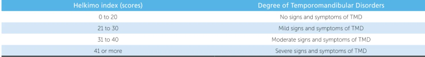
The Helkimo anamnesis index (AI) is based on a questionnaire where the individual reports the presence of symptoms of TMD. The results can generate three diferent levels of dysfunction: no symptoms; mild, moderate, or severe symptoms. The Helkimo clinical index (CI) considers the functional evaluation of the stomatognathic system. According to the presence and/ or severity of clinical signs, individuals are assigned scores ranging from 0, 1 or 5 points. The following as-pects are observed: Range of mouth opening and lateral movements of the jaw; restricted jaw function; pain on palpation of masticatory muscles, TMJ and neck poste-rior muscles. The signs are also classiied as none, mild, moderate or severe. The third index is called Helkimo occlusal index (OI) and is obtained by analyzing the oc-clusion of the individual regarding the number of teeth, number of teeth in occlusion and occlusal interference between the RC and MHI positions. According to the data obtained for each item, scores 0, 1 or 5 are assigned once again. The sum of the three indices generates the Helkimo dysfunction index (DI) (Table 2).
However, this tool, although widely used, is not able to diagnose and classify TMD, it only shows its signs and symptoms. There are limitations in using the DI, irst because it gives equal importance to all the symptoms, it does not separate the muscular TMD from articular TMD, its categorization by points does not promote a continuous variable, reducing its speci-icity. Some symptoms are ignored, such as the type of joint noises and when they occur, and some muscle regions. Even though this index is able to document the signs and symptoms of TMD in the population, the organization of data from these indices seems not to beneit other areas of Epidemiology, for example, in understanding TMD etiology.29 As an example of
how the index might be lawed, if a person has more than 15 episodes of headache per month, and she/he is very tense and present pain on palpation of the poste-rior muscles: He/she would be classiied as presenting with moderate TMD, without even having a single peculiar sign or symptom of TMD — i.e., the person might not even present TMD.
Bevilaqua-Grossi et al29 suggested that a way to
© 2013 Dental Press Journal of Orthodontics 155 Dental Press J Orthod. 2013 Jan-Feb;18(1):150-7
special article
Leite RA, Rodrigues JF, Sakima MT, Sakima T
would be to determine the frequency and intensity of signs and symptoms of TMD. The authors suggest us-ing the questionnaire proposed by Fonseca30 (Table 3)
and a clinical examination assessing the range of mouth opening and the tenderness of masticatory muscles and TMJ to palpation. According to the authors, Fonseca questionnaire30 is a simple questionnaire, without
pre-tension to diagnose TMD, but it can be a useful tool in observing the symptoms reported by patients. Not only the frequency of symptoms should be checked, but also its severity, aiming to identify those patients that require treatment for TMD. Three studies con-ducted in Brazil and reviewed in the present article used the Fonseca anamnesis questionnaire in order to discriminate patients who would present TMD, fol-lowed by physical examination.
Since 1992, to facilitate the conduction of clinical research, researchers in epidemiological and clinical studies or aiming to determine samples in random-ized and controlled trials, use a classiication scheme called the Research Diagnostic Criteria for Temporo-mandibular Disorders (RDC/TMD) which diagnoses the presence of TMD. The RDC/TMD is a tool for clinical diagnostic criteria, measurable and reproduc-ible, that aims at identifying subgroups of patients with TMD. The RDC/TMD classiies the most common types of TMD into three subgroups: Disorders of the masticatory muscles (myofascial pain), TMJ internal derangement (disk displacement), and degenerative diseases of the TMJ (arthralgia, arthritis and osteoar-thritis). The use of the RDC depends on anamnesis and physical examination data, making use of ques-tionnaires, surveys and speciications.31 The study by
Katzberg et al20 used this tool to diagnose disk
dis-placement with reduction in its sample.
Thus, none of the studies associating TMD and Orthodontics diagnosed TMD, they only observed the presence of signs and symptoms. Therefore, one cannot conclude from these studies whether the
TMD would be a condition that motivates indi-viduals to seek treatment for their functional prob-lems. There is a large disparity between the signs and symptoms of TMD (which can be present in up to 68% of the population)32 and TMD diagnosis
(8-15% of women and 3-10% of men).33
Another diiculty in analyzing the signs and symp-toms of TMD in the cited studies is the episodic or loating character of the appearance of these symptoms observed in long-term studies. The prevalence varied among the analysis performed on diferent occasions. Krenemak et al12 showed in their sample that 90% of
patients who developed signs and symptoms of TMD,
Helkimo index (scores) Degree of Temporomandibular Disorders
0 to 20 No signs and symptoms of TMD
21 to 30 Mild signs and symptoms of TMD 31 to 40 Moderate signs and symptoms of TMD 41 or more Severe signs and symptoms of TMD
Table 2 - Degree of temporomandibular disorder according to Helkimo dysfunction Index.
1 Do you have diiculty to open your mouth?
2 Do you feel diiculty to move your jaw sideways? To the right? To the left? To both sides? 3 Do you have muscle fatigue or pain when chewing? 4 Do you often have headaches? 5 Do you feel pain in the neck or torticollis?
6 Do you have ear ache or in the temporomandibular joints region (TMJ)?
7 Have you noticed if you have TMJ sounds when chewing or when you open your mouth?
8 Have you noticed if you have any habits like pushing and/or grinding teeth, chewing gum, biting lip or pencil, nail biting? 9 Do you feel that your teeth do not it together well? 10 Do you consider yourself a tense or nervous person?
Categories of severity of TMD symptoms
Ratings threshold to classify the categories No signs and symptoms 0 to 15 Mild signs and symptoms 20 to 40 Moderate signs and symptoms 45 to 65 Severe signs and symptoms 70 to 100
Table 3 - Fonseca30 questionnaire for anamnesis of temporomandibular
disorder.
© 2013 Dental Press Journal of Orthodontics 156 Dental Press J Orthod. 2013 Jan-Feb;18(1):150-7
Relationship between temporomandibular disorders and orthodontic treatment: A literature review
specialarticle
ater two years maintained or improved the situation, while 10% worsened. While Mohlin et al27 showed that
25% of patients at the end of 19 years of follow-up, had complete remission of signs and symptoms of TMD. The signs and symptoms appear to improve with time, except for joint noises, which increased ater 2 years of follow-up.23 Still, Owen22 reported that 2.6% of the
pa-tients developed signs and symptoms of TMD during orthodontic treatment. Egermark, Carlsson and Mag-nusson9 in a 17 years follow-up study, showed that 1%
of the sample required TMD clinical care per year. The studies associating signs and symptoms of TMD with orthodontic treatment showed discrepant results. Some studies have found positive efects of orthodontic treatment on the signs and symptoms of TMD, howev-er, none showed statistically signiicant results.9,11,12,14,23
All studies cited in this literature review reported that orthodontic treatment did not provide risk to the de-velopment of signs and symptoms of TMD, regardless of the technique used for treatment, whether or not the extraction of premolars was performed, and the type of malocclusion previously presented by the pa-tient.8,9,11-25,27 Some long-term studies concluded that
the orthodontic treatment would not be preventive or a treatment modality for TMD.9,15,27 Henrikson and
Nilmer24 suggested that due to the luctuating character
of the signs and symptoms of TMD, and as orthodontic treatment is not efective in treating TMD, a conserva-tive and reversible approach should be adopted in the treatment of TMD, which agrees with the guidelines of the American Academy of Orofacial Pain.2
Some articles also mentioned the relationship between malocclusion and signs and symptoms of TMD. There was no statistically signiicant association between malocclusions and signs and symptoms of
TMD.18,21,25,27 However there was a trend that patients
with Class II malocclusion with overbite or moderate to severe overjet,22 absence of anterior guidance,25
uni-lateral crossbite and diference between CR and MHI9
could present a greater number of signs and symptoms of TMD. Still, Corotti-Valle26 found in their sample a
signiicant association between severity of symptoms of TMD and interference in the balance side.
CONCLUSIONS
© 2013 Dental Press Journal of Orthodontics 157 Dental Press J Orthod. 2013 Jan-Feb;18(1):150-7
special article
Leite RA, Rodrigues JF, Sakima MT, Sakima T
1. Durso BC, Azevedo LR, Ferreira JTL. Inter-relação Ortodontia X Disfunção da articulação temporomandibular. J Bras Ortodon Ortop Facial. 2002;7(38):155-60.
2. Okeson JP. Dor orofacial: guia para avaliação, diagnóstico e tratamento. São Paulo: Quitenssence; 1998.
3. Siqueira JTT. Disfunção temporomandibular: classificação e abordagem clínica. In: Siqueira JTT, Teixeira MJ. Dor orofacial: diagnóstico, terapêutica e qualidade de vida. Curitiba: Ed. Maio; 2001. p. 373-404.
4. Okeson JP. Current diagnostic classification schema and assessment of patients with temporomandibular disorders. Oral Surg Oral Med Oral Pathol Oral Radiol Endod. 1997;83:61-4.
5. McNamara Jr JA, Seligman DA, Okeson JP. Occlusion, orthodontic treatment, and temporomandibular disorders: a review. J Orofacial Pain. 1995;9(1):73-90. 6. Selaimen CMP, Jeronymo JC, Brilhante DP, Lima EM, Grossi PK, Grossi
ML. Occlusal risk factors for temporomandibular disorders. Angle Orthod. 2007;77(3):471-7.
7. Pellizoni SEP, Salioni MA, Juliano Y, Guimarães AS, Alonso LG. Temporomandibular joint disc position and configuration in children with functional unilateral posterior crossbite: a magnetic resonance imaging evaluation. Am J Orthod Dentofacial Orthop. 2006;129(6):785-93. 8. Conti A, Freitas M, Conti P, Henriques J, Janson G. Relationship between signs
ans symptoms of temporomandibular disorders and orthodontic treatment: a cross-sectional study. Angle Orthod. 2003;73(4):411-7.
9. Egermark I, Carlsson GE, Magnusson T. A prospective long-term study of signs and symptoms of tempomandibular disorders in patients who received orthodontic treatment in childhood. Angle Orthod. 2005;75(4):645-50. 10. Malterud K. Qualitative research: standards, challenges, and guidelines. Lancet.
2001;11:483-8.
11. Kremenak CR, Kinser DD, Harman HA, Menard CC, Jakobsen JR. Orthodontic risk factors for temporomandibular disorders (TMD) I: premolar extractions. Am J Orthod Dentofacial Orthop. 1992;101(1):13-20.
12. Kremenak CR, Kinser DD, Melcher TJ, Wright GR, Harrison SD, Ziaja RR, et al. Orthodontics as a risk factor for temporomandibular disorders (TMD) II. Am J Orthod Dentofacial Orthop. 1992;101(1):21-7.
13. Hirata RH, Heft MW, Hernandez B, King GJ. Longitudinal study of signs of temporomandibular disorders (TMD) in orthodontically treated and nontreated groups. Am J Orthod Dentofacial Orthop. 1992;101(1):35-40.
14. Egemark I, Thilander B. Craniomandibular disorders with special reference to orthodontic treatment: an evaluation from childhood to adulthood. Am J Orthod Dentofacial Orthop. 1992;101(1):28-32.
15. Wadhwa L, Utreja A, Tewari A. A study of clinical signs and symptoms of temporomandibular dysfunction in subjects normal occlusion, untreated, and treated malocclusions. Am J Orthod Dentofacial Orthop. 1993;103(1):54-61. 16. O Reilly M, Rinchuse DJ, Close J. Class II elastics and extractions and
temporomandibular disorders: a longitudinal prospective study. Am J Orthod Dentofacial Orthop. 1993;103(5):459-63.
17. Beattie JR, Paquette DE, Johnston LE. The functional impact of extraction and nonextraction treatments: a long-term comparison in patients with borderline, equally susceptible Class II malocclusions. Am J Orthod Dentofacial Orthop. 1994;105:444-9.
REFERENCES
18. Egemark I, Ronnerman A. Temporomandibular disorders in the active phase of orthodontic treatment. J Oral Rehabil. 1995;22:613-8.
19. Lima DR. Estudo de prevalência de disfunção craniomandibular segundo o índice de Helkimo tendo como variáveis: sexo, faixa etária e indivíduos tratados ou não ortodonticamente [dissertação]. São José dos Campos (SP): Universidade Estadual Paulista; 1995.
20. Katzberg RW, Westesson PL, Tallents RH, Drake CM. Orthodontics and temporomandibular joint internal derangement. Am J Orthod Dentofacial Orthop. 1996;109(5):515-20.
21. Lagerström L, Egemark I, Carlsson GE. Signs and symptoms of
temporomandibular disorders in 19-year-old individuals who have undergone orthodontic treatment. Swead Dental J. 1998;22(5-6):177-86.
22. Owen AH. Unexpected temporomandibular joint findings during fixed appliance therapy. Am J Orthod Dentofacial Orthop. 1998;113(6):625-31.
23. Henrikson T, Nilner M, Kurol J. Signs of temporomandibular disorders in girls receiving orthodontic treatment. A prospective and longitudinal comparison with untreated Class II malocclusions and normal occlusion subjects. Eur J Orthod. 2000;22:271-81.
24. Henrikson T, Nilner M. Temporomandibular disorders and the need for stomatognathic treatment in orthodontically treated and untreated girls. Eur J Orthod. 2000;22(3):283-92.
25. Valle KM. Estudo comparativo da oclusão e da sua relação com as Disfunções Temporomandibulares (DTM) em jovens com e sem tratamento ortodôntico [Dissertação]. Bauru (SP): Universidade de São Paulo; 2000.
26. Corotti-Valle KM. Avaliação da prevalência da disfunção temporomandibular (DTM) em pacientes tratados das más oclusões de Classe III, submetidos a tratamento ortodôntico e orto-cirúrgico [tese] Bauru (SP): Universidade de São Paulo; 2004.
27. Mohlin BO, Derweduwen K, Pilley R, Kingdon A, Shaw WC, Kenealy P. Malocclusion and temporomandibular disorder: a comparison of adolescents with moderate to severe dysfunction with those without signs and symptoms of temporomandibular disorder an their further development to 30 years of age. Angle Orthod. 2004;74(3):319-27.
28. Helkimo M. Studies on function and dysfunction of the masticatory system. II. Index for anamnestic and clinical dysfunction and occlusal state. Swed Dental J. 1974;67:101-21.
29. Bevilaqua-Grossi D, Chaves TC, de Oliveira AS, Monteiro-Pedro V. Anamnestic index severity and signs and symptoms of TMD. Cranio. 2006;24(2):112-8. 30. Fonseca DM. Diagnóstico pela anamnese da disfunção craniomandibular. Rev
Gaúcha Odontol. 1994;42:23-8.
31. Dworkin SF, Le Resche L. Research diagnostic criteria for temporomandibular disorders. J Craniomandib Disord. 1992;6(4):301-55.
32. Pedroni CR, Oliveira AS, Guaratini MI. Prevalence study of signs and symptoms of temporomandibular disorders in university students. J Oral Rehabil. 2003;30:283-9.