Evaluation of superficial microhardness
in dental enamel with different
eruptive ages
Abstract: This study evaluated the supericial microhardness of enamel in teeth at different posteruptive ages (before eruption in the oral cavity, 2-3 years after eruption, 4-10 years after eruption and more than 10 years after eruption). The study sample was composed of 134 specimens of human enamel. One fragment of each tooth was obtained from the lat-test central portion of the crown to produce specimens with 3 x 3 mm. The enamel blocks were minimally lattened out and polished in order to obtain a lat surface parallel to the base, which is fundamental for mi-crohardness testing. Mimi-crohardness was measured with a mimi-crohardness tester and a Knoop diamond indenter, under a static load of 25 g applied for 5 seconds. Comparison between the supericial microhardness ob-tained for the different groups was performed by analysis of Student’s t
test. The results demonstrated that supericial microhardness values have a tendency to increase over the years, with statistically signiicant differ-ence only between unerupted enamel and that with more than 10 years after eruption. According to the present conditions and methodology, it was concluded that there were differences between the supericial micro-hardness of specimens at different eruptive ages, revealing an increasing mineralization. However, this difference was signiicant only between unerupted specimens and those with more than 10 years after eruption.
Descriptors: Dental enamel; Hardness; Tooth eruption; Tooth, unerupted.
Dafna Geller Palti(a)
Maria Aparecida de Andrade Moreira Machado(b)
Salete Moura Bonifacio da Silva(c) Ruy Cesar Camargo Abdo(d) José Eduardo de Oliveira Lima(b)
(a) Graduate student; (b)Associate Professors; (c)PhD, Professor; (d)Chair Professor
– Department of Pediatric Dentistry, Orthodontics and Public Oral Health, School of Dentistry of Bauru, University of São Paulo, Bauru, SP, Brazil.
Corresponding author:
Dafna Geller Palti
Departamento de Odontopediatria Faculdade de Odontologia da USP Al. Octávio Pinheiro Brisola, 9-75 Bauru - SP - Brazil
CEP: 17012-901
E-mail: [email protected]
Introduction
Amongst the problems of oral health, dental car-ies still constitutes one of the biggest challenges of dentistry.1 Its understanding as a multifactorial pro-cess leaded researchers to pursue a broader compre-hension of it and a search for several methods for its prevention.
Dental caries is a dynamic process that initiates as a located enamel lesion, provoked by alternating periods of demineralization and remineralization. It is caused by intimate contact of the enamel with dental bioilm which is inluenced by the oral envi-ronment.2,3 As an effect of the complexity of this en-vironment, several factors such as the surface of the enamel, the saliva and the presence of dental bio-ilm, among others, modulate the severity and the development of caries lesions in an individual.
When a tooth erupts in the oral cavity, the enam-el has not yet undergone post-eruptive maturation, and presents special characteristics that render it more susceptible to demineralization.4-7 At this point, the enamel is more porous,5,8-10 with larger carbonated apatite concentration11,12 and a greater percentage of impurities (sodium, magnesium, etc) in its composition.13 As a consequence, enamel crystals become more soluble in the oral environ-ment. Thus, at eruption, the mineral structures of the enamel are vulnerable to variations of the oral environment.14 The saliva, since it presents calcium, phosphorus and luorine ions – which are the main mineral components of the crystalline structure of the tooth – naturally protects the enamel, favoring the maturation and rendering it less soluble.
Previous studies have shown the different char-acteristics of the enamel that has not yet undergone post-eruptive maturation from those of enamel that has undergone this type of dynamic in the oral cav-ity.5,8-12,15 However, the nature of this maturation is not well understood. Furthermore, studies have dem-onstrated that immature enamel is more susceptible to demineralization than mature enamel,6,7,12,13,16 probably due to post-eruptive maturation.
Taking those reasons into account, the purpose of this study was to evaluate the supericial micro-hardness of human enamel with different eruptive ages.
Material and Methods
This study was approved by the Research and Ethics Committee, School of Dentistry of Bauru, University of São Paulo (Proc. n. 144/2005).
Collection and grouping of the teeth
Permanent caries-free teeth extracted from pa-tients from a luoridated area (0.70 mgF/L) with widely different ages were used. They were collected in the emergency center, School of Dentistry of Ba-uru, University of São Paulo (FOB-USP), and were divided into 4 groups: Group I - unerupted third mo-lars with incomplete root formation; Group II - pre-molars extracted for orthodontics reasons with 2-3 years after eruption and with incomplete root forma-tion; Group III - premolars extracted for orthodon-tics reasons with 4-10 years after eruption and with complete root formation; Group IV - teeth extracted for periodontal or surgical reasons with more than 10 years after eruption. The teeth were stored in a stop-pered bottle with a 0.1% thymol neutral solution.
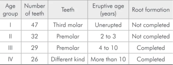
Teeth with caries, areas of hypomineralization or attrition affecting the enamel and those damaged during extraction were excluded from the study. The teeth inally selected for the study are shown in Table 1.
Specimen preparation
In order to produce specimens with 3 x 3 mm, one fragment of each tooth was obtained from the lattest central portion of the buccal aspect of the crown, which was sectioned with a precision sec-tioning machine (Isomet Low Speed Saw; Buehler, Lake Bluff, IL, USA) and two double-faced dia-mond discs (Diadia-mond wafering blade, series 15 HC diamond, arbor size ½ inch, Isomet 1000; Buehler,
Table 1 - The 134 teeth used in this study grouped accord-ing to their eruptive age.
Age group
Number
of teeth Teeth
Eruptive age
(years) Root formation
I 47 Third molar Unerupted Not completed
II 32 Premolar 2 to 3 Not completed
III 29 Premolar 4 to 10 Completed
Lake Bluff, IL, USA), separated by a 3-mm spacer adapted to it.
The dentinal aspects of the blocks were initially lattened with silicon carbide paper grit 320 (Bue-hler, Lake Bluff, IL, USA). Then, the enamel blocks were minimally lattened out (600 and 1,200 grades of Al2O3 paper; Buehler, Lake Bluff, IL, USA) and polished with felt paper wet by 1 µm diamond spray (Buehler), resulting in the removal of about 100 µm in depth of the enamel which was controlled with a micrometer.
Microhardness tests
To investigate the mineral content of the speci-mens, microhardness tests were performed. The mi-crohardness of the specimens was measured with a microhardness tester (HMV- 2000; Shimadzu Cor-poration, Tokyo, Japan) and a Knoop diamond in-denter, under a static load of 25 g applied for 5 sec-onds. Five indents were made on each fragment with a distance of 100 µm.
Statistical analysis
Comparison of the mean values of the post-erup-tive age groups was performed by applying Student’s
t test. The signiicance level was set at 5%.
Results
The values of supericial microhardness found in human enamel with different eruptive ages, un-erupted, 2-3 years of eruption, 4-10 years of erup-tion and with more than 10 years of eruperup-tion, are shown in Table 2.
The values of supericial microhardness presented an increasing trend over the years. Signiicant
differ-ence was found only between unerupted enamel and that with more than 10 years of eruption (ρ < 0.05).
Discussion
In academic literature, there is no pattern to be considered for age groups in studies regarding post-eruptive maturation. This lack of standardization in studies4-10,12,13,15-18 generates dificulties for the com-parison of results and its trustworthiness.
Therefore, in this study, the collected human teeth presented different post-eruptive ages, and were divided in four groups: Group I - unerupted; Group II - 2-3 years in the oral environment which did not present completely formed roots; Group III - 4-10 years in the oral environment with completely formed roots; and Group IV - more than 10 years in the oral environment. We opted for these age inter-vals due to the fact that each one of them represents different characteristics, as follows.
The unerupted tooth has never been exposed to the oral environment, its enamel is completely min-eralized, and it has not yet undergone post-eruptive maturation.
The 2-3 years interval group includes teeth in the period of eruption or with complete eruption, which already hadbeen exposed to the oral environ-ment. This is a critical period for the development of caries lesions. Since teeth remain long periods in infra-occlusion and children are unable to perform their teeth brushing properly, there is a greater risk of accumulation of dental bioilm. However, teeth in eruption are exposed to dental bioilm during several months before functional occlusion occurs. Accord-ing to Ekstrand et al.19 (2003), the time required for the irst and second permanent molars to erupt rang-es, respectively, from 5 to 32 and 9 to 45 months.
The 4-10 years interval group presents teeth that already are in occlusal contact and have been ex-posed to the oral environment for a longer period of time.3
The last group, which contains tooth specimens with more than 10 years of eruption, represents those teeth that already are in the oral environment for a much longer period of time.
In the present study, the values of supericial microhardness of the different eruptive age groups Table 2 - Mean microhardness values (KHN) and standard
deviation for the human enamel of the studied groups.
Eruptive
age Unerupted 2-3 years 4-10 years
More than 10 years
N 47 32 29 26
Mean* 375.79a 384.72ab 383.55ab 390.69b
Standard
Deviation 21.62 14.92 16.42 11.90
presented an increasing trend with the increase in age. Signiicant difference was found only between unerupted enamel specimens and those with more than 10 years of eruption. As known, a direct re-lation exists between the values of hardness and the percentage of mineral volume.20,21 This inding can be related to the exposure of the enamel to the oral-salivary environment14 and to normal dynamic processes of demineralization and remineralization during and after the eruptive process.3
The results of the present study are in accordance with the indings of Crabb5 (1976), who observed, through relected light, polarized light and scanning electron microscopy, a deined outer white zone with a honeycomb aspect. He concluded that unerupted or just-after-eruption teeth show a greater suscep-tibility to demineralization than erupted teeth, as a function of the supericial layer characteristics of the enamel. Brudevold et al.15 (1982), albeit following a different methodology, found similar results. They demonstrated that post-eruptive maturation of the enamel involves a reduction in permeability. Fur-thermore, Fejerskov et al.8 (1984), through scanning electron microscopy, observed that the surface of the enamel at the time of eruption presented many vari-eties of irregularities in its surface, as well as great inter-crystalline spaces, which render the tooth more susceptible to aggression. Still in 1984, Thylstrup et al.22 compared unerupted with just-after-eruption teeth. They found small differences between them. The most important was the size of the inter-crys-talline spaces, which were smaller in erupted teeth that were exposed to the oral environment.
Driessens et al.13 (1985) also have a study which agrees with the present results. They suggested that
post-eruptive maturation occurs in minerals of the supericial layer of the enamel, thus producing a greater resistance to caries lesions. Schulte et al.9 (1999) and Ten Bosch et al.10 (2000) observed an in-crease in electric resistance with the inin-crease of the post-eruptive ages of the teeth. This characteristic is directly related to a reduction in the porosity of the enamel due to post-eruptive maturation.
It can be suggested that this increasing trend of supericial microhardness could be related to a tendency to be less susceptible to caries over the years.3,5-10,12,14-16 Unerupted teeth or with incomplete post-eruptive maturation are more liable to suffer demineralization.6,7,16 However, the fact that the
su-pericial layer of the enamel was polished and lat-tened, as well as the fact that different kinds of teeth were used, could have inluenced the results of the present study.
A better knowledge of post-eruptive matura-tion, as well as of the natural processes occurring in the enamel soon after eruption, would be an asset to clinical dentists, pediatric dentists and to public health through the deinition of preventive strat-egies, as it would help to recognize the periods of higher susceptibility for the development of new caries lesions.
Conclusion
The present study revealed differences between the supericial microhardness of specimens with dif-ferent eruptive ages. The results also indicated an increasing mineralization behavior of enamel. How-ever, signiicant difference was found only between unerupted enamel specimens and those with more than 10 years of eruption.
References
1. Lima JEO. Um plano de prevenção para consultório odontope-diátrico. Rev Gaúcha Odontol. 1992;40(6):395-9.
2. Dowd FJ. Saliva and dental caries. Dent Clin North Am. 1999;43(4):579-97.
3. Fejerskov O, Nyvad B, Kidd EAM. Características clínicas e histológicas da cárie dentária. In: Fejerskov O, Kidd EAM. Cárie Dentária: a doença e seu tratamento clínico. São Paulo: Santos; 2005. p. 72-96.
4. Baker OD. Posteruptive changes in dental enamel. J Dent Res. 1966;45(Suppl 3):503-11.
5. Crabb HSM. The porous outer enamel of unerupted human premolars. Caries Res. 1976;10(1):1-7.
7. Woltgens JHM, Bervoets TJM, Witjes F, Driessens FCM. Effect of post-eruptive age on Ca and P loss from human enamel during demineralization in vitro. Arch Oral Biol. 1981;26(9):721-5.
8. Fejerskov O, Josephsen K, Nyvad B. Surface ultrastructure of unerupted mature human enamel. Caries Res. 1984;18(4):302-14.
9. Schulte A, Gente M, Pieper K. Posteruptive changes of electri-cal resistance values in fissure enamel of premolars. Caries Res. 1999;33(3):242-7.
10. Ten Bosch JJ, Fennis-le Y, Verdonschol EH. Time-depen-dent decrease and seasonal variation of the porosity of re-cently erupted sound dental enamel in vivo. J Dent Res. 2000;79(8):1556-9.
11. Cury JA. Uso do flúor e controle da cárie como doença. In: Baratieri LN. Odontologia restauradora: fundamentos e pos-sibilidades. São Paulo: Santos; 2002. p. 31-68.
12. Gängler P, Norén JG, Hoyer I, Bjarnason S, Kraft U, Odelius H et al. Reactivity of young and old human enamel to demin-eralization. Scand J Dent Res. 1993;101(6):345-9.
13. Driessens FCM, Heijligers HJM, Borggreven JMPM, Woltgens JHM. Posteruptive maturation of tooth enamel studied with the electron microprobe. Caries Res. 1985;19(4):390-5. 14. Fanning RJ, Dix FNJ, Shaw JH, Sognnaes RF. Salivary
con-tribution to enamel maturation and caries resistance. J Am Dent Assoc. 1954;49(6):668-71.
15. Brudevold F, Aasenden R, Bakhos Y. A preliminary study of posteruptive maturation of teeth in situ. Caries Res. 1982;16(3):243-8.
16. Woltgens JHM, Bervoets TJM, Witjes F, Driessens FCM. Changes in the comparison of the enamel of human premolar teeth shortly after eruption. Arch Oral Biol. 1981;26(9):717-9.
17. Aasenden R. Post-eruptive changes in the fluoride concen-trations of human tooth surface enamel. Arch Oral Biol. 1975;20(5-6):359-63.
18. Kidd EAM, Richards A, Thylstrup A, Fejerskov O. The sus-ceptibility of “young” and “old” human enamel to artificial caries in vitro. Caries Res. 1984;18(3):226-30.
19. Ekstrand KR, Christiansen J, Christiansen MEC. Time and duration of eruption of first and second permanent molars: a longitudinal investigation. Community Dent Oral Epidemiol. 2003;31(5):344-50.
20. Arends J, Ten Bosch JJ. Demineralization and remineralization evaluation techniques. J Dent Res. 1992;71(1):924-33. 21. Featherstone JDB, ten Cate JM, Shariati M, Arends J.
Com-parison of artificial caries-like lesions by quantitative mi-croradiography and microhardness profiles. Caries Res. 1983;17(5):385-91.