REVIEW
Variable clinical expression in patients with a
germline
MEN1
disease gene mutation: clues to a
genotype–phenotype correlation
Cornelis J. Lips,
IKoen M. Dreijerink,
IJo W. Ho¨ppener
IIIUniversity Medical Center Utrecht, Department of Internal Medicine & Endocrinology, Utrecht, The Netherlands.IIUniversity Medical Center Utrecht,
Department of Metabolic & Endocrine Diseases, Utrecht, The Netherlands.
Multiple endocrine neoplasia type 1 is an inherited endocrine tumor syndrome, predominantly characterized by
tumors of the parathyroid glands, gastroenteropancreatic tumors, pituitary adenomas, adrenal adenomas, and
neuroendocrine tumors of the thymus, lungs or stomach. Multiple endocrine neoplasia type 1 is caused by germline
mutations of the multiple endocrine neoplasia type 1 tumor suppressor gene. The initial germline mutation, loss of
the wild-type allele, and modifying genetic and possibly epigenetic and environmental events eventually result in
multiple endocrine neoplasia type 1 tumors. Our understanding of the function of the multiple endocrine neoplasia
type 1 gene product, menin, has increased significantly over the years. However, to date, no clear genotype–
phenotype correlation has been established. In this review we discuss reports on exceptional clinical presentations
of multiple endocrine neoplasia type 1, which may provide more insight into the pathogenesis of this disorder and
offer clues for a possible genotype–phenotype correlation.
KEYWORDS:
Multiple Endocrine Neoplasia type 1; MEN1; Menin; Genotype–Phenotype Correlation; Clinical
Expression.
Lips CJ, Dreijerink KM, Ho¨ppener JW. Variable clinical expression in patients with a germlineMEN1disease gene mutation: clues to a genotype– phenotype correlation. Clinics. 2012;67(S1):49-56.
E-mail: [email protected]
Tel.:+31 703240428
INTRODUCTION
’’It is an old experience that through her errors, Nature
often grants us unexpected insights into her secrets
which are otherwise a closed domain‘‘, William Harvey,
1657.
Multiple endocrine neoplasia type 1 (MEN1) is an
inherited endocrine tumor syndrome, characterized
pre-dominantly by tumors of the parathyroid glands,
gastro-enteropancreatic tumors, pituitary adenomas, adrenal
adenomas, and neuroendocrine tumors of the thymus,
lungs or stomach. MEN1 is caused by germline mutations
of the
MEN1
tumor suppressor gene. It appears that in the
MEN1 syndrome, clinical expression differs between
families. This may be the result of the specific
MEN1
gene
mutation in a family (genotype–phenotype correlation). As
a rule, the development of a tumor depends on a series of
genetic events (multistep tumorigenesis). Thus, additional
co-segregating modifying factors such as germline
muta-tions in other genes are likely to play a role in the
interfamilial variability of MEN 1. Moreover, clinical
expression can also vary between individual members of
the same family, possibly because of additional genetic or
epigenetic factors. To date, a clear correlation between
genetic events and the variable clinical expression of MEN1
has not been established (1–5). Further understanding of the
genetic aspects of MEN1 and the pathogenesis of
MEN1-related tumors could enable more tailored clinical screening
and treatment strategies.
In this review, we discuss recent reports on aberrant
clinical expression of MEN1, which may allow us a glimpse
into the pathogenesis of this intriguing disorder.
In 1903, Erdheim described the case of an acromegalic
patient with a pituitary adenoma and three enlarged
parathyroid glands. Fifty years later, Underdahl et al.
reported eight patients with a syndrome of pituitary-,
parathyroid-, and pancreatic islet adenomas. In 1954,
Wermer found that the syndrome was transmitted as a
dominant trait (6). In 1968, Steiner et al. introduced the term
‘‘multiple endocrine neoplasia’’ (MEN) to describe
disor-ders featuring combinations of endocrine tumors; they
designated the Wermer syndrome as MEN1 and the
Sipple syndrome as MEN2. Gorlin subdivided type 2 into
A and B. Then, in 1975, Khairi (7) suggested that type 2B be
called type 3; however, this was not generally accepted.
More recently, in 2006, families with multiple endocrine
tumors but without
MEN1
or
RET
(MEN2
)
gene mutations
were identified (8). This related syndrome is referred to as
MEN4.
Copyrightß2012CLINICS– This is an Open Access article distributed under the terms of the Creative Commons Attribution Non-Commercial License (http:// creativecommons.org/licenses/by-nc/3.0/) which permits unrestricted non-commercial use, distribution, and reproduction in any medium, provided the original work is properly cited.
DISCOVERY OF THE
MEN1
GENE
In positional cloning, gene mapping precedes, and
even-tually leads to, gene identification. The first step is mapping
the gene to a specific chromosomal region by linkage analysis.
The second step is identifying the correct gene among all
possible candidate genes within that particular chromosomal
region. After the gene has been identified, it is possible to
study its (patho)physiologic function.
In 1996, two groups independently identified the
MEN1
gene on chromosome 11q13 (9,10). To date, more than 1336
different
MEN1
gene mutations (both germline and
spora-dic) have been reported in the literature (4). Most of these
mutations are clearly inactivating, in accordance with the
notion that the
MEN1
gene is a tumor suppressor gene.
There are no mutational hot spots in the
MEN1
gene.
FUNCTION OF THE
MEN1
-GENE PRODUCT
The
MEN1
gene product, menin, functions as an adaptor
protein that is involved in interactions with multiple protein
partners.
Men1
null mutant mice have indicated that menin is
essential for viability (11). Menin is involved in
neuroendo-crine cell development and function. Later on, it is active in
many cellular processes, including gene transcription
regula-tion, DNA replicaregula-tion, DNA repair, and signal transduction.
Menin target genes that are important for development
and proliferation, including homeobox domain (HOX)
genes, the
CDKN2C
and
CDKN1B
cyclin-dependent kinase
inhibiting genes, the human telomerase (
hTERT
) gene, and
nuclear receptor target genes (12–15).
As a transcriptional co-regulator, menin may function as a
co-activator or co-repressor by recruiting histone-modifying
enzymatic activity (12,15,16). As a co-activator, menin is
involved in the regulation of histone methylation by recruiting
the mixed-lineage leukaemia (MLL) proteins MLL1 and MLL2
(12,17). In this way, menin can bind to nuclear receptors and
activate nuclear receptor-mediated gene transcription (12,15).
By tethering histone deacetylase activity to genes, menin can
serve as a repressor of transcription (18).
In order to understand the role of menin as a tumor
suppressor protein and as a co-factor of MLL fusion proteins,
the structural basis had to be revealed. Recently, the crystal
structure of menin in
Nematostella vectensis
was reported (19).
Knowledge about the three-dimensional structure may
elucidate the interactions essential to the function of menin.
It appears that the Leucine, Leucine, Tryptophan, Leucine,
Leucine (LLWLL) amino acids nuclear receptor interaction
motif of menin is well-conserved and is located in an
alpha-helix. In general, modeling gene mutations into this structure
will be helpful in determining the effects on protein function.
Inactivation of the
MEN1
gene results in predisposition to
tumor formation (see Figure 1, Table 1).
ABERRANT CLINICAL EXPRESSION OF MEN1
A
MEN1
gene mutation may be completely detrimental to
gene function. It may also result in a protein product with
some residual function. An aberrant menin protein may be
impaired in its function by several mechanisms: menin can
interact with many different proteins. Possibly, germline
mutations in the
MEN1
gene selectively affect menin
binding to its partners, leading to distinct phenotypes.
The type of missense mutation (e.g. replacement of
arginine with glycine) may have a differential effect on the
function of menin (38): in-frame or missense mutations
differ from frameshift/nonsense mutations (39), whereas
missense and in-frame mutations may affect the interactions
of a menin domain with transcription factors such as JunD,
Smad3 and NFkappaB and nuclear receptors (1), or impair
sensitization to apoptosis from caspase-3, p53 or p21 (40).
A
MEN1
gene missense mutation may result in protein
instability, and enhanced and early proteolytic degradation
via the ubiquitin–proteasome pathway (41).
It was generally assumed that, in contrast to MEN2, in
MEN1 there is no clear genotype–phenotype correlation
(1,3–5). However, several reports challenge this assumption.
Familial aberrant expression
1. MEN1 Burin.
Four large kindreds from the Burin
peninsula/Fortune Bay area of Newfoundland with
promi-nent features of prolactinomas, in addition to carcinoids, and
parathyroid tumors (referred to as MEN1Burin) have been
described, and they show linkage to 11q13, the same locus as
that of MEN1. Haplotype analysis with 16 polymorphic
markers now reveals that representative affected individuals
from all four families share a common haplotype over a
2.5 Mb region. A nonsense mutation in the
MEN1
gene has
been found to be responsible for the disease in the affected
members in all four of the MEN1-Burin families. This suggests
that either a common ancestral mutation in the
MEN1
-Burin
gene or a modifying gene on 11q13 is responsible for this
prolactinoma variant of MEN1 (42–45).
2. Familiar isolated hyperparathyroidism and
MEN1
gene
missense mutations.
Familial isolated primary
hyperpa-rathyroidism (FIHP) is an autosomal dominant disorder that
can represent an early stage of either MEN1 caused by an
allelic variant of the
MEN1
gene, or of hyperparathyroidism–
jaw tumor (HPT-JT) syndromes; alternatively, the condition
can be caused by an allelic variant of the hyperparathyroidism
2 (
HRPT2
) gene, or caused by a mutation at another locus.
Interestingly, the major proportion of
MEN1
gene germline
mutations that have been found in FIHP are seemingly mild
missense mutations or in-frame deletions (46–55). In MEN1,
roughly 80% of patients harbor nonsense mutations.
3. Predominant mutations in MEN1 pancreatic
neu-roendocrine tumors.
Schaaf performed mutation
analy-sis of the
MEN1
gene in tumors from 306 patients with
MEN1, and found that patients with gastroenteropancreatic
tumors more often had truncating mutations, very probably
leading to completely inactivated menin (56).
4. Mild/late onset versus malignant phenotypes.
To
date, several disease-related
MEN1
gene intron mutations
have been reported. These intron mutations may affect
mRNA splicing and cause mild phenotypes, with late, and
relatively low, penetrance of the disease (57–59). However,
clinical expression at a young age may occur. This may be
explained by interpatient variations in gene transcription
and translation of the
MEN1
gene.
Two recent case reports described families with a high
penetrance of malignant neuroendocrine tumors of the
pancreas (60,61). Both these families carried germline
mutations that completely abolish menin function.
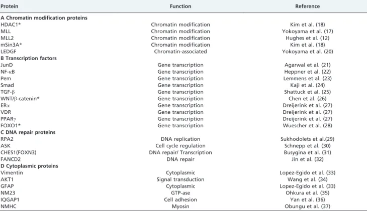
Table 1 -
Menin-interacting proteins.
Protein Function Reference
A Chromatin modification proteins
HDAC1* Chromatin modification Kim et al. (18)
MLL Chromatin modification Yokoyama et al. (17)
MLL2 Chromatin modification Hughes et al. (12)
mSin3A* Chromatin modification Kim et al. (18)
LEDGF Chromatin-associated Yokoyama et al. (20)
B Transcription factors
JunD Gene transcription Agarwal et al. (21)
NF-kB Gene transcription Heppner et al. (22)
Pem Gene transcription Lemmens et al. (23)
Smad Gene transcription Kaji et al. (24)
TGF-b Gene transcription Shattuck et al. (25)
WNT/b-catenin* Gene transcription Chen et al. (26)
ERa Gene transcription Dreijerink et al. (27)
VDR Gene transcription Dreijerink et al. (27)
PPARc Gene transcription Dreijerink et al. (27)
FOXO1* Gene transcription Wuescher et al. (28)
C DNA repair proteins
RPA2 DNA replication Sukhodolets et al.(29)
ASK Cell cycle regulation Schnepp et al. (30)
CHES1(FOXN3) DNA repair/ Transcription Busygina et al. (31)
FANCD2 DNA repair Jin et al. (32)
D Cytoplasmic proteins
Vimentin Cytoplasmic Lopez-Egido et al. (33)
AKT1 Signal transduction Wang et al. (34)
GFAP Cytoplasmic Lopez-Egido et al. (33)
NM23 GTP-ase Ohkura et al. (35)
IQGAP1 Cell adhesion Yan et al. (36)
NMHC Myosin Obungu et al. (37)
The earliest manifestation of MEN1 was a pituitary
adenoma in a 5-year-old boy who had a missense mutation
leading to a H139D substitution in the
MEN1
protein (62).
Functional analysis of the mutant protein revealed severely
reduced protein stability (41), reduced binding to JunD (16),
reduced binding to the estrogen receptor alpha and absent
histone-methylation recruiting capacity. Thus, functional
analysis of this potentially mild missense
MEN1
gene
mutation shows that the protein product is, in fact, completely
inactivated.
5. Metabolic effects of aberrant expression of the
MEN1
gene.
In
MEN1
disease-gene carriers, all vitamin D
receptors (VDRs) and peroxisome proliferator-activated
receptors (PPARs)-
c
are expressed but are probably less
activated because of impaired menin function.
A. PPAR
c
2 is a transcription factor that plays a key role in
adipocyte differentiation. Polymorphisms in this gene may
contribute to the variability in body mass index and insulin
sensitivity in the general population. PPAR
c
is the receptor
for the thiazolidinediones, which act as PPAR
c
agonists and
lower the blood glucose levels in patients with type 2 diabetes
mellitus by increasing insulin sensitivity. Individuals with
dominant-negative PPAR
c
gene mutations manifest a
syn-drome that combines lipodystrophy with features of the
metabolic syndrome, including insulin resistance, type 2
diabetes, hepatic steatosis, dyslipidemia, hypertension and
(in women) polycystic ovary syndrome. In patients with
MEN1, decreased activation of PPAR may result in insulin
resistance and weight gain (63).
B. Vitamin D receptors (VDRs) are found in a large
number of tissues beyond the classic target tissues gut, bone
and kidney. These tissues include endocrine glands such as
pituitary, parathyroid glands, pancreatic islets, etc.
Lourenc¸o et al. discussed the increased bone loss pattern
found in patients with MEN1 compared with that of patients
with sporadic primary HPT (64). Besides increased bone loss,
resistance to vitamin D may be associated with insulin
resistance and beta cell dysfunction, leading to increased risk
for type 2 diabetes in patients with MEN1 (65,66).
Effect of gender
The prevalence and probability of pancreatic tumors
among patients with MEN1 were higher in males than
in females. This difference was due to the differential
occurrence of gastrinomas. The prevalence and probability
of developing pituitary adenomas were significantly greater
in females. Thymic tumors are found nearly exclusively in
male MEN1 patients (67).
The difference in clinical expression between the genders
may be explained by the difference in transcription
regulation of estrogen and androgen receptors. Menin can
act as a co-activator of nuclear hormone receptors including
estrogen (ER
a
) and possibly androgen (AR) receptors. A
defect in the
MEN1
gene, together with gender-specific
differences in concentrations of the hormones involved and
tissue-specific distribution of their receptors, may contribute
to the observed gender-specific differences in prevalence of
prolactinoma and gastrinoma.
Additional genetic effects
1. Loss of heterozygosity; the AIP gene.
In accordance
with the tumor suppressor function of menin,
MEN1-associated tumors exhibit loss of the wild-type allele. This
second hit occurs as a somatic mutation and often involves
deletion of a larger chromosomal region containing multiple
genes [loss of heterozygosity (LOH)].
The gene encoding the aryl hydrocarbon receptor
inter-acting protein (AIP) is located on 11q13, in the vicinity of the
MEN1
gene. Recently, it was found that inactivating
mutations in the
AIP
gene are the underlying cause of
low-penetrance pituitary adenoma predisposition. The
finding of a truncated gene and LOH indicates that
AIP
acts as a tumor suppressor gene (68,69). In northern Finland,
AIP
-germline mutations accounted for 16% of cases of
acromegaly in young patients. The tumor suppressor genes
AIP
and
MEN1
are located 3 Mb apart. Concomitant
deletions of these genes may underlie predisposition to
MEN1 and pituitary adenoma. To what extent deletion of
the
AIP
gene is present in MEN1 tumors has yet to be
established. Inactivation of this gene in animal models
may reveal a potential causative role in MEN1-associated
tumors.
2. Genetic predisposition for other diseases.
Genetic
predisposition for other diseases may contribute to
enhancement of tumor formation in patients with MEN1
(70). For instance, normally the vitamin D receptor on
parathyroid cells inhibits production and release of
parathyroid hormone (PTH). In families with inactivating
mutations in the gene encoding VDR, this is associated with
end-organ resistance to calcitriol.
In the parathyroid glands of patients with MEN1, there
exists a decreased activation of the VDR. An additional
defect in the VDR or calcium receptor may contribute to
hyperactivity, hyperplasia, and adenoma (71).
3. Additional, somatic mutations involved in acceleration
of tumor growth.
3a). Data from other familial neuroendocrine tumor
syndromes.
How can we identify acquired mutations that
are responsible for acceleration of tumor growth in MEN1?
Clues for modifier genes may be found in other familial
neuroendocrine tumor syndromes such as MEN2 and
MEN4 (the latter is also referred to as MENX). Which are
their traditional pathogenetic pathways and are these
involved in aberrant clinical MEN1 expression? Overlap
between MEN1 and MEN2 and additional genetic events
have to be explored (e.g. p18/p27 knock-out mice develop
both MEN1- and MEN2-associated tumors) (72,73).
Phenotypic overlap between MEN1- and MEN2-like
syndromes was identified in the rat and named MENX.
The syndrome is caused by a germline inactivating
mutation in the
CDKN1B
gene encoding p27
Kip1(8).
p27
Kip1has a key role in cell cycle regulation and is
involved in differentiation, apoptosis, and angiogenesis.
Subsequently, germline mutations in the
CDKN1B
gene
were identified in the germline of a MEN1-like family. In
these patients, germline mutations of the
MEN1
gene could
not be detected (8). However, only the menin-coding region
and splice junctions were analyzed. The patients were also
negative for
RET
gene mutations (MEN2). Mutations in
CDKN1B
and related genes, but without
MEN1
gene
mutations, are a rare cause of MEN1-like phenotype (74–
76). As a consequence of mutations in the p27 gene, a novel
human MEN syndrome was recognized and named MEN4.
In mice, the
Cdkn2c
gene encoding p18
Ink4cwas shown to
collaborate with menin in suppression of neuroendocrine
tumor development (77). Whether occurrence of somatic
mutations in p18
Ink4cand/or p27
Kip1accelerates tumor
transgenic MEN2 mice and human patients with MEN2,
inactivating mutations in p18 will promote tumor growth
(72,73,78,79). Reduced expression of
CDKN1B
, but not
CDKN2C
, has been observed in parathyroid adenomas from
patients with MEN1.
3b). Clues from sporadic parathyroid adenomas,
pituitary tumors, and pancreatic NETs
i) Sporadic Parathyroid adenoma
.
A high rate of
so-matic
MEN1
gene mutations is seen in sporadic parathyroid
adenomas. There exists an interaction between menin and
the transforming growth factor (TGF)-beta/Smad signaling
pathway.
In vitro
experimentation has demonstrated that
the presence of menin is required for TGF-beta to effectively
inhibit parathyroid cell proliferation and PTH production
(80).
ii) Pituitary tumors
.
The pituitary tumor transforming
gene (PTTG; securin) was the first transforming gene found to
be highly expressed in pituitary tumor cells, and seems to play
an important role in the process of oncogenesis. Cell signaling
abnormalities have been identified in pituitary tumors, but their
genetic basis is unknown. Both Raf/mitogen activated protein
kinase kinase (MEK)/extracellular signal-regulated kinase
(ERK) and phosphoinositide 3-kinase(PI3K)/Akt/mammalian
target of rapamycin (mTOR) pathways are over-expressed
and/or over-activated in pituitary tumors (81). These pathways
share a common root, including initial activation by a tyrosine
kinase receptor.
Pit-1 is a direct transcriptional target of VDR. Recruitment
of histone deacetylase 1 is involved in the repressive effect
of VDR on Pit-1 expression (82).
There is a critical role for the growth factor activin in
regulating inhibition of pituitary cell growth and Pit-1/PRL
expression through the Smads and menin (83). Alterations in
the activin/TGF-beta downstream signaling pathways may
be critical steps towards tumor formation and progression
(84). To date, the occurrence of additional TGF-beta, Pit-1,
PTTG, or VDR mutations in MEN1-associated tumors has not
been published.
iii) Pancreatic neuroendocrine tumors
.
In nonfamilial
pancreatic neuroendocrine tumors (PanNETs) the most
commonly mutated genes specify proteins implicated in
chromatin remodeling: 44% of the tumors had somatic
inactivating mutations in
MEN1.
Clinically, mutations in the
MEN1
gene were associated with better prognosis. Also,
mutations in genes in the mTOR pathway were found in
14% of the tumors, a finding that could potentially be used
to stratify patients for treatment with mTOR inhibitors (85).
Loss of menin expression is associated with
over-expression of the Raf/MEK/ERK and P13K/AKT/mTOR
pathways in pancreatic tumors (34). Intact menin has an
essential role in WNT/
b
-catenin signaling, and inhibits
mouse pancreatic islet tumor proliferation (26). Menin
regulates subcellular localization of
b
-catenin via
nuclear-cytoplasmic shuttling. Loss of menin leads to Wnt/
b
-catenin
signaling activation. Expression of p27 was found to be
repressed in pancreatic islet cell tumors (86).
3c) Pathways in multistep carcinogenesis.
It appears
that interaction of components of the PI3K/AKT pathway is
involved in neuroendocrine tumor formation (87,88).
Deregulation of the PI3K/AKT pathway in neuroendocrine
tumors can occur through a range of processes (see Figure 2),
including gain of function by oncogenic mutations of PI3K
signalling, loss of function of the tumor suppressor PTEN
through gene deletion, mutation, micro-RNA expression or
epigenetic silencing, upstream activation through receptor
tyrosine kinase (RTK) signaling, and/or downstream loss of
the tumor suppressors p18 and p27.
A combination of a mutation in the
MEN1
disease gene
with other specific mutations of genes in the PI3K/AKT
pathway may be associated with acceleration of tumor
growth.
In addition, inactivated or absent menin promotes RAS
expression; activating mutations of RAF, MEK, or ERK may
accelerate cell proliferation (89).
Ecogenetic factors
Common environmental factors may interact with genetic
predisposition to MEN1 and contribute to enhancement of
tumor formation (70). In the parathyroid glands of patients
with MEN1, a diet low in vitamin D or calcium may result
in tumor growth. In the lactotropic cells of the pituitary
gland, estrogenic or neuroleptic drugs may stimulate cell
division. In the gastrin-producing cells of the stomach,
presence of achlorhydria or use of histamine-H2 receptor or
proton pump blockers may promote tumor growth (90).
Lifestyle factors such as smoking and exposure to radiation
may have deleterious effects on menin function and tumor
growth, as with many types of cancer.
CONCLUDING REMARKS
Careful observation of patients and collaboration between
disciplines, including molecular endocrinology, has opened
new directions in the management of MEN1 syndrome, and
has promoted development of novel target-directed
ther-apy. Since 1980, life expectancy and quality of life have
improved considerably.
By contrast, thymic tumors and duodenopancreatic
tumors, including nonsecreting pancreatic tumors increase
the risk of death (91). Rare, but aggressive, adrenal tumors
may also cause early death. In
MEN1
disease gene carriers
in MEN1 families, most deaths were related to the disease,
and probably resulted from additional mutations.
It is possible that consecutive and specific pathogenetic
pathways are involved in MEN1 tumor formation. We
presume that, through the inherited germline mutation
resulting in organ-specific cell division, the patient is
rendered vulnerable to additional, somatic mutations in these
organs. These mutations may occur spontaneously or may be
triggered by life style and/or environment. Predisposition
and expression of other genetic diseases may also be involved.
A complex genotype–phenotype relationship may be present.
Unfortunately, for the majority of the patients it is not
currently possible to predict the course of the disease.
Germline mutations that result in complete inactivation of
the gene product apparently cause more severe disease.
Consequently, extensive periodical clinical examination
has to be performed in all carriers of the
MEN1
disease gene.
In the near future, tumor gene-expression profiles and high
throughput sequencing may permit more insight into
additional genetic and epigenetic events that cause
progres-sion of tumor development. Functional analysis of
MEN1
AUTHOR CONTRIBUTIONS
Lips CJ conceived and designed the study and was also responsible for the manuscript writing and preparation of figures and table. Dreijerink KM searched the literature for important contents, provided assistance to the manuscript writing and to the molecular aspects of the menin protein. Ho¨ppener JW provided assistance to the study design, manuscript writing and final version of the manuscript.
REFERENCES
1. Wautot V, Vercherat C, Lespinasse J, Chambe B, Lenoir GM, Zhang CX, et al. Germline mutation profile of MEN1 in multiple endocrine neoplasia type 1: search for correlation between phenotype and the functional domains of the MEN1 protein. Hum Mutat. 2002;20(1):35-47, http://dx.doi.org/10.1002/humu.10092.
2. Kouvaraki MA, Lee JE, Shapiro SE, Gagel RF, Sherman SI, Sellin RV, et al. Genotype-phenotype analysis in multiple endocrine neoplasia type 1. Arch Surg. 2002;137(6):641-7, http://dx.doi.org/10.1001/arch-surg.137.6.641.
3. Turner JJ, Leotlela PD, Pannett AA, Forbes SA, Bassett JH, Harding B, et al. Frequent occurrence of an intron 4 mutation in multiple endocrine neoplasia type 1. J Clin Endocrinol Metab. 2002;87(6):2688-93, http://dx.doi.org/ 10.1210/jc.87.6.2688.
4. Lemos MC, Thakker RV. Multiple endocrine neoplasia type 1 (MEN1): analysis of 1336 mutations reported in the first decade following identification of the gene. Hum Mutat. 2008;29(1):22-32, http:// dx.doi.org/10.1002/humu.20605.
5. Conte-Devolx B, Niccoli P. Groupe d’e´tude des Tumeurs Endocrines. Clinical characteristics of multiple endocrine neoplasia. Bull Acad Natl Med. 2010; 194(1):69-78.
6. Wermer P. Genetic aspects of adenomatosis of endocrine glands. Am J Med. 1954;16:363–71, http://dx.doi.org/10.1016/0002-9343(54)90353-8.
7. Khairi MRA, Dexter RN, Burzynski NJ, Johnston CC. Mucosal neuroma, pheochromocytoma and medullary thyroid carcinoma: multiple endo-crine neoplasia-type III. Medicine. 1975;54(2):89-112.
8. Pellegata NS, Quintanilla-Martinez L, Siggelkow H, Samson E, Bink K, Ho¨fler H, et al. Germ-line mutations in p27Kip1 cause a multiple endocrine neoplasia syndrome in rats and humans. Proc Natl Acad Sci USA. 2006; 103: 15558-63. Erratum in: Proc Natl Acad Sci U S A. 2006; 103(50):19213, http://dx.doi.org/10.1073/pnas.0603877103
9. Chandrasekharappa SC, Guru SC, Manickam P, Olufemi SE, Collins FS, Emmert-Buck MR, et al. Positional cloning of the gene for multiple endocrine neoplasia type 1. Science. 1997;276(5311):404–7, http:// dx.doi.org/10.1126/science.276.5311.404.
10. Lemmens I, van der Ven WJ, Kas K, Zhang CX, Giraud S, Wautot V, et al. Identification of the MEN1 gene. The European consortium on MEN1. Hum Mol Genet. 1997;6(7):1177-83.
11. Zhang HL, Li WY, Zhang CP, Zhu YX, Wu L, Long HM, et al. Differentially expressed genes in Men1 knockout and wild type embryoid bodies for pancreatic islet development. Mol Med Report. 2011;4(2):301-5, http://dx.doi.org/10.3892/mmr.2011.409. Epub 2011 Jan 3.
12. Hughes CM, Rozenblatt-Rosen O, Milne TA, Copeland TD, Levine SS, Lee JC, et al. Menin associates with a trithorax family histone methyltransferase complex and with the hoxc8 locus. Mol Cell. 2004; 13(4):587-97, http://dx.doi.org/10.1016/S1097-2765(04)00081-4. 13. Milne TA, Hughes CM, Lloyd R, Yang Z, Rozenblatt-Rosen O, Dou Y,
et al. Menin and MLL cooperatively regulate expression of cyclin-dependent kinase inhibitors. Proc Natl Acad Sci USA. 2005;102(3):749– 54, http://dx.doi.org/10.1073/pnas.0408836102.
14. Lin SY, Elledge SJ. Multiple tumor suppressor pathways negatively regulate telomerase. Cell. 2003;113(7):881–9, http://dx.doi.org/10.1016/ S0092-8674(03)00430-6.
15. Dreijerink KM, Ho¨ppener JW, Timmers, Lips CJ. Mechanism of disease: MEN 1-relation to chromatin modifications and transcription regulation. Nat Clin Pract Endocr Metab. 2006;2(10):562–70, http://dx.doi.org/ 10.1038/ncpendmet0292.
16. Agarwal SK, Guru SC, Heppner C, Erdos MR, Collins RM, Park SY, et al. Menin interacts with the AP1 transcription factor JunD and represses JunD-activated transcription. Cell. 1999;96:143–52, http://dx.doi.org/ 10.1016/S0092-8674(00)80967-8.
17. Yokoyama A, Wang Z, Wysocka J, Sanyal M, Aufiero DJ, Kitabayashi I, et al. Leukemia proto-oncoprotein MLL forms a SET1-like histone methyltransferase complex with menin to regulate Hox gene expression. Mol Cell Biol. 2004;24(13):5639–49, http://dx.doi.org/10.1128/ MCB.24.13.5639-5649.2004.
18. Kim H, Lee JE, Cho EJ, Liu JO, Youn HD. Menin, a tumor suppressor, represses JunD-mediated transcriptional activity by association with an mSin3A-histone deacetylase complex. Cancer Res. 2003;63(19):6135-9. 19. Murai MJ, Chruszcz M, Reddy G, Grembecka J, Cierpicki T. Crystal
structure of Menin reveals the binding site for mixed lineage Leukemia (MLL) protein. J Biol Chem. 2011; 9;286(36):31742-8.
20. Yokoyama A, Cleary ML. Menin critically links MLL proteins with LEDGF on cancer-associated target genes. Cancer Cell. 2008;14(1):36-46, http://dx.doi.org/10.1016/j.ccr.2008.05.003.
21. Agarwal SK, Impey S, McWeeney S, Scacheri PC, Collins FS, Goodman RH, et al. Distribution of menin-occupied regions in chromatin specifies a broad role of menin in transcriptional regulation. Neoplasia. 2007;9(2):101-7, http://dx.doi.org/10.1593/neo.06706.
22. Heppner C, Bilimoria KY, Agarwal SK, Kester M, Whitty LJ, Guru SC, et al. The tumor suppressor protein menin interacts with NF-kappaB proteins and inhibits NF-kappaB-mediated transactivation. Oncogene. 2001;20(36):4917-25, http://dx.doi.org/10.1038/sj.onc.1204529. 23. Lemmens IH, Forsberg L, Pannett AA, Meyen E, Piehl F, Turner JJ, et al.
Menin interacts directly with the homeobox-containing protein Pem. Biochem Biophys Res Commun. 2001;286(2):426-31, http://dx.doi.org/ 10.1006/bbrc.2001.5405.
24. Kaji H, Canaff L, Lebrun JJ, Goltzman D, Hendy GN. Inactivation of menin, a Smad3-interacting protein, blocks transforming growth factor type beta signaling. Proc Natl Acad Sci USA. 2001;98(7):3837-42, http:// dx.doi.org/10.1073/pnas.061358098.
25. Shattuck TM, Costa J, Bernstein M, Jensen RT, Chung DC, Arnold A. Mutational analysis of Smad3, a candidate tumor suppressor implicated in TGF-beta and menin pathways, in parathyroid adenomas and enteropan-creatic endocrine tumors. J Clin Endocrinol Metab. 2002;87(8):3911-4, http://dx.doi.org/10.1210/jc.87.8.3911.
26. Chen G, A J, Wang M, Farley S, Lee LY, Lee LC, Sawicki MP. Menin promotes the Wnt signaling pathway in pancreatic endocrine cells. Mol Cancer Res. 2008;6(12):1894–907.
27. Dreijerink KM, Mulder KW, Winkler GS, Ho¨ppener JW, Lips CJ, Timmers HT. Menin links estrogen receptor activation to histone H3K4 trimethylation. Cancer Res. 2006;66(9):4929–35, http://dx.doi.org/ 10.1158/0008-5472.CAN-05-4461.
28. Wuescher L, Angevine K, Hinds T, Ramakrishnan S, Najjar SM, Mensah-Osman EJ. Am J Phsyiol Endocrinol Metab. 2011;301(3):E474-83.
29. Sukhodolets KE, Hickman AB, Agarwal SK, Sukhodolets MV, Obungu VH, Novotny EA, et al. The 32-kilodalton subunit of replication protein A interacts with menin, the product of the MEN1 tumor suppressor gene. Mol Cell Biol. 2003;23(2):493-509, http://dx.doi.org/10.1128/ MCB.23.2.493-509.2003.
30. Schnepp RW, Hou Z, Wang H, Petersen C, Silva A, Masai H, et al. Functional interaction between tumor suppressor menin and activator of S-phase kinase. Cancer Res. 2004;64(18):6791-6.
31. Busygina V, Kottemann MC, Scott KL, Plon SE, Bale AE. Multiple endocrine neoplasia type 1 interacts with forkhead transcription factor CHES1 in DNA damage response. Cancer Res. 2006;66(17):8397-403, http://dx.doi.org/10.1158/0008-5472.CAN-06-0061.
32. Jin S, Mao H, Schnepp RW, Sykes SM, Silva AC, D’Andrea AD, et al. Menin associates with FANCD2, a protein involved in repair of DNA damage. Cancer Res. 2003;63(14):4204-10.
33. Lopez-Egido J, Cunningham J, Berg M, Oberg K, Bongcam-Rudloff E, Gobl A. Menin’s interaction with glial fibrillary acidic protein and vimentin suggests a role for the intermediate filament network in regulating menin activity. Exp Cell Res. 2002;278(2):175-83, http:// dx.doi.org/10.1006/excr.2002.5575.
34. Wang Y, Ozawa A, Zaman S, Prasad NB, Chandrasekharappa SC, Agarwal SK, et al. The tumor suppressor protein menin inhibits AKT activation by regulating its cellular localization. Cancer Res. 2011;71(2): 371–82, http://dx.doi.org/10.1158/0008-5472.CAN-10-3221.
35. Ohkura N, Kishi M, Tsukada T, Yamaguchi K. Menin, a gene product responsible for multiple endocrine neoplasia type 1, interacts with the putative tumor metastasis suppressor nm23. Biochem Biophys Res C o m mu n . 2 0 0 1; 2 82 ( 5 ) : 1 2 0 6 - 1 0 , h t t p : / / d x . d o i .o r g / 1 0 . 1 0 0 6 / bbrc.2001.4723.
36. Yan J, Yang Y, Zhang H, King C, Kan HM, Cai Y, et al. Menin interacts with IQGAP1 to enhance intercellular adhesion of beta-cells. Oncogene. 2009;28(7):973-82, http://dx.doi.org/10.1038/onc.2008.435.
37. Obungu VH, Lee Burns A, Agarwal SK, Chandrasekharapa SC, Adelstein RS, Marx SJ. Menin, a tumor suppressor, associates with nonmuscle myosin II-A heavy chain. Oncogene. 2003;22(41):6347-58, http://dx.doi.org/10.1038/sj.onc.1206658.
38. Hou R, Manwaring LP, Moley JF, Whelan AA. A novel missense mutation in the MEN1 gene in a patient with multiple endocrine neoplasia type 1. Endocr Pract. 2011;17(3):e63–7.
39. Vierimaa O, Ebeling TM, Kyto¨la¨ S, Bloigu R, Eloranta E, Salmi J, et al. Multiple endocrine neoplasia type 1 in Northern Finland; clinical features and genotype phenotype correlation. Eur J Endocrinol. 2007;157(3):285–94, http://dx.doi.org/10.1530/EJE-07-0195.
40. Bazzi W, Renon M, Vercherat C, Hamze Z, Lacheretz-Bernigaud A, Wang H, et al. MEN1 missense mutations impair sensitization to apoptosis induced by wild-type menin in endocrine pancreatic tumor cells. Gastroenterology. 2008;135(5):1698–1709, http://dx.doi.org/ 10.1053/j.gastro.2008.07.031.
41. Yaguchi H, Ohkura N, Takahashi M, Nagamura Y, Kitabayashi I, Tsukada T. Menin missense mutants associated with multiple endocrine neoplasia type 1 are rapidly degraded via the ubiquitin-proteasome pathway. Mol Cell Biol. 2004;24(15):6569–80, http://dx.doi.org/10.1128/ MCB.24.15.6569-6580.2004.
42. Olufemi SE, Green JS, Manickam P, Guru SC, Agarwal SK, Kester MB, et al. Common ancestral mutation in the MEN1 gene is likely responsible for the prolactinoma variant of MEN1 (MEN1-Burin) in four kindreds from Newfoundland. Hum Mutat. 1998;11(4):264–69, http://dx.doi.org/ 10.1002/(SICI)1098-1004(1998)11:4,264::AID-HUMU2.3.0.CO;2-V. 43. Kong C, Ellard S, Johnston C, Farid NR. Multiple endocrine neoplasia
type 1Burin from Mauritius: a novel MEN1 mutation. J Endocrinol Invest. 2001;24(10):806–10.
44. Hao W, Skarulis MC, Simonds WF, Weinstein LS, Agarwal SK, Mateo C, et al. Multiple endocrine neoplasia type 1 variant with frequent prolactinoma and rare gastrinoma. J Clin Endocrinol Metab. 2004;89(8):3776–84, http://dx.doi.org/10.1210/jc.2003-031511. 45. Agarwal SK, Ozawa A, Mateo CM, Marx SJ. The MEN1 gene and
pituitary tumours. Horm Res. 2009;71(Suppl. 2):131–8, http:// dx.doi.org/10.1159/000192450.
46. Teh BT, Esapa CT, Houlston R, Grandell U, Farnebo F, Nordenskjo¨ld M, et al. A family with isolated hyperparathyroidism segregating a missense MEN1 mutation and showing loss of the wild-type alleles in the parathyroid tumors. Am J Hum Genet. 1998;63(5):1544–9, http:// dx.doi.org/10.1086/302097.
47. Honda M, Tsukada T, Tanaka H, Maruyama K, Yamaguchi K, Obara T, et al. A novel mutation of the MEN1 gene in a Japanese kindred with familial isolated primary hyperparathyroidism. Eur J Endocrinol. 2000;142(2):138–43, http://dx.doi.org/10.1530/eje.0.1420138.
48. Kassem M, Kruse TA, Wong FK, Larsson C, Teh BT. Familial isolated hyperparathyroidism as a variant of multiple endocrine neoplasia type 1 in a large Danish pedigree. J Clin Endocrinol Metab. 2000;85(1):165–7, http://dx.doi.org/10.1210/jc.85.1.165.
type 1 variant? Eur J Endocrinol. 2001;145(2):155–60, http://dx.doi.org/ 10.1530/eje.0.1450155.
50. Villablanca A, Wassif WS, Smith T, Ho¨o¨g A, Vierimaa O, Kassem M, et al. Involvement of the MEN1 gene locus in familial isolated hyperparathyr-oidism. Eur J Endocrinol. 2002;147(3):313–22, http://dx.doi.org/ 10.1530/eje.0.1470313
51. Simonds WF, James-Newton LA, Agarwal SK, Yang B, Skarulis MC, Hendy GN, et al. Familial isolated hyperparathyroidism: clinical and genetic characteristics of 36 kindreds. Medicine (Baltimore) 2002;81:1–26. 52. Pannett AA, Kennedy AM, Turner JJ, Forbes SA, Cavaco BM, Bassett JH, et al. Multiple endocrine neoplasia type 1 (MEN1) germline mutations in familial isolated primary hyperparathyroidism. Clin Endocrinol (Oxf). 2003;58:639–46, http://dx.doi.org/10.1046/j.1365-2265.2003.01765.x. 53. Hannan FM, Nesbit MA, Christie PT, Fratter C, Dudley NE, Sadler GP,
Thakker RV. Familial isolated primary hyperparathyroidism caused by mutations of the MEN1 gene. Nat Clin Pract Endocrinol Metab. 2008;4:53–8, http://dx.doi.org/10.1038/ncpendmet0718.
54. Miyauchi A, Sato M, Matsubara S, Ohye H, Kihara M, Matsusaka K, et al. A family of MEN1 with a novel germline missense mutation and benign polymorphisms. Endocr J. 1998;45:753–9, http://dx.doi.org/10.1507/ endocrj.45.753.
55. Ukita C, Yamaguchi M, Tanaka T, Shigeta H, Nishikawa M A novel missense mutation of the MEN1 gene in a multiple endocrine neoplasia type 1 patient associated with carcinoid syndrome. Intern Med. 2003;42:1112–6, http://dx.doi.org/10.2169/internalmedicine.42.1112. 56. Schaaf L, Pickel J, Zinner K, Hering U, Ho¨fler M, Goretzki PE, et al.
Developing effective screening strategies in multiple endocrine neoplasia type 1 (MEN 1) on the basis of clinical and sequencing data of German patients with MEN 1. Exp Clin Endocrinol Diabetes. 2007;115:509–17, http://dx.doi.org/10.1055/s-2007-970160.
57. Carrasco CA, Gonza´lez AA, Carvajal CA, , Campusano C, Oestreicher E, Arteaga E, et al. Novel intronic mutation of MEN1 gene causing familial isolated primary hyperparathyroidism. J Clin Endocrinol Metab. 2004;89:4124–9, http://dx.doi.org/10.1210/jc.2003-032101.
58. Dreijerink KM, van Beek AP, Lentjes EG, Post JG, van der Luijt RB, Canninga-van Dijk MR, et al. Acromegaly in a multiple endocrine neoplasia type 1 (MEN1) family with low penetrance of the disease. Eur J Endocrinol. 2005;153:741–6, http://dx.doi.org/10.1530/eje.1.02022. 59. Drori-Herishanu L, Horvath A, Nesterova M, Patronas Y, Lodish M, Bimpaki E, et al. An Intronic mutation is associated with prolactinoma in a young boy, decreased penetrance in his large family, and variable effects on MEN1 mRNA and protein. Horm Metab Res. 2009;41:630–4, http://dx.doi.org/10.1055/s-0029-1216358.
60. Raef H, Zou M, Baitei EY, Al-Rijjal RA, Kaya N, Al-Hamed M, et al. A novel deletion of the MEN1 gene in a large family of multiple endocrine neoplasia type 1 (MEN1) with aggressive phenotype. Clin Endocrinol (Oxf). 2011; May 31, http://dx.doi.org/10.1111/j.1365-2265.
61. Hasani-Ranjbar S, Amoli MM, Ebrahim-Habibi A, Gozashti MH, Khalili N, Sayyahpour FA, et al. A new frameshift MEN1 gene mutation associated with familial malignant insulinomas. Fam Cancer. 2011;10:343–8, http://dx.doi.org/10.1007/s10689-010-9412-z.
62. Stratakis CA, Schussheim DH, Freedman SM, Keil MF, Pack SD, Agarwal SK, et al. Pituitary macroadenoma in a 5-year-old: an early expression of multiple endocrine neoplasia type 1. J Clin Endocrinol Metab. 2000;85:4776–80, http://dx.doi.org/10.1210/jc.85.12.4776.
63. van Wijk JP, Dreijerink KM, Pieterman CR, Lips CJ, Zelissen PM, Valk GD. Increased prevalence of impaired fasting glucose in MEN1 gene mutation carriers. Clin Endocrinol (Oxf). 2011; Jul 4. http://dx.doi.org/ 10.1111/j.1365-2265.2011.04166.x.
64. Lourenc¸o DM Jr, Toledo RA, Mackowiac II, Coutinho FL, Cavalcanti MG, Correia-Deur JE, et al. MEN1 type 1 in Brazil: MEN1 founding mutation, clinical features, and bone mineral density profile. Eur J Endocrinol 2008;159:259–74.
65. Pittas AG, Lau J, Hu FB, Dawson-Hughes B. The role of vitamin D and calcium in type 2 diabetes. A systematic review and meta-analysis. J Clin Endocrinol Metab. 2007;92:2017–29, http://dx.doi.org/10.1210/jc.2007-0298.
66. Kayaniyil S, Vieth R, Retnakaran R. Association of vitamin D with insulin resistance an beta cell dysfunction in subjects at risk for diabetes. Diabetes Care 2010;33:1379–81
67. Goudet P, Bonithon-Kopp C, Murat A, Ruszniewski P, Niccoli P, Menegaux F, et al. Gender-related differences in MEN1 lesion occurrence and diagnosis.A 734-case cohort study from the GTE (Groupe d’etude des Tumeurs Endocrines). Eur J Endocrinol. 2011;165:97–105, http:// dx.doi.org/10.1530/EJE-10-0950.
68. Karhu A, Aaltonen LA. Susceptibility to pituitary neoplasia related to MEN-1, CDKN1B and AIP mutations: an update. Hum Mol Genet. 16, spec No 1R73-9. (2007). Hum Mutat. 2002;20:35–47.
69. Georgitsi M, Helio¨vaara E, Paschke R, Kumar AV, Tischkowitz M, Vierimaa O, et al. Large genomic deletions in AIP in pituitary adenoma predisposition. J Clin Endocrinol Metab. 2008;93:4146–51, http:// dx.doi.org/10.1210/jc.2008-1003.
70. Laconi E. The evolving concept of tumor microenvironments. Bioessays. 2007;29:738–44, http://dx.doi.org/10.1002/bies.20606.
71. Dreijerink KM, Varier RA, van Nuland R, Broekhuizen R, Valk GD, van der Wal JE, et al. Regulation of vitamin D receptor function in MEN1-related parathyroid adenomas. Mol Cell Endocrinol. 2009;313:1–8, http://dx.doi.org/10.1016/j.mce.2009.08.020.
72. van Veelen W, van Gasteren CJ, Acton DS, Franklin DS, Berger R, Lips CJ, et al. Synergistic effect of oncogenic RET and loss of p18 on medullary thyroid carcinoma development. Cancer Res. 2008;68:1329–37, http:// dx.doi.org/10.1158/0008-5472.CAN-07-5754.
73. Van Veelen W, Klompmaker R, Gloerich M, van Gasteren CJ, van Kalkhoven E, Berger R, et al. P18 is a tumor suppressor gene involved in human medullary thyroid carcinoma and pheochromocytoma develop-ment. Int J Cancer. 2009;124:339–45, http://dx.doi.org/10.1002/ijc.23977. 74. Agarwal SK, Mateo CM, Marx SJ. Rare germline mutations in cyclin-dependent kinase inhibitor genes in multiple endocrine neoplasia type 1 and related states. J Clin Endocrinol Metab. 2009;94:1826–34. Comment in: J Clin Endocrinol Metab. 2009;94:1518–20, http://dx.doi.org/10.1210/ jc.2008-2083
75. Owens M, Stals K, Ellard S, Vaidya B. Germline mutations in the CDKN1B gene encoding p27 Kip1 are a rare cause of multiple endocrine neoplasia type 1. Clin Endocrinol (Oxf). 2009;70:499–500, http:// dx.doi.org/10.1111/j.1365-2265.2008.03363.x.
76. Igreja S, Chahal HS, Akker SA, Gueorguiev M, Popovic V, Damjanovic S, et al. Assessment of p27 (cyclin-dependent kinase inhibitor 1B) and aryl hydrocarbon receptor-interacting protein (AIP) genes in multiple endocrine neoplasia (MEN1) syndrome patients without any detectable MEN1 gene mutations. Clin Endocrinol (Oxf). 2009;70:259–64, http:// dx.doi.org/10.1111/j.1365-2265.2008.03379.x.
77. Bai F, Pei XH, Nishikawa T, Smith MD, Xiong Y. p18Ink4c, but not p27Kip1, collaborates with Men1 to suppress neuroendocrine organ tumors. Mol Cell Biol. 2007;27:1495–504, http://dx.doi.org/10.1128/ MCB.01764-06.
78. Molatore S, Pellegata NS. The MENX syndrome and p27: relationships with multiple endocrine neoplasia. Prog Brain Res. 2010;182:295–20, http://dx.doi.org/10.1016/S0079-6123(10)82013-8.
79. Marinoni I, Pellegata NS. p27kip1: a new multiple endocrine neoplasia gene? Neuroendocrinology. 2011;93:19–28, http://dx.doi.org/10.1159/ 000320366.
80. Davenport, Agha A. The role of menin in parathyroid tumorigenesis. Adv Exp Med Biol. 2009;668:79–86, http://dx.doi.org/10.1007/978-1-4419-1664-8_8.
81. Dworakowska D, Grossman AB. The pathophysiology of pituitary adenomas. Best Pract Res Clin Endocrinol Metab. 2009;23:525–41, http://dx.doi.org/10.1016/j.beem.2009.05.004.
82. Seoane S, Perez-Fernandez R. The vitamin D receptor represses transcription of the pituitary transcription factor Pit-1 gene without involvement of the retinoid X receptor. Mol Endocrinol. 2006;20:735–48, http://dx.doi.org/10.1210/me.2005-0253.
83. Lacerte A, Lee EH, Reynaud R, Canaff L, de Guise C, Devost D, et al. Activin inhibits pituitary prolactin expression and cell growth through Smads, Pit-1 and menin. Mol Endocr. 2004;18:1558–69, http:// dx.doi.org/10.1210/me.2003-0470.
84. Lebrun JJ. Activin, TGF-beta and menin in pituitary tumorigenesis. Adv Exp Med Biol. 2009;668:69–78, http://dx.doi.org/10.1007/978-1-4419-1664-8_7.
85. Jiao Y, Shi C, Edil BH, de Wilde RF, Klimstra DS, Maitra A, et al. DAXX/ ATRX, MEN1, and mTOR pathway genes are frequently altered in pancreatic neuroendocrine tumors. Science. 2011;331:1199–203. Comment in: Science. 2011;331: 1145–6, http://dx.doi.org/10.1126/science.1200609. 86. Ishida E, Yamada M, Horiguchi K, Taguchi R, Ozawa A, Shibusawa N, et al. Attenuated expression of menin and p27 (Kip1) in an aggressive case of multiple endocrine neoplasia type 1 (MEN1) associated with an atypical prolactinoma and a malignant pancreatic endocrine tumor. Endocr J. 2011;58:287–96, http://dx.doi.org/10.1507/endocrj.K10E-158. 87. Cully M, You H, Levine AJ, Mak TW. Beyond PTEN mutations: the PI3K
pathway as an integrator of multiple inputs during tumorigenesis. Nat Rev Cancer. 2006;6:184–92, http://dx.doi.org/10.1038/nrc1819. 88. Pitt SC, Chen H, Kunnimalaiyaan M. Inhibition of phosphatidylinositol
3-kinase/Akt signaling suppresses tumor cell proliferation and neu-roendocrine marker expression in GI carcinoid tumors. Ann Surg Oncol. 2009;16:2936–42, http://dx.doi.org/10.1245/s10434-009-0591-5 89. Kim YS, Burns AL, Goldsmith PK, Heppner C, Park SY,
Chandrasekharappa SC, et al. Stable overexpression of MEN1 sup-presses tumorgenicity of RAS. Oncogene. 1999;18:5936–42, http:// dx.doi.org/10.1038/sj.onc.1203005
90. Marx SJ, Simonds WF. Hereditary hormone excess: genes, molecular pathways, and syndromes. Endocr Rev. 2005;26:615–61, http:// dx.doi.org/10.1210/er.2003-0037.
91. Goudet P, Murat A, Binquet C, Cardot-Bauters C, Costa A, Ruszniewski P, et al. Risk factors and causes of death in MEN1 disease. A GTE (Groupe d’Etude des Tumeurs Endocrines) cohort study among 758 patients. World J Surg. 2010;34:249–55.