Cop
yright
© ABE&M t
odos os dir
eit
os r
eser
vados
.
Application of genetic testing
to deine the surgical approach
in a sporadic case of multiple
endocrine neoplasia type 1
Aplicação de teste genético para deinição da conduta cirúrgica num caso esporádico de neoplasia endócrina múltipla do tipo 1
Cesar Luiz Boguszewski1, Leila Caroline Bianchet1, Salmo Raskin2, Luiz M. Nomura3, Luis Alencar Borba4, Teresa Cristina Santos Cavalcanti5
SUMMARY
We report the use of a genetic test for therapeutic decision making in a case of primary hyperparathyroidism associated with Cushing’s disease (CD). A 20-year-old woman was evaluated for gradual weight gain, asthenia, muscle pain, and hypertension. Biochemical and radiologic tests confirmed CD and she underwent transsphenoidal surgery. Immu-nohistochemistry of the microadenoma was positive for adrenocorticotropic hormone (ACTH). On follow-up, hypercalcemia with high parathyroid hormone (PTH) levels was detected, associated with nephrolithiasis and low bone mineral density in the spine and hip. Parathyroid scintigraphy showed tracer uptake in the inferior region of the left thyroid lobe, and cervical ultrasound showed a heterogeneous nodule in the same area, sugges-tive of a parathyroid adenoma (PA). Genetic testing detected mutation in the MEN 1 gene and total parathyroidectomy with the implantation of a fragment of one gland in the fo-rearm was performed. Pathology showed a PA and 3 normal parathyroid glands, without hyperplasia, despite the diagnosis of MEN 1. This case illustrates the role of genetic testing in defining the therapeutic approach for sporadic MEN 1. Arq Bras Endocrinol Metab. 2010;54(8):705-10
SUMÁRIO
Relatamos o uso de teste genético para decisão terapêutica em um caso de hiperparati-reoidismo primário associado com doença de Cushing (DC). Uma jovem de 20 anos foi avaliada por ganho de peso gradual, astenia, mialgias e hipertensão. Os exames com-plementares confirmaram DC e ela foi submetida à cirurgia transesfenoidal. A análise imuno-histoquímica do microadenoma foi positiva para hormônio adrenocorticotrófico (ACTH). No seguimento, a paciente apresentou hipercalcemia com níveis elevados de hormônio de paratireoide (PTH), nefrolitíase e densidade mineral óssea baixa em coluna e fêmur. A cintilografia de paratireoide mostrou captação do traçador em região inferior do lobo esquerdo da tireoide e a ecografia cervical revelou nódulo heterogêneo na mes-ma área, sugestivo de adenomes-ma da paratireoide (AP). O teste genético detectou mutação no gene MEN 1 e ela foi submetida à paratireoidectomia total com implante de fragmento de uma das glândulas no antebraço. A patologia confirmou AP e as outras três glândulas normais, sem hiperplasia, apesar do diagnóstico de MEN 1. Esse caso ilustra a importância do teste genético para definir a abordagem terapêutica em um caso esporádico de MEN 1. Arq Bras Endocrinol Metab. 2010;54(8):705-10
1 Endocrine Division (SEMPR), Department of Internal Medicine, Hospital das Clínicas, Universidade Federal do Paraná (UFPR), Curitiba, PR, Brazil 2 Genetika Laboratory, Curitiba, PR, Brazil 3 Oncology Division, Nossa Senhora das Graças Hospital, Curitiba, PR, Brazil
4 Department of Neurosurgery, UFPR, Curitiba, PR, Brazil 5 Department of Pathology, UFPR, Curitiba, PR, Brazil
Correspondence to:
Cesar Luiz Boguszewski Departamento de Clínica Médica, SEMPR, Universidade Federal do Paraná
Av. Agostinho Leão Junior, 285 80030-110 − Curitiba, PR, Brazil [email protected]
Received on Jul/31/2010 Accepted on Oct/27/2010
INTRODUCTION
M
ultiple endocrine neoplasia type 1 (MEN 1) is an autosomal dominant disorderCop
yright
© ABE&M t
odos os dir
eit
os r
eser
vados
.
prevalence of 0.01-2.5 cases per 1,000 individuals (1-3). The presentation of MEN 1 occurs within the context of previously identiied kindred, in a newly ascertained individual with advanced disease who might be the proband of new kindred, or an example of a de novo mutation. In the latter case, it might be challenged to establish the diagnosis. The familial forms are the most common and require a irst de-gree relative that presents the tumor in at least one of the three most frequently affected tissues. The sporadic forms account for 8%-14% of the affected patients and it is usually diagnosed in advanced stages. Comparing tumors in the same tissues, they usual-ly appear one to two decades earlier in the familiar forms compared to the sporadic ones (1-3).
The MEN 1 gene was identiied in 1997 and con-sists of 10 exons encoding a 610-amino acid pro-tein known as menin (4). Menin is predominantly a nuclear protein that has roles in transcriptional regulation, genome stability, cell division, and pro-liferation (5). MEN 1 mutations are scattered in and around the open reading frame of menin and may include nonsense, missense and donor-splice muta-tions, as well as small deletions and small insertions. These mutations usually predict protein absence or truncation (the “irst hit”), which in combination with a somatic or postnatal loss of the other copy of MEN 1 (the second hit, frequently involving the loss of a large segment or all of chromosome 11) in one cell, initiate neoplastic clonal expansion from that cell (1).
Pituitary adenoma occurs in about one third of MEN 1 cases, whereas the frequency of MEN 1 in cases of apparently sporadic pituitary tumor is usual-ly below 5% (1,6,7). Cushing’s syndrome in MEN 1 can be caused by a pituitary adenoma, an ectopic tu-mor or rarely by an adrenal adenoma (1,6-9). Cush-ing’s disease due to ACTH oversecretion accounts for only 5% of the pituitary tumors involved in MEN 1 (6). On the other hand, hyperparathyroidism (HPT) is the most common hormonal manifestation of MEN 1 (1,2), although only 1%-5% of the cases of primary HPT are related to the syndrome (10). In comparison with sporadic cases, HPT in MEN 1 has an earlier age of presentation and a different pathol-ogy, characterized by a multiglandular disease (1-3). Enteropancreatic neuroendocrine tumors such as gastrinomas, insulinomas and glucagonomas, occur
in about 60% of the patients; they are usually mul-tiple, can secrete various hormones and can be ma-lignant, although a proportion of these tumors are silent and do not produce symptoms (1-3).
Genetic testing has been advocated to identify MEN 1 carriers of the MEN 1 families for earlier
detection of tumors. Analysis of the MEN 1 gene has also been recommended for therapeutic decision making such as in cases of apparently sporadic tu-mors (1). The aim of the present Case Report was to demonstrate the importance of genetic testing for therapeutic decision making in a sporadic case of MEN 1, manifested by Cushing’s disease and subse-quent primary HPT.
SUBJECT AND METHODS
Case report
A 20-year-old female patient was initially evaluated in May, 2008 due to a progressive weight gain of 20 kg (representing 37% of her previous body weight) during the two previous years, especially in the last 9 months. In addition, she had complaints of asthe-nia, localized muscle pain in the lower limbs, and referred previous detection of high blood pressure. She had been taking oral hormonal contraceptives for two years. The positive indings in her physi-cal examination were a mild hypertension (130/90 mmHg), body mass index (BMI) of 30.8 kg/m2 and
a centripetal body fat distribution. Moon face, red-purple striae and “buffalo hump” were not observed. Laboratorial investigation tests revealed urinary free cortisol of 489.6 μg/24 hours (reference range 42-473 μg), serum cortisol level of 21.2 µg/dL (585 nmol/L) after 1 mg-dexamethasone (normal sup-pression below 5 µg/dL or 140 nmol/L), and serum ACTH level of 43.8 ρg/mL (9.6 pmol/L)
insuf-Cop
yright
© ABE&M t
odos os dir
eit
os r
eser
vados
.
iciency two weeks after surgery, with serum cortisol levels < 1 µg/dL (< 27.6 nmol/L), and prednisone was initiated. Glucocorticoid replacement therapy was gradually withdrawn, her symptoms improved, she lost 13 kg and her serum and urinary cortisol levels normalized.
Two months after the transsphenoidal surgery, a blood test showed calcium levels of 11 mg/dL (2.85 mmol/L). Subsequent evaluation conirmed hypercalcemia in association with high serum lev-els of PTH (367.5 ng/L; reference range 1-84 ng/L). Phosphatemia and calciuria were normal, and 25OH-vitamin D levels were low (11.3 ng/mL or 28.2 nmol/L). Parathyroid scintigraphy showed tracer uptake in the topography of the inferior re-gion of the left thyroid lobe, and a cervical ultra-sound showed a solid, irregular, and heterogeneous nodule of 21x9x10 mm in the same area, sugges-tive of a parathyroid adenoma. Other relevant ind-ings were a bilateral nephrolithiasis in the abdominal ultrasonography and a low bone mineral density at the lumbar spine (0.759 g/cm2; Z-score = -2.4 SD),
femoral neck (0.564 g/cm2; Z-score = -2.6 SD),
and total femur (0.716 g/cm2; Z-score = -1.9 SD).
When the primary HPT was conirmed, just af-ter the diagnosis and treatment for Cushing’s dis-ease, the existence of a MEN 1 was strongly consid-ered, but in a sporadic form, since her family history was negative for typical MEN 1 manifestations. In this case, as the disease is multiglandular,
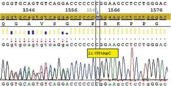
parathy-roidectomy with the implantation of a fragment of one gland in the forearm would be indicated. On the other hand, if the diagnosis of MEN was not conirmed, adenomectomy would probably be the best choice (11,12). At this time, mutational analy-sis was performed by sequencing the entire coding region of the MEN 1 gene (Genetika Laboratory, Curitiba, Brazil). A frameshift c1561dupC muta-tion at the exon 10 of the MEN 1 gene was found, predicting a truncated form of menin which was identiied (Figure 1). On July, 2009, with the diag-nosis of sporadic MEN 1, the patient was submitted to total parathyroidectomy with immediate implan-tation of a small fragment of one parathyroid gland in the nondominant forearm. Pathology conirmed the presence of a parathyroid adenoma, while the remaining three parathyroid glands were histologi-cally normal, with no evidence of hyperplasia or neoplasia (Figure 2). One year after surgery, she has maintained normal calciuria and serum levels of calcium (last result = 9.5 mg/dL or 2.46 mmol/L) and PTH (last result = 56.6 ng/L) within the nor-mal range on treatment with 1.500 mg of calcium per day and 10.000 IU of vitamin D per week. The only complication encountered during follow-up was the cardiologic diagnosis of Wolff-Parkinson-White syndrome, for which a catheter ablation was performed. Thus far, there has been no clinical, bio-chemical or radiologic evidence of any enteropan-creatic neuroendocrine tumor.
Cop
yright
© ABE&M t
odos os dir
eit
os r
eser
vados
.
radic tumor combinations, such as parathyroid and somatotroph or corticotroph, as in this reported young female patient, may have unexpectedly low frequency of MEN 1 mutation (14). The reasons for that include two kinds of tumors with high natural incidence in older subjects developed by chance, an-other familial tumor syndrome with low penetrance, as previously observed in familial acromegaly with primary hyperparathyroidism by mutation of another gene, or a somatic mutation during early embryonic stages (14). Taken together, the data demonstrate that despite the high clinical suspicion of MEN 1 in our patient, the coincident presentation of Cush-ing’s disease and primary hyperparathyroidism could not be completely ruled out, especially with a para-thyroid scintigraphy showing a pattern compatible with only one hyperfunctioning gland.
The likelihood of MEN 1 mutation is higher with younger onset age for the tumor or with tumor multiplicity in that organ. The frequency of MEN 1 germline mutation with a tumor, presumed to be
sporadic based on family evaluations, is speculated as follows: parathyroid adenoma (1%), gastrinoma (5%), prolactinoma (1%), foregut carcinoid (2%), lipoma (0.1%), and angioibroma (1%) (1). These estimates show the importance of establishing the genetic diagnosis of our patient before the decision for the surgical approach for her primary HPT: a negative test would indicate more conservative man-agement with resection of the parathyroid adenoma, while the positive test for MEN 1 would favor a complete removal of the parathyroid glands (11,12). The latter approach was chosen with the inding of a c1561dupC mutation at the exon 10 of the MEN 1 gene in our patient. In this heterozygous frameshift mutation, an additional cytosine occurs at position 1561, resulting in a stop codon. Lemos and Thakker (15) reported on 1,336 mutations described in the irst decade following the identiication of the MEN 1 gene, which are scattered in and around its open reading frame. MEN 1 mutations include nonsense, missense, donor-splice, small deletions and small in-sertions, and more than 70% of them are predicted to generate truncated forms of menin (1,2,15).
From a practical point of view, MEN 1 is deined in a patient with two of the three main MEN 1-re-lated endocrine tumors (parathyroid adenomas, en-tero-pancreatic endocrine tumors, and pituitary tu-mor). Familial MEN 1 is similarly deined as at least
Figure 2. Histopathology slide demonstrating parathyroid gland with sheet-like arrangement of monotonous parathyroid cells (hematoxylin and eosin 20X).
DISCUSSION
Early identiication of the manifestations, improve-ments in diagnostic techniques and therapeutic ap-proaches has had a favorable impact on the morbidi-ty and mortalimorbidi-ty associated with MEN syndromes. In the classical MEN 2A – which is clinically character-ized by the presence of medullary thyroid carcinoma (MTC), bilateral pheochromocytoma and primary HPT; in MEN 2B, where MTC is associated with pheochromocytoma and mucosal neuroma; and in familial MTC, genetic testing detects nearly 100% of RET proto-oncogene mutation carriers and is now considered the standard of care for all irst degree relatives of patients with newly diagnosed MTC. Each variant of MEN 2 results from a different RET mutation, with a good genotype-phenotype corre-lation, allowing the design of good guidelines for timing of prophylactic thyroidectomy and extent of surgery based on risk levels (13).
spo-Cop
yright
© ABE&M t
odos os dir
eit
os r
eser
vados
.
one MEN 1 case plus at least 1 irst degree relative with one of the three main MEN 1-related endo-crine tumors (1). However, the expression of one or more of the less common tumors of MEN 1 can occur by chance, unlike syndromic variants that oc-cur repeatedly in MEN 2, especially in sporadic cases (13). This was the situation of our patient, whose initial manifestation was Cushing’s Disease at 20 years of age. Anterior pituitary adenoma is the irst clinical inding of MEN 1 in up to 25% of sporadic cases, but only less than 5% of these tumors are cor-ticotropinomas (1,2,6). In fact, Cushing’s disease is a rare disease even in the general population, with an incidence of 5-10 cases per million, per year. In 1993, Gaitan and cols. (16) reported on a similar case of a woman with Cushing’s disease and primary HPT, who had a daughter with Cushing’s disease and another daughter and two other relatives with primary HPT. In contrast, our patient had a negative family history for any tumor related to MEN. Pro-lactinomas represent 25% of the pituitary adenomas in MEN 1, followed by nonfunctioning adenomas in 10% and somatotropinomas in 5% of the cases (1,2,6). As corticotropinomas are very rare, sched-ules to screen for tumor expression in the carriers of MEN 1 mutation include annual determination of prolactin and IGF-1 levels and an MRI every 3-5 years, beginning at 5 years of age, but they do not usually recommend for a biochemical evaluation of Cushing’s syndrome (1).
The diagnosis of primary HPT in our case was initially suspected by the detection of asymptomatic hypercalcemia. This emphasizes the importance of measuring serum calcium in patients with pituitary tumors, especially those at younger ages and who have prolactinomas, as a simple test for detection of MEN 1 (7). For carriers of MEN 1 mutation, deter-mination of serum calcium and PTH is advised to start at 8 years of age (1). Primary HPT is the most common endocrinopathyin MEN 1, reaching nearly 100% of penetrance by the age of 50 years (1-3). In contrast, MEN 1 representsless than 5% of all cases of primary HPT (10). In MEN 1, HPT is most frequently asymptomatic, but when symptoms are present, they are similar to those observed in other forms of HPT (1-3). Compared to sporadic parathyroid adenomas, HPT in MEN 1 starts at an earlier age (25 versus 55 years), lack gender preference (1:1 versus 3:1 female/ male ratio), and differs in pathology, with the presence
of multiglandular disease, which justiies a most aggres-sive surgical approach (1-3,11-12). However, despite the diagnosis of MEN 1, the pathologic study in our case did not reveal a multiglandular disease. It is believed that the development of multiglandular parathyroid disease in MEN 1 is a question of time, and the age of our patient may explain the absence of hyperplasia in the other three parathyroid glands (17). Our patient presented no speciic symptoms of HPT, but subsequent investigation demonstrated bi-lateral nephrolithiasis and low bone mineral density at the lumbar spine and femoral neck. In Brazil, a re-cent study of 36 cases from 8 unrelated MEN 1 families with uncontrolled HPT found that bone mineral and urolithiasis-related renal complications in this popula-tion was early-onset, frequent, extensive, severe and progressive, which are in agreement with our indings (18). Moreover, the hypercortisolism in our patient was also a contributor to the impairment of her bone mass.
Our patient has no clinical, biochemical or radiolog-ical evidence of any enteropancreatic neuroendocrine tumor so far. The prevalence of these tumors in MEN 1-affected individuals varies in different clinical series from 30%-75% and approaches 80% in necropsy series (1-3). Biochemical screening for these tumors includes fasting glucose, gastrin, insulin, proinsulin, glucagon, and chromogranin A. In suspected cases, additional tests and more detailed investigation may be indicated. Search for insulinoma in carriers is recommended to start at 5 years of age, whereas for gastrinoma, foregut carcinoid and other enteropancreatic tumors the age to begin testing is 20 years of age, with annual evaluation of the biochemical screening (1).
hy-Cop
yright
© ABE&M t
odos os dir
eit
os r
eser
vados
.
pertrophy as well as alterations in the properties of cardiac ion channels, such as voltage-gated sodium channel (19,20). To our knowledge, there have been no reports on this cardiac abnormality in pa-tients with MEN 1, indicating the fortuitous nature of such association.
In summary, this report illustrates the impor-tance of genetic testing in a case of sporadic MEN 1 to deine the best therapeutic approach for HPT and to optimize patient follow-up.
Disclosure: no potential conlict of interest relevant to this ar-ticle was reported.
REFERENCES
1. Brandi ML, Gagel RF, Angeli A, Bilezikian JP, Beck-Peccoz P, Bordi C, et al. Guidelines for diagnosis and therapy of MEN type 1 and type 2. J Clin Endocrinol Metab. 2001;86:5658-71.
2. Hoff AO, Hauache OM. Multiple endocrine neoplasia type 1 (MEN 1): clinical, biochemical and molecular diagnosis and treatment of the associated disturbances. Arq Bras Endocrinol Metab. 2005;49:735-46.
3. Burgess J. How should the patient with multiple endocrine neoplasia type 1 (MEN 1) be followed? Clin Endocrinol (Oxf). 2010;72:13-6.
4. Chandrasekharappa SC, Guru SC, Manickam P, Olufemi SE, Collins FS, Emmert-Buck MR, et al. Positional cloning of the gene for mul-tiple endocrine neoplasia-type 1. Science. 1997;276(5311):404-7. 5. Hendy GN, Kaji H, Canaff L. Cellular functions of menin. Adv Exp
Med Biol. 2009;668:37-50.
6. Vergès B, Boureille F, Goudet P, Murat A, Beckers A, Sassolas G, et al. Pituitary disease in MEN type 1 (MEN1): data from the France-Belgium MEN1 multicenter study. J Clin Endocrinol Me-tab. 2002;87:457-65.
7. Corbetta S, Pizzocaro A, Peracchi M, Beck-Peccoz P, Faglia G, Spa-da A. Multiple endocrine neoplasia type 1 in patients with recog-nized pituitary tumours of different types. Clin Endocrinol (Oxf). 1997;47:507-12.
8. Rix M, Hertel NT, Nielsen FC, Jacobsen BB, Hoejberg AS, Brixen K, et al. Cushing’s disease in childhood as the first manifestation of multiple endocrine neoplasia syndrome type 1. Eur J Endocri-nol. 2004;151:709-15.
9. Alzahrani AS, Al-Khaldi N, Shi Y, Al-Rijjal RA, Zou M, Baitei EY, et al. Diagnosis by serendipity: Cushing syndrome attributable to cortisol-producing adrenal adenoma as the initial manifestation of multiple endocrine neoplasia type 1 due to a rare splicing site MEN1 gene mutation. Endocr Pract. 2008;14:595-602.
10. Skandarajah A, Barlier A, Morlet-Barlat N, Sebag F, Enjalbert A, Conte-Devolx B, et al. Should routine analysis of the MEN1 gene be performed in all patients with primary hyperparathyroidism under 40 years of age? World J Surg. 2010;34:1294-8.
11. Lew JI, Solorzano CC. Surgical management of primary hyperpa-rathyroidism: state of the art. Surg Clin North Am. 2009;89:1205-25. 12. Waldmann J, López CL, Langer P, Rothmund M, Bartsch DK. Sur-gery for multiple endocrine neoplasia type 1-associated primary hyperparathyroidism. Br J Surg. 2010;97(10):1528-34.
13. Raue F, Frank-Raue K. Genotype-phenotype relationship in multi-ple endocrine neoplasia type 2. Implications for clinical manage-ment. Hormones (Athens). 2009;8:23-8.
14. Hai N, Aoki N, Shimatsu A, Mori T, Kosugi S. Clinical features of multiple endocrine neoplasia type 1 (MEN1) phenocopy without germline MEN1 gene mutations: analysis of 20 Japanese spora-dic cases without MEN1. Clin Endocrinol (Oxf). 2000;52:509-18. 15. Lemos MC, Thakker RV. Multiple endocrine neoplasia type 1
(MEN1): analysis of 1336 mutations reported in the first decade following identification of the gene. Hum Mutat. 2008;29:22-32. 16. Gaitan D, Loosen PT, Orth DN. Two patients with Cushing’s
dise-ase in a kindred with multiple endocrine neoplasia type 1. J Clin Endocrinol Metab. 1993;76:1580-2.
17. Doherty GM, Lairmore TC, DeBenedetti MK. Multiple endocrine neoplasia type 1 parathyroid adenoma development over time. World J Surg. 2004;28:1139-42.
18. Lourenço DM Jr, Coutinho FL, Toledo RA, Montenegro FL, Cor-reia-Deur JE, Toledo SP. Early-onset, progressive, frequent, exten-sive and severe bone mineral and renal complications in multiple endocrine neoplasia type 1-associated primary hyperparathyroi-dism. J Bone Miner Res. 2010;25(11):2382-91.
19. Light PE. Familial Wolff-Parkinson-White Syndrome: a disease of glycogen storage or ion channel dysfunction? J Cardiovasc Elec-trophysiol. 2006;17(Suppl 1):S158-61.