CASE REPORT
A rare benign ovarian tumour
Marta Morna Palmeiro, Teresa Margarida Cunha, Ana Luisa Loureiro, Gonçalo Esteves
Department of Radiology, Instituto Português de Oncologia de Lisboa Francisco Gentil, Lisbon, Portugal
Correspondence to Dr Marta Morna Palmeiro, palmeiro.marta@gmail.com
Accepted 16 February 2016
To cite:Morna Palmeiro M, Cunha TM, Loureiro AL,
et al.BMJ Case Rep
Published online: [please includeDay Month Year] doi:10.1136/bcr-2015-214101
SUMMARY
Sclerosing stromal tumour (SST) of the ovary is an extremely rare and benign ovarian neoplasm, accounting for 6% of the sex cord stromal ovarian tumours subtype. Usually, it is found during the second and third decades of life. Patients commonly present with pelvic pain, a palpable pelvic mass or menstrual irregularity. We report a case of a 20-year-old woman reporting of mild pelvic pain, with normal laboratory data. On imaging examinations, a large right adnexal tumour was found, with features suggesting an ovarian sex cord tumour. The patient underwent right salpingo-oophorectomy, diagnosing a SST of the ovary. This paper also reviews the literature, and emphasises the typical pathological and imaging characteristics of these rare benign ovarian lesions, and their impact, in a conservative surgery.
BACKGROUND
Sclerosing stromal tumours (SSTs) of the ovary are rare benign neoplasms, corresponding to 6% of all sex cord stromal tumours.1 2The majority of these neoplasms affect patients in the second and third decades of life.2
SSTs show unique pathological and radiological features that distinguish them from other ovarian stromal tumours.3The presentation is unspecific in
most patients and frequently related to an adnexal mass.4
These typical characteristics of SSTs are essential to enhance the benign nature of these neoplasms and to strength the possibility of a conservative surgery with fertility sparing.
CASE PRESENTATION
A 20-year-old woman, recently married, presented to us with a 6-week history of intermittent mild pelvic pain. There was no relevant previous medical history.
On initial examination, the gynaecological evalu-ation was normal, but on digital rectal examinevalu-ation, an anterior and partially mobile mass was felt, bulging the anterior rectal wall.
The patient was initially investigated in an out-patient clinic with ultrasound, which detected a solid lesion with a long axis of 80 mm in the right adnexal region, but it was not possible to determin-ate if the origin was adnexal or exophytic uterine.
INVESTIGATIONS
Laboratory findings, including tumour markers, such as CA-125 and α-fetoprotein, and human
chorionic gonadotropin, were normal.
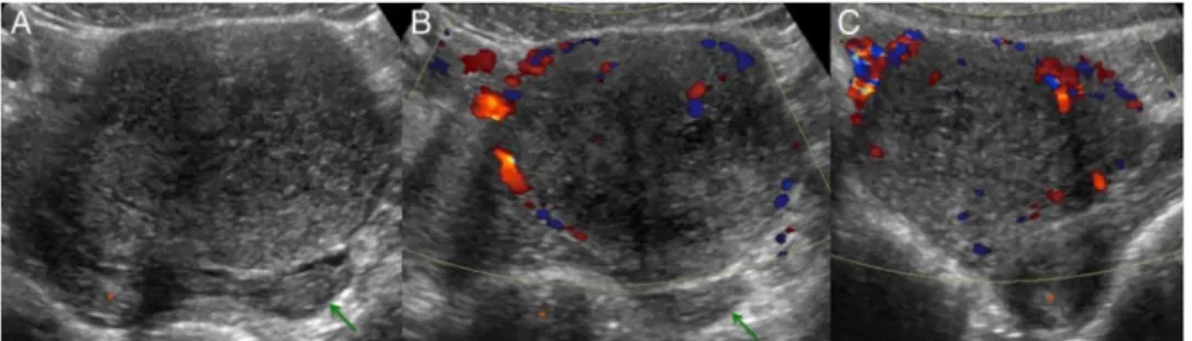
A new gynaecological ultrasound was carried out with transabdominal and transvaginal evaluation, identifying a solid hypoechoic tumour with regular borders, measuring 78 mm of major axis, probably in the dependence of the right ovary (figure 1A). Colour Doppler examination revealed the presence of multiple vessels inside the lesion, more numer-ous at the periphery and extending to its central portion (figure 1B, C). The uterus and the left ovary were normal. A small amount of ascites was present.
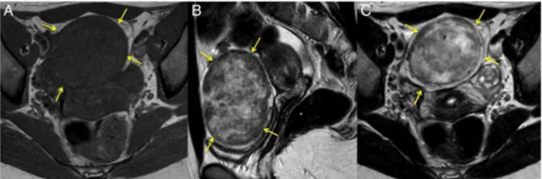
To further characterise this lesion, MR was per-formed, revealing an ovoid solid tumour associated with the right ovary, with regular contours, measur-ing 80 mm of major axis. On T1-weighted images, this lesion was homogeneous, with low signal intensity (arrows) (figure 2A). On T2-weighted images, this mass was heterogeneous, with predom-inant areas of hypointensity and small areas with hyperintensity (figure 2B,C). On T1-weighted and T2-weighted images, the lesion revealed a thin, low-intensity rim surrounding the mass (figure 2A–C). Dynamic contrast-enhanced MRI demonstrated an avid early enhancement with pseudolobular pattern, starting on the peripheral portion of the
Figure 1 (A) Transabdominal ultrasound shows a solid hypoechoic tumour with regular borders, measuring 78×55×70 mm (159 cc), in the dependence of the right ovary. (B) Transabdominal and (C) transvaginal colour Doppler examination revealed the presence of multiple vessels inside the tumour, more numerous on the periphery and extending to the central portion. The uterus (*) and left ovary (arrow) were normal. A small amount of ascites was present.
lesion (figure 3A, B). Over time, the lesion showed a progressive centripetal and prolonged enhancement, except in small-scattered areas of cyst and cleft (figure 3C, D). The lesion did not show restricted diffusion (figure 4). On the Adnex MR Scoring System, this lesion was classified with a score of 2, with features suggesting a sex cord stromal tumour type, most likely a SST. The uterus and the left ovary were normal. A small amount of pelvic ascites was found (figure 2B).
DIFFERENTIAL DIAGNOSIS
The differential diagnosis of SSTs includes other sex cord stromal tumours including fibromas, thecomas, steroid cell tumours (lipid cell tumours), metastases and malignant epithelial ovarian tumours. Fibromas and thecomas usually show low signal intensity on T2-weighted imaging, and slow and pro-longed enhancement on dynamic contrast MRI. Lipoid cell tumours commonly manifest as a small mass with hyperintense areas on T1-weighted images due to abundant intracellular lipid. Ovarian metastases and malignant epithelial tumours usually occur in older patients, and on dynamic enhanced MRI these lesions commonly show restricted diffusion and display more aggressive behaviour.
TREATMENT
The patient underwent right salpingo-oophorectomy with intraoperative consultation.
OUTCOME AND FOLLOW-UP
Macroscopic examination of the surgical specimen described the complete substitution of the right ovary by a capsulated tumour, with firm consistency, presenting yellowish nodular peripheral components and an oedematous central portion, with 76 mm of longest axis (figure 5). The right fallopian tube was normal.
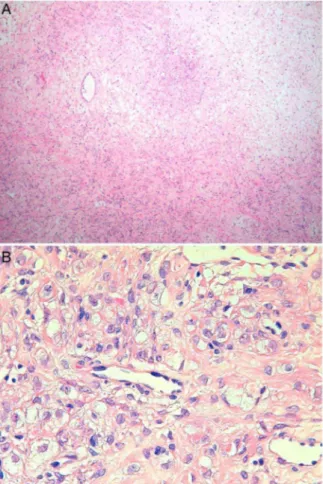
Microscopic examination revealed a pseudolobular pattern in which cellular nodules were separated by areas of densely col-lagenous or oedematous connective tissue that contained fewer cells (figure 6A). The cellular nodules were an admixture of collagen-producing spindle cells and lipid-containing round or ovoid cells without atypia and with prominent thin-walled vessels (figure 6B).
The asciticfluid did not contain malignant cells.
Thefinal pathological diagnosis was SST of the right ovary. The patient recovered from surgery without complications and stayed asymptomatic after 10 months of follow-up.
DISCUSSION
Eight percent of ovarian neoplasms are due to ovarian sex cord stromal tumours. These tumours are classified as granulosa stromal cell tumours, Sertoli’s stromal tumours, steroid cell tumours (lipid cell tumours) and other types. Ovarian granulosa stromal cell tumours are subdivided as granulosa cell tumours, thecomas, fibromas and SSTs.3 5 SSTs represent only 6% of ovarian sex cord stromal tumours.1 2
SSTs are a particularly benign subtype of ovarian sex cord stromal tumour, described by Chalvardjian and Scully in 1973,6 with specific and individual pathological and radio-logical features that distinguish them from other stromal tumours.3
Around 80% of SSTs affect young women in the second and third decades of life, unlike the other types of stromal tumours, which are more common in thefifth and sixth decades.2 6
Usually, SSTs present as pelvic pain, menstrual irregularity and non-specific symptoms associated with the ovarian mass. Although these tumours are usually hormonally inactive, they may have oestrogenic and, infrequently, androgenic effects.4 7 They may, rarely, present with ascites.8
Figure 2 MRIs revealed an ovoid solid tumour associated with the right ovary, with regular contours, measuring 8 cm of major axis. (A) On axial T1-weighted MRIs, the tumour was homogeneous hypointense (arrows). On (B) sagittal and (C) axial T2-weighted MRIs, this mass was
heterogeneous with predominant areas of hypointensity and small areas with hyperintensity (arrows). On both, T1-weighted and T2-weighted images, the lesion revealed a thin, low-intensity rim surrounding the mass. A small amount of pelvic ascites was found (*). The uterus and left ovary were normal.
Figure 3 Axial dynamic contrast-enhanced MRIs demonstrated avid early enhancement with pseudolobular pattern, starting on the peripheral portion of the lesion (A and B). Over time, the tumour showed a progressive centripetal and prolonged enhancement, except in small scattered areas of the cysts and clefts (C and D).
2 Morna Palmeiro M,et al.BMJ Case Rep2016. doi:10.1136/bcr-2015-214101
In the revised literature, the majority of SSTs were unilateral lesions, and all were benign.9
Histologically, SSTs were described by Chalvardjian and Scully as lesions with a characteristic pseudolobular pattern, constituted by heterogeneous cellular nodules composed of an admixture of collagen-producing spindle cells and lipid-containing round or ovoid cells, without atypia, showing intra-nodular collagenous sclerosis—‘sclerosing stromal tumour’. Oedematous and collagenous hypocellular areas andfibrosis are found between these cellular nodules. SSTs also demonstrate a typical exuberant vascularisation.1 3 10
On ultrasound, SSTs appear as large round masses, predomin-antly solid, with small central hypoechoic areas, in the affected adnexal region. Doppler evaluation shows numerous vessels, with peripheral predominance directing to the lesion’s inner portion, assuming a‘spoke-wheel’aspect.5
On MRI, SSTs appear as large and well-circumscribed solid heterogeneous masses. On T2-weighted images, these tumours are heterogeneous, showing predominant isointense to hypoin-tense solid components, and small hyperinhypoin-tense cystic areas. On T1-weighted images, they appear homogeneously hypointense. On T2-weighted and T1-weighted images, a thin hypointense peripheral line is seen, which represents the stretched ovarian cortex. On dynamic contrast-enhanced images, these masses demonstrate an early and avid peripheral enhancement with centripetal distribution over time, reflecting the peripheral rich cellularity and vascularity, and the inner collagenous hypocellu-larity typical of these lesions.3 8 11 12
Since SSTs of the ovary are rare benign tumours that occur mainly in young women, it is essential to be aware of their char-acteristic imaging features in order to perform a conservative surgery with fertility sparing.
Very few studies have been published about long-term follow-up and fertility outcomes of postsurgery SSTs. Those studies that are available emphasise the benign nature of these neoplasms, the impact on a fertility sparing surgery and the absence of reported SST recurrences.13–15
Learning points
▸ Sclerosing stromal tumours (SSTs) of the ovary are benign
and extremely rare ovarian neoplasms.
▸ The majority of SSTs occur in the second and third decades
of life, with unspecific symptoms.
▸ SSTs have distinctive histological features that reflect on
characteristic imagingfindings.
▸ The familiarity with typical imaging presentation of SSTs
allows the accurate preoperative diagnosis of these benign tumours and enables a less invasive surgery with fertility preservation.
Figure 4 On diffusion study, the lesion showed hypointensity on high bvalues (b=1000) (A), and hyperintensity on ADC map (B),
demonstrating no restricted diffusion.
Figure 5 Macroscopic examination of the surgical specimen described a capsulated tumour, withfirm consistency, presenting yellowish nodular peripheral components and an oedematous central part, 76 mm on its largest dimension.
Figure 6 (A) Microscopic examination (H&E ×10) revealed a pseudolobular pattern in which cellular nodules were separated by areas of densely collagenous or oedematous connective tissue that contained fewer cells. (B) On microscopic examination (H&E ×40), the cellular nodules were an admixture of collagen-producing spindle cells and lipid-containing round or ovoid cells without atypia, and with prominent thin-walled vessels.
Morna Palmeiro M,et al.BMJ Case Rep2016. doi:10.1136/bcr-2015-214101 3
Acknowledgements The authors wish to thank Dr Joaninha Costa Rosa, an experienced pathologist at our institution, who contributed by performing the pathological diagnosis of this rare benign ovarian tumour.
Contributors All the authors contributed to this case report. MMP carried out the bibliographic research and wrote the case report text. TMC was responsible for the patient examinations and revised the case report. All the authors chose and processed the case report images, and revised the case report. GE performed the pathological analysis and diagnosis of the tumour, and provided the corresponding histological images and subtitles.
Competing interests None declared. Patient consent Obtained.
Provenance and peer reviewNot commissioned; externally peer reviewed.
REFERENCES
1 Matsubayashi R, Matsuo Y, Doi J,et al. Sclerosing stromal tumor of the ovary: radiologicfindings.Eur Radiol1999;9:1335–8.
2 Kaygusuz EI, Cesur S, Cetiner H,et al. Sclerosing stromal tumour in young women: clinicopathologic and immunohistochemical spectrum.J Clin Diagn Res
2013;7:1932–5.
3 Kim JY, Jung KJ, Chung DS,et al. Sclerosing stromal tumor of the ovary: MR-pathologic correlation in three cases.Korean J Radiol2003;4:194–9. 4 Iravanloo G, Nozarian Z, Sarrafpour B,et al. Sclerosing stromal tumor of the ovary.
Arch Iranian Med2008;11:561–2.
5 Lee MS, Cho HC, Lee YH,et al. Ovarian sclerosing stromal tumors: gray scale and color Doppler sonographicfinding.J Ultrasound Med2001;20:413–17. 6 Khanna M, Khanna A, Manjari M. Sclerosing stromal tumor of ovary: a case report.
Case Rep Pathol2012;2012:592836.
7 Marwah N, Sankala M, Sansanwal P,et al. Sclerosing stromal tumor of the ovary: a case report.Int J Healthc Biomed Res2015;3:91–4.
8 Jung SE, Rha SE, Lee JM,et al. CT and MRIfindings of sex cord-stromal tumor of the ovary.AJR Am J Roentgenol2005;185:207–15.
9 Akbulut M, Colakoglu N, Soysal ME,et al. Sclerosing stromal tumor of the ovary: report of a case and review of the literature.Aegean Pathol J
2004;1:84–9.
10 Peng HH, Chang TC, Hsueh S. Sclerosing stromal tumor of ovary.Chang Gung Med J2003;26:444–8.
11 Jung SE, Lee JM, Rha SE,et al. CT and MR imaging of ovarian tumors with emphasis on differential diagnosis.Radiographics2002;22:1305–25. 12 Loureiro A, Cunha TM, Félix A. Sclerosing stromal tumor of the ovary. EURORAD
2013; case 10870.
13 Haroon S, Zia A, Idrees R,et al. Clinicopathological spectrum of ovarian sex cord-stromal tumors; 20 years’retrospective study in a developing country.
J Ovarian Res2013;6:87.
14 Grechi G, Clemente N, Tozzi A,et al. Laparoscopic treatment of sclerosing stromal tumor of the ovary in a woman with Gorlin-Goltz syndrome: a case report and review of the literature.J Minim Invasive Gynecol2015;22:892–5.
15 Abd A, Hafez E. Sclerosing stromal tumor of the ovary: a rare entity with distinctive features.Case Rep Clin Pathol2014;1:5–7.
Copyright 2016 BMJ Publishing Group. All rights reserved. For permission to reuse any of this content visit http://group.bmj.com/group/rights-licensing/permissions.
BMJ Case Report Fellows may re-use this article for personal use and teaching without any further permission.
Become a Fellow of BMJ Case Reports today and you can: ▸ Submit as many cases as you like
▸ Enjoy fast sympathetic peer review and rapid publication of accepted articles ▸ Access all the published articles
▸ Re-use any of the published material for personal use and teaching without further permission
For information on Institutional Fellowships contact consortiasales@bmjgroup.com
Visit casereports.bmj.com for more articles like this and to become a Fellow
4 Morna Palmeiro M,et al.BMJ Case Rep2016. doi:10.1136/bcr-2015-214101