Cop
yright
© ABE&M t
odos os dir
eit
os r
eser
vados
.
Ovarian intratumoral
21-hydroxylase deiciency in a
postmenopausal hirsute woman
Deiciência de 21-hidroxilase ovariana intratumoral na virilização da mulher pós-menopausa
Selma B. Souto1,2, Pedro V. Baptista3, Filomena Barreto4,
Pedro F. Sousa5, Daniel C. Braga1,2, Davide Carvalho1,2
SUMMARY
Virilising ovarian tumours are a rare cause of hyperandrogenism in women, accounting for less than 5% of all ovarian neoplasms. It occurs most often in – and postmenopausal women. We report a case of a 64 year-old woman with signs of virilisation that had started 3 years before. Blood hormone analysis revealed increased levels of testosterone, and 17-hydroxyprogestero-ne. The tetracosactin test revealed 21-hydroxylase deiciency. Radiological imaging demonstra-ted a nodule in her left ovary. The patient was submitdemonstra-ted to bilateral laparoscopic oophorectomy, and histopathological examination revealed a luteoma of the left ovary. Postoperative serum testosterone level and 17-hydroxyprogesterone returned to normal levels in one month. Virilism regressed within six months. Our patient also showed an elevation in 17-OHP serum levels. Normalization of 17-OHP after oophorectomy suggests a case of intratumoral 21-hydroxylase deiciency. To our knowledge, this is the irst description of ovarian intratumoral 21-hydroxylase deiciency in a postmenopausal woman. Arq Bras Endocrinol Metab. 2012;56(9):672-6
SUMÁRIO
Tumores ovarianos virilizantes são uma causa rara de hiperandrogenismo em mulheres, con-tabilizando menos de 5% de todos as neoplasias ovarianas. Esses tumores ocorrem mais co-mumente em mulheres em peri ou pós-menopausa. Relatamos aqui o caso de uma mulher de 64 anos de idade com sintomas de virilização que começaram 3 anos antes. O peril hormonal revelou níveis aumentados de testosterona e de 17-hidroxiprogesterona (17-OHP). O teste de tetracosactin demonstrou deiciência de 21-hidroxilase. Exames radiológicos mostraram um nódulo no ovário esquerdo. A paciente foi submetida à ooforectomia laparoscópica bilateral e o exame histopatológico revelou um luteoma no ovário esquerdo. A concentração sérica de testosterona e de 17-hidroxiprogesterona após a cirurgia retornou aos níveis normais em um mês. A virilização regrediu em 6 meses. Nossa paciente também revelou uma elevação dos níveis séricos de 17-OHP. A normalização da 17-OHP após a ooforectomia sugere um caso de deiciência de 21-hidroxilase intratumoral. Esta é a primeira descrição de deiciência de 21-hi-droxilase intratumoral em uma mulher na pós-menopausa. Arq Bras Endocrinol Metab. 2012;56(9):672-6 1 Department of Endocrinology,
Diabetes and Metabolism, Centro Hospitalar São João, Porto, Portugal 2 University of Porto, School of Medicine, Porto, Portugal 3 Department of Gynaecology and Obstetrics, Centro Hospitalar São João, Porto, Portugal 4 Department of Pathology, Centro Hospitalar São João, Porto, Portugal 5 Department of Radiology, Centro Hospitalar São João, Porto, Portugal
Correspondence to:
Selma B. Souto
Department of Endocrinology, Diabetes and Metabolism, Centro Hospitalar São João, Porto, Portugal
School of Medicine, University of Porto, Portugal Alameda Prof. Hernâni Monteiro 4200-319 – Porto, Portugal [email protected]
Received on Jan/10/2012 Accepted on Mar/16/2012
INTRODUCTION
V
irilising ovarian tumours are a rare cause of hyper-androgenism in women, accounting for less than 5% of all ovarian neoplasms, and those that are malig-nant, for less than 10% of all ovarian cancers (1). It oc-curs most often in peri- and postmenopausal women (2). The spectrum of ovarian neoplasms covers an ex-tremely wide range of tumours. The best recognizedones are surface epithelial cell tumours. Among the less common variants, lipid or steroid cell tumours compri-se an important category, despite accounting for only 0.1% of all ovarian tumours (3).
tu-Cop
yright
© ABE&M t
odos os dir
eit
os r
eser
vados
.
mour occurs in postmenopausal women in 80% of ca-ses; estrogenic manifestations occur in 60% of patients, and only 12% of tumours are androgenic. Stromal lu-teoma accounts for approximately 20% of all ovarian steroid cell tumours, and is characterized by a location within the ovarian stroma and absence of Reinke’s crys-tals. Tumours are small, almost always < 3 cm in diame-ter, solid, circumscribed, and centred in the ovary pro-per. They are usually grayish white or yellow but may have red or brown areas. They are composed of large polygonal lutein-type cells that proliferate diffusely or are arranged in cords or nests. Mitoses are uncommon. Nodules composed entirely of lutein cells that are 1 cm or larger are arbitrarily classiied as stromal luteomas, whereas smaller nodules are considered to be stromal hyperthecosis.
In most cases, hormonal abnormalities found in patients with virilising ovarian tumours include increa-sed serum testosterone levels in the presence of nor-mal levels of serum dehydroepiandrosterone-sulfate (DHEA-S) (5).
This paper focuses on a case of a virilising ovarian tumour diagnosed in a 64-year-old postmenopausal woman. The patient was found to have a steroid-cell ovarian tumour, stromal luteoma type, which proved to be the aetiology of the virilisation in this patient.
CASE REPORT
A 64-year-old married woman, Jehovah’s witness, who complained of progressive hirsutism, was sent to the Endocrinology Department. She had a history of essen-tial hypertension, obesity, primary infertility, and obs-tructive sleep apnoea. She was medicated with lisinopril 5 mg/day and simvastatin 20 mg/day.
She reported hirsutism for three years, initially in-volving the face, and later her back and abdomen. In the six months before the appointment, she developed androgenetic alopecia, worsening of hirsutism, breast atrophy and deepening of the voice. She did not have vaginal bleeding or systemic symptoms. Familial clinical history was irrelevant.
Physical examination showed hirsutism of the face, back and abdominal area (score > 8, Ferriman--Gallwey scale). Gynaecological examination was nor-mal, without adnexal masses or enlargement of the clitoris. Blood pressure was 120/80 mmHg. She had no features of Cushing syndrome. Haemoglobin, hae-matocrit, fasting plasma glucose, serum electrolytes,
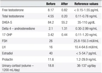
cholesterol, triglycerides, calcium, phosphorus, renal and liver function were normal. Endocrine evaluation revealed hyperandrogenism, with a marked increase in total serum testosterone concentration (4.55 ng/mL), while concentrations of DHEA-S and delta 4-androste-nedione were within the normal range (Table 1). She also had high serum levels of 17-hydroxyprogesterone (17OHP) (3.42 ng/mL). Serum prolactin and thyroid stimulating hormone (TSH) were within the normal range. Serum luteinizing hormone (LH), follicle sti-mulating hormone (FSH), and estradiol concentrations were within the normal range for menopause. Normal urinary cortisol levels ruled out subclinical hypercorti-solism. Tetracosactin test was performed, pointing out a deiciency in 21-hydroxylase (Table 2).
Transvaginal ultrasound suggested a vascularised node in the stroma of the left ovary. The other ovary was normal; endometrial stripe was linear and there was no luid in the peritoneal cavity. The pelvic and abdo-minal computerized axial tomography (CT) (Figure 1),
showed a solid nodule in the left ovary with 16 mm in diameter, and morpho-dimensional normality of the adrenal glands. No other pathological indings were detected, namely, ascites or lymph node enlargement. Magnetic resonance imaging (MRI) was also performed, conirming the left ovarian solid nodule (Figure 2).
Table 1. Laboratory evaluation before surgery, and two months after surgery
Before After Reference values
Free testosterone 8.17 0.82 < 0.15-1.55 pg/mL
Total testosterone 4.55 0.20 0.11-0.78 ng/mL
DHEA-S 84.2 55.2 35-110 μg/dL
Delta 4 – androstenedione 2.1 1.31 0.30-2.99 ng/mL
17-OHP 3.42 0.44 0.11-1.20 ng/mL
FSH 26 25.8-150.3 mUI/mL
LH 16 10.4-64.6 mUI/mL
Estradiol 40 < 5-54.7 pg/mL
Prolactin 11.6 1.2-29.9 ng/mL
Urinary cortisol (volume – 1200 mL/day)
18.8 36-137 ug/day
DHEA-S: dehydroepiandrosterone sulfate; 17-OHP: 17-hydroxyprogesterone; FSH: follicle stimulating hormone; LH: luteinizing hormone.
Table 2. Tetracosactin test
0’ 60’
17-OHP (ng/mL) 10.7 12.5
Cortisol (µg/dL) 14.3 35.2
Cop
yright
© ABE&M t
odos os dir
eit
os r
eser
vados
.
A diagnostic laparoscopy was proposed. The uterus was normal, as well as the right ovary. The left ovary was slightly enlarged. There were no adhesions, ascites, or any evidence of disseminated disease. Bilateral lapa-roscopic oophorectomy was performed, as previously discussed with the patient.
The histopathologic examination showed, in the left ovary, a tumour composed of large polygonal lutein-type cells that proliferated diffusely or were arranged in cords or nests. The stroma was usually sparse, sometimes hya-linized; mitoses were uncommon. It was also possible to see corpus albicans in the remaining ovarian tissue. The diagnosis was determined as stromal luteoma. Peritoneal washing and diaphragmatic cytology were negative.
There were no postoperative complications. Two months after surgery, androgen levels had normalized, including 17-OHP (Table 1). The patient maintained hypertension after surgery. Six months later, hirsutism had improved signiicantly (score 3, Ferriman-Gallwey scale).
Figure 1. CT scan shows a 16-mm enhancing nodule on the left ovary.
Figure 2. (A) Axial T1-weighted MR image shows a well-deined, ovoid solid mass with low signal intensity. (B) Axial T2-weighted MR image shows the
mass with high signal intensity. (C, D) fat-suppressed T1-weighted image before and after gadolinium injection demonstrates the mass is well-enhanced.
A
C
B
D
Cop
yright
© ABE&M t
odos os dir
eit
os r
eser
vados
.
loss of the female body contour. This is followed by hirsutism, acne, clitoral enlargement, increased libido, sterility, enlargement of the larynx, deepening of the voice, and temporal alopecia (8). On the other hand, some of these tumours have little or no androgenic ac-tivity and may, in fact, have estrogenic effects (8).
Our patient had worsening hirsutism and developed signs of virilisation, such as androgenetic alopecia, breast atrophy and deepened voice, six months before diagnosis.
Although testosterone levels above 200 ng/dL raise the suspicion of an androgen-producing tumour, 20% of patients fail to reach this level (6). Detecting the source of an androgen-producing tumour is an exclu-sion process. DHEA-S levels above 700 ng/dL should raise the suspicion of an adrenal source.
In this case, high free and total serum testosterone with normal DHEA-S pointed out the presence of an ova-rian virilising neoplasm. Our patient also revealed an ele-vation of the 17-OHP serum levels, and the tetracosactin test conirmed a deiciency in 21-hydroxylase. A differen-ce was noted in basal 17-OHP level between the measure in the irst month and the one carried out 2 months later (initial - 3.42 ng/mL, 2 months later - 10.7 ng/mL).
The normalization of 17-OHP after oophorectomy suggests a case of intratumoral 21-hydroxylase deiciency. Biochemical evidence for partial 21-hydroxylase deiciency is a common inding in patients with an adrenal incidenta-loma, even in the absence of a congenital adrenal hyperpla-sia history (9). A case of a virilising tumour in a 6 year old girl with high levels of 17OHP that normalized after tu-mour resection was previously reported (9). To our know-ledge, this is the irst description of an ovarian intratumoral 21-hydroxylase deiciency in a postmenopausal woman.
The diagnosis of virilising tumours of the ovary is often dificult and challenging, especially when dealing with small tumours, not detectable by gynaecological examination. In a woman with signs of virilisation, it is essential to perform a careful gynaecological examina-tion, measure serum androgens, and perform pelvic and abdominal CT scan to exclude an ovarian or adrenal an-drogen-producing tumour. Nevertheless, transvaginal ultrasound is the most sensitive method for the detec-tion of ovarian tumours. However, the accuracy of the ultrasound examination is always dependent on the skill of the sonographer and the quality of the equipment.
Virilising ovary tumours are primarily treated surgi-cally and have a generally good prognosis.
Disclosure: no potential conlict of interest relevant to this article was reported.
Figure 3. Stromal luteoma and adjacent ovarian parenchyma.
Figure 4. The tumour is composed of uniform polygonal cells with eosinophilic cytoplasm. Reinke’s Crystals were not identiied.
DISCUSSION
Cop
yright
© ABE&M t
odos os dir
eit
os r
eser
vados
.
REFERENCES
1. Stegner HE, Loning T. [Endocrine-active tumors of the ovary]. Pa-thologe. 2003;24(4):314-22.
2. Takemori M, Nishimura R, Hasegawa K. Ovarian thecoma with ascites and high serum levels of CA125. Arch Gynecol Obstet. 2000;264(1):42-4.
3. Mehdi G, Ansari HA, Sherwani RK, Rahman K, Akhtar N. Ovarian steroid cell tumour: correlation of histopathology with clinicopa-thologic features. Patholog Res Int. 2011: p. 987895.
4. Roth LM, Czernobilsky B. Perspectives on pure ovarian stromal neoplasms and tumor-like proliferations of the ovarian stroma. Am J Surg Pathol. 2011;35(3):e15-33.
5. Shenker Y, Shenker Y, Malozowski SN, Ayers J, Grekin RJ, Ba-rkan AL. Steroid secretion by a virilizing lipoid cell ovarian tu-mor: origins of dehydroepiandrosterone sulfate. Obstet Gynecol. 1989;74(3 Pt 2):502-6.
6. David G, Garder DS, ed. Greenspan’s basic & clinical endocrino-logy. 8.ed., Mc Graw Hill. 2007. p. 534.
7. Wang PH, Chao HT, Lee RC, Lai CR, Lee WL, Kwok CF, et al. Steroid cell tumors of the ovary: clinical, ultrasonic, and MRI diagnosis--a case report. Eur J Radiol. 1998;26(3):269-73.
8. Salim S, Shantha GPS, Patel AD, Kumar AA, Ganeshram P, Mehra N, et al. Virilizing ovarian steroid cell tumor in a 40 year old South Indian female: a case report. Cases J. 2009;2:7521.