Sex cord-stromal tumors of the ovary: a comprehensive review and
update for radiologists
Mariana Horta
Teresa Margarida Cunha
O
varian sex cord-stromal tumors are uncommon neoplasms that represent approxi-mately 7% of all ovarian tumors (1). These tumors comprise a heterogeneous group and are formed by diverse cell types that arise from the primitive sex cords or stro-mal cells (1, 2). The strostro-mal cells include theca cells, ibroblasts, and Leydig cells whereas the gonadal primitive sex cords include granulosa cells and Sertoli cells (3). These cell types may be present separately or admixed and display diferent degrees of diferentiation (4).As some of the constituent cells of these types of tumors are engaged in ovarian steroid hormone production (e.g., androgens, estrogens, and corticoids), sex cord-stromal tumors are commonly associated with various hormone-mediated syndromes and exhibit a wide spectrum of clinical features. Tumors formed from ovarian cells (e.g., granulosa cells and theca cells) are often hyperestrogenic, whereas those comprising testicular cell types (e.g., Sertoli and Leydig cells) are usually hyperandrogenic. However, many tumors are nonfunc-tioning, and those comprising female cells may produce androgens and vice-versa (4).
Tumors that induce hyperandrogenicity may present with virilization signs (e.g., hirsut-ism, acne, irregular menstrual periods, male-pattern baldness, loss of female fat distribution, and hoarse voice), whereas tumor subtypes associated with abnormal estrogen production may present with hyperestrogenicity (e.g., isosexual precocity in children, abnormal uterine bleeding, endometrial hyperplasia, and carcinoma).
The association of ovarian sex cord-stromal tumors with typical clinical syndromes is not the only characteristic distinguishing these tumors from the more common ovarian epithe-lial neoplasms. Although sex cord-stromal tumors afect patients throughout a wide age range, the majority tend to present in younger patients and as a low-grade disease (stage I) that usually follows a nonaggressive clinical course. Therefore, the primary treatment is surgical and the prognosis is generally favorable. Furthermore, ovarian sex cord-stromal tu-mors may exhibit characteristic radiologic features with which radiologists should become familiar. Conversely, recognition of the spectrum of the ultrasonography (US), computed tomography (CT), and magnetic resonance imaging (MRI) appearances as well as clinico-pathologic features of ovarian sex cord-stromal tumors may assist radiologists to narrow From the Department of Radiology (M.H. mariana.
[email protected]), Centro Hospitalar Lisboa Ocidental, Lisbon, Portugal; Institute of Anatomy (M.H.), Faculdade de Medicina da Universidade de Lisboa; the Department of Radiology (T.M.C.), Instituto Português de Oncologia de Lisboa Francisco Gentil, Lisbon, Portugal.
Received 14 September 2014; revision requested 25 November 2014; revision received 28 December 2014; accepted 19 January 2015.
Published online 4 June 2015. DOI 10.5152/dir.2015.34414
Diagn Interv Radiol 2015; 21: 277–286
© Turkish Society of Radiology 2015
A B D O M I N A L I MAG I N G
R E V I E W
ABSTRACT
the diferential diagnosis when facing ovar-ian tumors.
World Health Organization classiication of sex-cord stromal tumors
The World Health Organization (WHO) classiication of sex cord-stromal tumors has recently been revised in 2014 (5) (Ta-ble 1). In the current revision, these tu-mors were regrouped into the following clinicopathologic entities: pure stromal tu-mors, pure sex cord tutu-mors, and mixed sex cord-stromal tumors.
The “pure stromal tumors” category com-prises entities from the previous thecoma-i-broma group, which had been classiied un-der the granulosa-stromal cell tumor group division, as well as entities from the previous steroid cell group division (6) (Table 2).
In the recent classiication, luteinized thecoma is no longer considered a separate entity from the thecoma group, and there-fore this term should only be used when as-sociated with sclerosing peritonitis. Instead, a separate histopathologic entity titled “lu-teinized thecoma associated with scleros-ing peritonitis” now exists.
A distinct rare ovarian neoplasm, which was recently named “microcystic stromal tumor,” was added to this category whereas the denomination “stromal tumor with mi-nor sex cord elements” is not encompassed by this classiication.
Moreover, in terms of steroid cell tumors, the term “stromal luteoma,” which was used to designate small steroid tumors conined to the ovarian cortex, has also been discarded.
The subdivision of pure sex cord tumors now comprises tumors that were previous-ly categorized separateprevious-ly, including adult
granulosa cell tumors (GCTs), juvenile GCTs, Sertoli cell tumors, and sex cord tumors with annular tubules.
The diferent types with respect to Serto-li-Leydig cell tumor (SLCT) diferentiation as well as sex cord-stromal tumors, not other-wise speciied are categorized as “mixed sex cord-stromal tumors.” Stromal-Leydig cell tu-mor and gynandroblastomas are no longer considered in the current classiication.
Pure stromal tumors
Fibroma, cellular ibroma, and ibrosarcoma
Fibromas are almost always endocrine-in-ert tumors composed of spindle stromal cells that produce a collagenous stroma (5, 7, 8). These are undeniably the most com-mon sex cord-stromal tumors, representing 4% of all ovarian neoplasms (4, 5, 9) (Fig. 1). Fibromas can present at any age, although the mean age of occurrence is in the late forties, and are associated with nevoid
bas-al carcinoma syndrome (5). Fibromas range in size from small to large lesions. Small lesions are frequently asymptomatic, but women can present with pelvic discomfort or acute abdominal pain due to ovarian torsion as the size increases. Fibromas can mimic malignancy when present in the classic Meigs’ syndrome (hydrothorax, asci-tes, and benign ovarian tumor), which typ-ically disappears after tumor removal (3, 4, 10, 11). The tumor size is known to correlate with the presence of ascites.
Hypercellular ibromas are classiied as either cellular ibromas or ibrosarcomas. Cellular ibromas constitute 10% of all ovar-ian ibromas and have low malignant po-tential, exhibit mild nuclear atypia, and may present more than four mitotic igures per 10 high-powered ields (5). These generally exhibit the same clinical manifestations but tend to be larger than ibromas, thus poten-tially leading to necrosis and hemorrhage, particularly due to torsion (4, 5) (Fig. 2).
Table 1. WHO classiication scheme for ovarian sex cord-stromal tumors (2014) Pure stromal tumors
• Fibroma
• Cellular ibroma
• Thecoma
• Luteinized thecoma associated with sclerosing peritonitis
• Fibrosarcoma
• Sclerosing stromal tumor
• Signet-ring stromal tumor
• Microcystic stromal tumor
• Leydig cell tumor
• Steroid cell tumor
• Steroid cell tumor, malignant
Pure sex cord tumors
• Adult granulosa cell tumor
• Juvenile granulosa cell tumor
• Sertoli cell tumor
• Sex cord tumor with annular tubules
Mixed sex cord-stromal tumors
• Sertoli-Leydig cell tumors
- Well-diferentiated
- Moderately diferentiated with heterologous elements
- Poorly diferentiated with heterologous elements
- Retiform with heterologous elements
Main points
•
Ovarian sex cord-stromal tumors are infrequent tumors that difer from the more frequent epithelial neoplasms via strong associations with hormone-mediated syndromes, presentation in a broad age range, and the near-ubiquitous diagnosis of low-stage disease with a good outcome.•
The World Health Organization sex cord-stromal tumor classiication has recently been revised, and currently these tumors have been regrouped into the following clinicopathologic entities: pure stromal tumors, pure sex cord tumors, and mixed sex cord-stromal tumors.Ovarian ibrosarcomas are rare entities that follow a malignant clinical course and tend to exhibit moderate-to-severe nucle-ar atypia and mitotic igures (5, 12). These tumors usually present in postmenopausal
women as large unilateral masses, often with necrosis and hemorrhage (4, 5, 12).
Although diferent ovarian ibroma US patterns may be encountered, the US ap-pearance is usually a solid hypoechoic
at-tenuating ovarian mass (13). On CT, ibro-mas usually appear as homogeneous solid ovarian masses with delayed contrast en-hancement (3, 13) (Fig. 1a). Calciication may be present and widespread throughout the tumor (13). Given their predominant collag-enous and ibrous components, ibromas exhibit characteristic MRI features such as hypointensity on T1-weighted images and marked hypointensity on T2-weighted im-ages as well as weak and delayed enhance-ment after gadolinium administration (3) (Fig. 1b, 1c). Notwithstanding, edema and cystic degeneration may be encountered in ibromas; these appear as dispersed hy-perintense areas on T2-weighted images (8, 10, 13). These characteristics are most fre-quently observed in large lesions and ibro-sarcomas (8, 10, 13). Both predominantly cystic ibromas and ibromas with T1 and T2 hyperintensity/isointensity have also been described (9, 14).
Regarding the relationship between ovar-ian ibromas and the ovary, Oh et al.(9) re-ported that exophytic ibroma growth from the ovarian periphery without altering the normal form of the ipsilateral ovary is not un-common (46%) and that depicting the rest of the ipsilateral ovary in premenopausal wom-en is normal (83%). In addition, 67% of the 24 studied ibromas exhibited a border of T2 hypointensity or pseudocapsule around the outer ovarian margin that relected com-pression of the ovarian tissue (9).
The MRI-based diferential diagnosis in-cludes essentially ibrous ovarian tumors such as Brenner tumors and adenoibromas as well as pedunculated nondegenerative subserosal and broad ligament leiomyo-mas (15). Depiction of the tumor-feeding ovarian arteries or vascular signal voids between the uterus and leiomyoma might help to diferentiate these entities (16). Thomassin-Naggara et al. (17), in a retro-spective study that accessed the accuracy of dynamic MRI for diferentiating ovarian ibromas from uterine subserosal leiomy-omas, reported that the dynamic-contrast MRI enhancement of uterine leiomyomas was higher in terms of maximal enhance-ment and enhanceenhance-ment at 30, 60, and 90 seconds. However, no signiicant statistical diference existed in a delayed T1 post-con-trast sequence (17).
Fibroma may induce adnexal torsion. In such cases, hemorrhagic infarction or necro-sis may occur (3, 10). Hemorrhagic infarction may be diicult to deine because of the solid nature of the tumor. Nonetheless, an
Table 2. Former WHO classiication scheme for ovarian sex cord-stromal tumors (2003) Granulosa-stromal cell tumors
°
Granulosa cell tumor group • Adult granulosa cell tumor• Juvenile granulosa cell tumor
°
Thecoma-ibroma group • Thecoma, NOS- Typical
- Luteinized
• Fibroma
• Cellular ibroma
• Fibrosarcoma
• Stromal tumor with minor sex cord elements
• Sclerosing stromal tumor
• Signet-ring stromal tumor
• Unclassiied (ibrothecoma)
Sertoli-stromal cell tumors
°
Sertoli-Leydig cell tumors group (androblastoma) • Well-diferentiated• Of intermediate diferentiation
- variant with heterologous elements
• Poorly diferentiated (sarcomatoid)
- variant with heterologous elements
• Retiform
- variant with heterologous elements
°
Sertoli cell tumor°
Stromal-Leydig cell tumorSex cord-stromal tumors of mixed or unclassiied cell types
°
Sex cord tumor with annular tubules°
Gynandroblastoma°
Sex cord-stromal tumor, unclassiied Steroid cell tumors°
Stromal luteoma°
Leydig cell tumor group• Hilus cell tumor
• Leydig cell tumor, non-hilar type
• Leydig cell tumor, NOS
°
Steroid cell tumor, NOS • Well-diferentiated• Malignant
eccentric hyperintense area on T1-weighted images may allow this diagnosis (3).
Excepting a few cellular ibromas and i-brosarcomas, these types of tumors have typically benign courses and are curable via surgical excision (7).
Thecoma and luteinized thecoma associated with sclerosing peritonitis
Thecomas are composed of lipid-lad-en stromal cells that resemble theca cells, which usually encircle the ovari-an follicles, ovari-and exhibit estrogenic
ac-tivity in most cases (4, 7, 18) (Fig. 3). Thecomas account for 0.5%–1% of all pri-mary ovarian tumors (19). These tumors are more likely to occur in postmenopaus-al women and, with rare exceptions, are considered benign neoplasms (5). Afected
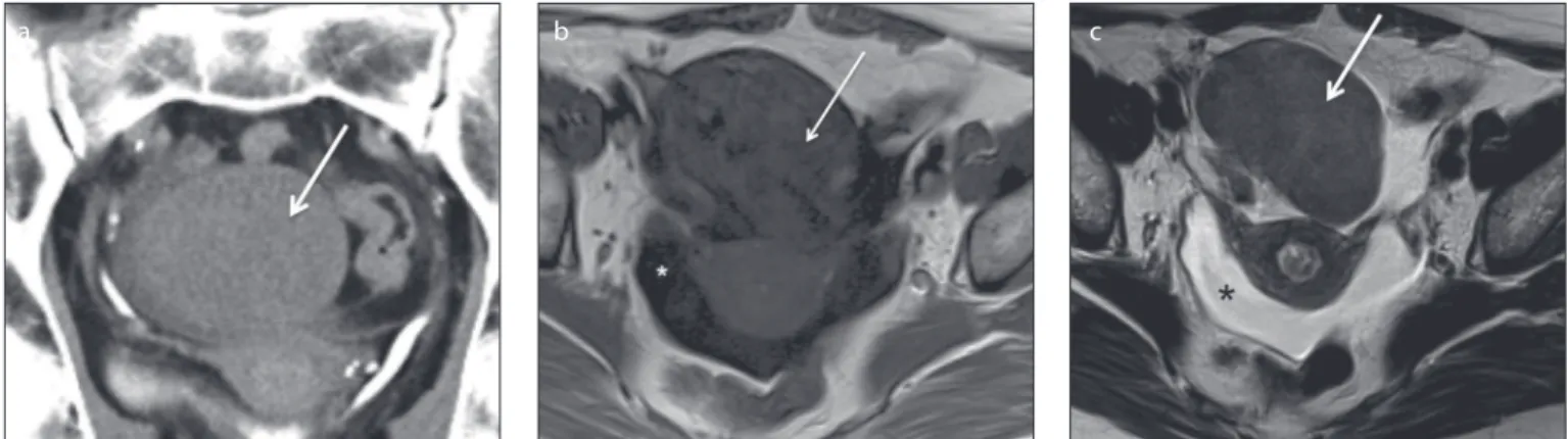
Figure 2. a–c. A 50-year-old female patient with a right ovarian cellular ibroma. Axial T1-weighted (a), T2-weighted (b) and gadolinium-enhanced fat-suppressed T1-weighted (c) images show a large, heterogeneous and well-deined tumor of the right ovary. The tumor features a large-centred cystic/necrotic area with T2 hyperintensity and T1 hypointensity (open arrows). The solid components exhibit an intermediate T2 signal and T1 isointensity to the muscle as well as avid contrast uptake (arrows).
a b c
a b c
Figure 1. a–c. A 77-year-old female patient with a right ovarian ibroma. Coronal delayed contrast-enhanced computed tomography image (a) shows a solid, well-deined, homogeneous right ovarian mass isodense to the uterus with very sparse contrast uptake (arrow). This ibroma is hypointense on both axial T1-weighted (b) and T2-weighted images (c). A small amount of ascites is observed in the pelvic recesses (asterisks).
women experience estrogen-related symp-toms such as uterine bleeding, endometrial hyperplasia, and endometrial carcinoma; the latter has been reported to occur in 21% of cases (4, 8) (Fig. 3).
Thecomas associated with androgen production may contain steroid-type cells (lutein cells) and were previously formally classiied as “luteinized thecomas” (5). This histopathologic classiication is no longer recommended and should only be used if there is an association with sclerosing peri-tonitis. However, luteinized thecoma associ-ated with sclerosing peritonitis is usually a bilateral hormonally inert tumor that occurs in younger women at an average age of 28 years (5). Thecomas that manifest in combi-nation with ibrous tissue may be classiied as ibrothecomas (5).
Generally, pure thecomas or thecomas with scanty ibrotic components do not have distinct US and CT appearances and mimic other solid ovarian tumors (13). A recent study by Zhang et al.(19) prospec-tively evaluated the MRI characteristics of 18 thecomas/ibrothecomas, namely the difusion-weighted imaging (DWI) features and apparent difusion coeicients (ADC) at 3.0 T, and correlated these with the fea-tures of other solid ovarian tumors and adnexal leiomyomas. The authors conclud-ed that most thecomas/ibrothecomas (61.1%) were homogenous masses and isointense to the myometrium on DWI-MRI and that the ADC values of thecomas/ ibrothecomas did not signiicantly difer from those of other ovarian solid tumors and leiomyomas (19). When compared with predominant ibrous tumors, pure thecomas tend to exhibit greater hyperin-tensity on T2-weighted images; more avid contrast-enhancement may be observed
because theca cells are greatly vascular-ized (13, 18) (Fig. 3).
Sclerosing stromal tumor
Sclerosing stromal tumor (SST) is a benign neoplasm that accounts for less than 5% of ovarian sex cord-stromal tumors (20) (Fig. 4). Unlike ibroma, thecoma, and adult GCTs, SSTs are more likely to occur in young wom-en; approximately 80% of reported cases are under 30 years of age (13, 21). Although SSTs most commonly occur after menarche, a few cases have been reported in premenarchal girls (22). Typically, SSTs manifest as unilateral masses. To our knowledge, only two cases of bilateral SSTs have been described: one in an 11-year-old premenarchal girl and the other in a pregnant woman with Gorlin syndrome after clomiphene therapy for infertility (21, 23).
Pelvic pain and menstrual irregularities are frequent symptoms (20, 24). A few hormon-ally active tumors that produced androgens and/or estrogens have been documented in the literature (18). SSTs of the ovary have also been associated with pregnancy (25). Although rare, ascites may be present (3).
Upon gross examination, SSTs are most-ly solid masses with yellowish foci, edema, and cystic areas (4). The histopathology is characterized by the presence of an ill-de-ined pseudolobular pattern in the cellular areas, which are separated by edematous ibrous areas. These nodular portions con-tain collagen-producing spindle-shaped cells and vacuolated lipid-containing lu-tein cells. Prominent vascularization is seen within these areas (4, 24). Both these typical features and the edema present in SSTs are thought to result from the expression of vascular permeability factor/vascular endo-thelial growth factor in lutein cells and its receptor fms-like tyrosine kinase 1 in small to middle-sized blood vessels (4, 24, 26).
The imaging indings of SSTs relect their characteristic histologic features. US com-monly reveals unilateral tumors with star-shaped hypoechoic areas enclosed by solid areas or solid tumors with medially located multiple small round or cleft-like hypoechoic areas (15, 24). Multilocular heterogeneous cystic masses and irregular septae have also been described (15). Color Doppler imaging reveals intratumoral blood vessels in the periphery and between the central cystic spaces. Low-resistance waveforms are visible on pulsed Doppler US (24). The described US features may mimic those of malignant ovarian tumors; therefore, further MRI-based radiologic evaluation is usually necessary.
On MRI, a heterogeneous solid tumor with iso- to hyperintensity is observed on T2-weighted imaging (Fig. 4a) (3). The pseudolobular areas in the outer part of the lesion commonly exhibit a spoke-wheel pattern and iso- to hypointensity on T2-weighted imaging in contrast with the hyperintense septa (15, 18) (Fig. 4a). Cystic areas are hyperintense on T2-weighted aging and hypointense on T1-weighted im-aging (15) (Fig. 4a, 4b). A thick, hypointense rim that outlines the tumor on T2-weighted imaging relects compression of the ovarian cortex by the slow-growing tumor (3). Dy-namic contrast-enhanced MRI and CT imag-es reveal early and avid peripheral contrast uptake, relecting prominent vasculature in the cellular areas, with centripetal progres-sion on late images (16, 18, 20) (Fig. 4c).
- To our knowledge, all ovarian SSTs de-scribed in literature were benign, did not recur, and were treated successfully via sur-gical excision.
Steroid cell tumor and Leydig tumor
Steroid cell tumors are very uncommon neoplasms that account for 0.1% of all
ovar-Figure 4. a–c. A 14-year-old female patient with a left ovarian sclerosing stromal tumor. Sagittal T2-weighted image (a) reveals a left ovarian mass with a pseudolobular spoke-wheel pattern characterized by intermediate-signal intensity of the outer nodules (open arrow) surrounding a central hyperintense cystic area (arrowhead). Axial T1-weighted image (b) shows a well-deined, slightly lobulated, smooth-bordered hypointense mass (arrow). Axial gadolinium-enhanced fat-suppressed T1-weighted image (c) shows early and avid contrast uptake by the solid portions of the tumor (arrows).
ian tumors (5). These are deined as ovarian neoplasms composed exclusively of cells resembling steroid-secreting cells without Reinke crystals (5). In contrast, intracyto-plasmatic Reinke crystals may characteristi-cally be present in Leydig cell tumors, which are formed from Leydig cells and represent approximately 20% of all steroid cell tumors (5) (Fig. 5). Approximately 80% of steroid tumors are steroid cell tumors, not
other-wise speciied (5) (Fig. 6). Formally, the term “stromal luteoma” was used to designate a small steroid cell tumor conined to the ovarian cortex; however, this designation was discarded in the most recent WHO clas-siication of ovarian tumors (5).
Steroid cell tumors occur at an average age of 43 years (5). The majority is an-drogenic, and patients exhibit virilizing symptoms (50% of cases). Occasionally,
these tumors are associated with estro-genic manifestations, and a few cases have been associated with hypercortisolism and progestational changes (3, 5, 7). Leydig tu-mors usually occur in older women, most of whom exhibit hyperandrogenicity (7). Estrogenic Leydig tumors are less frequent but may also occur. Whereas Leydig tu-mors are benign, approximately one-third of steroid cell tumors, not otherwise speci-ied, are clinically malignant (5).
During imaging evaluations, Leydig and steroid cell tumors appear as unilateral sol-id masses. Leydig tumors tend to be small (mean, 2.4 cm) and are reportedly isoechoic to the uterus on US and hypoattenuating on CT (5, 27, 28). The signal intensity on T2-weighted imaging difers depending on the amount of ibrous stroma (10, 28). Hyperintense areas may be observed on T1 and T2-weighted images; these relect the presence of lipid components (10) (Fig. 5). Small virilizing Leydig tumors are some-times diicult to identify on CT and trans-abdominal US. Transvaginal US with color Doppler and MRI are important tools for diagnosing these tumors, which often can only be identiied by depicting morpholog-ic changes within the ovary (13).
Few cases of steroid cell tumors, not other-wise speciied, have been reported in the lit-erature. These have been described as larger tumors (average size, 8.4 cm) that vary from solid masses to multilocular cystic masses with nodular walls (29, 30). On MRI, isointen-sity on T2-weighted imaging and avid con-trast uptake may be observed (18, 28) (Fig. 6).
Pure sex cord tumors
Adult and juvenile granulosa cell tumors
GCTs are low-grade malignant ovarian sex cord-stromal tumors that represent less
a b c
Figure 5. a–d. A 53-year-old female patient, who was being monitored for hirsutism, diagnosed with a right ovarian Leydig cell tumor. Axial T1-weighted image (a) shows an increased in size right ovary
(arrow). Axial T2-weighted image (b) demonstrates a hypointense, small solid lesion in the right ovary
(arrow). Axial gadolinium-enhanced T1-weighted image (c) demonstrates enhancement of the lesion.
Note that the hyperintense mass is well delineated against the ovarian stroma (arrow, c). The section surface of the right adnexal specimen (d) contains an ill-deined brown–yellow hilar tumor with a long axis of 10 mm (arrowheads).
c a
than 5% of all malignant ovarian tumors (3). Clinicopathologically, these tumors are divided into two histologic subtypes, adult and juvenile, of which the former accounts for 95% of the neoplasms (13, 16). The inci-dence of adult GCT peaks strikingly in early postmenopausal women, whereas the juve-nile form occurs predominantly in children and young women (<30 years) (13, 31).
Although GCTs are the most common es-trogen-producing tumors, a small subset is androgenic (13, 18, 31, 32). Women typically present with hyperestrogenicity, including vaginal bleeding and breast tenderness during the postmenopausal stage. During the reproductive years, women frequent-ly present with altered menstrual patterns ranging from amenorrhea to excessive uterine bleeding (4, 13, 31). In the pediatric population, isosexual pseudoprecocity is common (13).
Estrogen overproduction is also respon-sible for endometrial hyperplasia and con-comitant endometrial cancer, which ac-cording to the literature occur in 32%–85% and 3%–22% of cases, respectively (33, 34). Uterine cancer is nearly always a low-grade, low-stage endometrioid adenocarcinoma (35). Moreover, women with GCT have a higher risk of breast cancer development (36, 37). Unregulated inhibin production can cause infertility, and androgen secre-tion may induce virilizing symptoms in a small group of patients (31, 32).
Most patients have palpable pelvic mass-es upon clinical examination. Mass enlarge-ment and the compression of adjacent structures can occasionally cause abdom-inal symptoms (4). Mass rupture and con-sequent hemoperitoneum may be seen at presentation (3, 4, 13).
Despite the diferent ages of onset, clin-ical indings, and histologic characteristics, the adult and juvenile subtypes of GCT have similar imaging features (13). GCTs difer from epithelial ovarian neoplasms via predom-inant unilaterality and coninement to the ovary with no peritoneal seeding at the time of the diagnosis in most cases (3, 31). More-over, GCTs usually do not feature intracystic papillary projections and rarely contain intra-tumoral calciications (13, 31, 38).
GCTs are typically unilateral masses (av-erage size, 12 cm) and can macroscopically range from solid masses to multilocular cys-tic lesions with solid components lesions and exclusively cystic masses, although homogeneous solid lesions and unilocular cysts are less common (3, 4, 13, 18, 31) (Figs.
7–9). The imaging appearances of these tu-mors vary according to the gross patholog-ic tumor characteristpatholog-ics. US and CT imaging usually reveal multicystic masses with solid components and either irregular thickened or thin septations (38) (Fig. 9).
On MRI, the tumor usually presents a sponge-like appearance, indicating a multilocular cystic mass (18) (Fig. 8). On T2-weighted images, the solid tumor com-ponent is usually isointense but thickened septations may be hypointense (15, 38). The cystic portions may exhibit luid-luid levels and areas of intracystic hemorrhage are typically hyperintense on T1-weighted images (15, 38). Solid components tend to exhibit contrast uptake on gadolinium-en-hanced images, whereas cystic areas are nonenhancing (15).
Continuous estrogenic stimulation is responsible for endometrial thickening,
endometrial hemorrhage, and uterine en-largement (31). Because of the associated hormonal activity, the majority of GCTs are detected early and present as stage I dis-ease (>80%), leading to a reported ive-year survival rate exceeding 90% (32, 39).
Characteristically, recurrence exhibits a late pattern and has been reported in less than 40% of patients with stage I disease, of whom 56% had fatal outcomes (40). Given the long natural history and rarity of these tumors, information regarding factors that might be predictive of recurrence is limit-ed. Still, the disease stage at presentation has been shown to be the most important factor (40). Most juvenile GCT cases present with stage I disease and are less likely to re-cur after simple resection; however, the oc-casional clinically malignant tumors usually exhibit fast growth and early intra-abdomi-nal spread (13, 38).
Figure 8. a, b. A 40-year-old female patient with a right ovarian adult granulosa cell tumor. Axial T1-weighted (a) and fat-suppressed T2-weighted (b) images show a large, well-deined, multiloculated cystic tumor with septations of varying thicknesses. Axial T1-weighted image (a) depicts areas of hyperintensity within the cystic locules that relect hemorrhagic areas (arrow).
a b
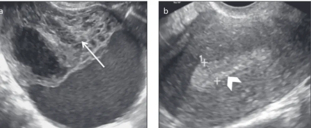
Figure 7. a, b. A 50-year-old postmenopausal female patient with a right ovarian adult granulosa cell tumor and proliferative endometrium who presented with metrorrhagia. Transvaginal US images (a, b) show a heterogeneous and apparently complex mass that is predominantly solid with focal cystic areas
(arrow, a). Note the thickness of the endometrium, which exceeded 8 mm (arrowhead, b).
Mixed sex cord-stromal tumors
Sertoli–Leydig cell tumor
SLCT is a rare sex cord-stromal tumor that accounts for approximately 0.5% of all ovarian neoplasms (3, 18) (Fig. 10). SLCT and SST occur predominantly in the same age group; approximately 75% of SLCTs are encountered in women younger than 30 years (13). However, few cases have been described in postmenopausal women (41, 42). For SLCT, nearly all cases are unilateral (98%) and 80% are restricted to the ovary at diagnosis (4, 18).
SLCT is the most common virilizing ovarian tumor, as approximately 30%–50% of these tumors produce androgens (testosterone and androgen precursors) and more than one-third of cases develop symptoms of vi-rilization (4, 15, 18). Notwithstanding, SLCT is a rare cause of virilization in premenopausal women. In this age group, other diferential diagnosis such as polycystic ovary syndrome and adrenal androgen-secreting tumors should be considered. In cases of andro-gen-secreting SLCTs, the laboratory data normally indicate elevated serum testoster-one levels but, in contrast to masculinizing adrenal tumors, normal or slightly elevated urinary 17-ketosteroid levels (4).
Many SLCTs are hormonally inactive and a small subgroup is estrogenic. Sudden abdominal pain and swelling are frequent symptoms of nonfunctioning tumors.
Very rarely, SLCT have been reported to associate with elevated alpha-fetoprotein serum levels; this is a typical feature of germ cell tumors such as yolk sac neoplasm. Al-though only approximately 30 cases have been described to date, SLCT is the most commonly reported ovarian nongerm cell tumor associated with elevated serum al-pha-fetoprotein levels (43, 44).
SLCT has a nonspeciic appearance. On US, these tumors usually present either as a distinct hypoechoic mass or a hetero-geneous mass that is primarily solid with multiple cystic spaces. Small virilizing SLCTs may be easily detected using color Doppler US rather than transvaginal US alone (13, 45, 46). On CT images, a soft-tissue atten-uating adnexal mass is usually seen (15). The solid tumor portions characteristically exhibit avid contrast uptake.
On MRI, the T2 signals of the solid
com-areas on T1-weighted images and hyperin-tense areas on T2-weighted images relect cystic areas. Cysts may also exhibit mild hy-perintensity on T1-weighted images (47). Striking homogeneous or heterogeneous contrast enhancement of the solid areas is observed on gadolinium-enhanced images.
The prognosis of SLCT is good, and 92% of tumors manifest as stage I (13). The most important prognostic factors are the stage and degree of histologic diferentiation (3, 13, 48). In a previous report by Sigismondi et al. (49), ive-year survival rates of 92.3% and 33.3% were reported for patients with stage I and stage ≥2 disease, respectively. In the same report, patients with well-diferen-tiated tumors had a ive-year survival rate of 100% versus 77.8% for patients with moder-ately and poorly diferentiated tumors.
In contrast to GCTs, which tend to recur late, malignant SLCTs usually recur early
in the pelvic and abdominal cavity, with a reported relapse rate of 71.4% within two years after diagnosis (49).
Conclusion
Ovarian sex cord-stromal tumors are in-frequent tumors that difer from the more frequent epithelial neoplasms via strong associations with hormone-mediated syn-dromes, presentation in a broad age range, and the near-ubiquitous diagnosis of low-stage disease with a good outcome. These tumors, which develop from cells arising from the primitive sex cords or stromal cells, comprise a diverse group. As a result, these tumors are currently subdivided as pure stromal tumors, pure sex cord tumors, or mixed sex cord-stromal tumors.
The radiologic appearances of these tumors vary along with their
morpholo-Figure 9. a, b. A 17-year-old female patient with a right ovarian juvenile granulosa cell tumor. A transabdominal US image (a) shows a multilocular cystic ovarian mass. An axial contrast-enhanced CT image (b), shows a multiloculated low attenuating mass with thin septations (arrow). Note the presence of abundant ascites (asterisks).
a b
gies. Notwithstanding, some radiologic features prevail in certain types of tumors. Fibromas typically present as solid hy-poechoic and homogenously isodense masses with characteristic hypointensity on T1-weighed images, strong hypointen-sity on T2-weighted images, and delayed enhancement after contrast administration. Thecomas and GCTs typically associate with hyperestrogenic states that may induce endometrial hyperplasia and endometrial carcinoma. Steroid cell tumors, Leydig tu-mors, and SLCTs are characteristic virilizing neoplasms. Leydig cell tumors are usually small tumors that may be diicult to de-pict via diferent imaging methods; there-fore, radiologists should note morphologic changes within the ovary, especially on MRI and transvaginal US with color Doppler. SSTs frequently manifest on US as unilat-eral tumors comprising star-shaped hy-poechoic areas enclosed by solid areas. On MRI, the pseudolobular solid areas exhibit a spoke-wheel pattern and display iso- to hypointensity on T2-weighted images and early and avid contrast uptake.
The rarity of sex cord-stromal tumors contributes to a low index of suspicion; therefore, a thorough knowledge of the clinicopathologic and radiologic indings of these tumors is important and allows radiologists to narrow the diferential diag-noses for ovarian tumors, thus facilitating surgical planning and the avoidance of in-appropriate treatments.
Conlict of interest disclosure
The authors declared no conlicts of interest.
References
1. Haroon S, Zia A, Idrees R, Memon A, Fatima S, Kayani N. Clinicopathological spectrum of ovari-an sex cord-stromal tumors; 20 years’ retrospec-tive study in a developing country. J Ovarian Res 2013; 6:87. [CrossRef]
2. Shim SH, Kim DY, Lee SW, et al. Laparoscopic management of early-stage malignant non-epi-thelial ovarian tumors: surgical and survival out-comes. Int J Gynecol Cancer 2013; 23:249–255.
[CrossRef]
3. Jung SE, Rha SE, Lee JM, et al. CT and MRI ind-ings of sex cord-stromal tumor of the ovary. Am J Roentgenol 2005; 185:207–215. [CrossRef]
4. Pratt J. Pathology of the ovary. 1st ed. Philadel-phia: Sounders, 2004; 197–226.
5. Kurman RJ, Carcangiu ML, Herrington CS, Young RH. Classiication of tumours of the ovary. In: WHO Classiication of Tumours, Volume 6. 4th
ed. Lyon: IARC, 2014; 44–56.
6. Tavassoli FA, Devilee P. Tumours of the ovary and the peritoneum. In: WHO Classiication of Tu-mours; Tumours of the Breast and Female Genital Organs. 3th ed. Lyon: IARC, 2003; 146–162.
7. Chen VW, Ruiz B, Killeen JL, Coté TR, Wu XC, Correa CN. Pathology and classiication of ovarian tumors. Cancer 2003; 97:2631–2642. [CrossRef]
8. Shinagare AB, Meylaerts LJ, Laury AR, Mortele KJ. MRI features of ovarian ibroma and ibrothe-coma with histopathologic correlation. AJR Am J Roentgenol 2012; 198:W296–303. [CrossRef]
9. Oh SN, Rha SE, Byun JY, et al. MRI features of ovar-ian ibromas: emphasis on their relationship to the ovary. Clin Radiol 2008; 63:529–535. [CrossRef]
10. Montoriol PF, Mons A, Da Ines D, Bourdel N, Tixier L, Garcier JM. Fibrous tumours of the ovary: aetiol-ogies and MRI features. Clin Radiol 2013; 68:1276– 1283. [CrossRef]
11. Yazdani S, Alijanpoor A, Sharbatdaran M, et al. Meigs’ syndrome with elevated serum CA125 in a case of ovarian ibroma /thecoma. Caspian J Intern Med 2014; 5:43–45.
12. Ray S, Biswas BK, Mukhopadhyay S. Giant prima-ry ovarian ibrosarcoma: Case report and review of pitfalls. J Cytol 2012; 29:255–257. [CrossRef]
13. Outwater EK, Wagner BJ, Mannion C, McLarney JK, Kim B. Sex cord-stromal and steroid cell tu-mors of the ovary. Radiographics 1998; 18:1523– 1546. [CrossRef]
14. Yen P, Khong K, Lamba R, Corwin MT, Gerscovich EO. Ovarian ibromas and ibrothecomas: sono-graphic correlation with computed tomography and magnetic resonance imaging: a 5-year single-institu-tion experience. J Ultrasound Med 2013; 32:13–18. 15. Hricak H. Fibrothecoma and Sclerosing Stromal
Tumor. In: Hricak H, Akin O, Sala E, Ascher SM, Levine D, Reinhold C, Eds. Diagnostic Imaging: Gynecology, 1st ed. Salt Lake City: Amirsys, 2007; 728–731.
16. Jung SE, Lee JM, Rha SE. CT and MR imaging of ovar-ian tumors with emphasis on diferential diagnosis. Radiographics 2002; 22:1305–1325. [CrossRef]
17. Thomassin-Naggara I, Daraï E, Nassar-Slaba J, Cortez A, Marsault C, Bazot M. Value of dynamic enhanced magnetic resonance imaging for distin-guishing between ovarian ibroma and subserous uterine leiomyoma. J Comput Assist Tomogr 2007; 31:236–242. [CrossRef]
18. Tanaka YO, Tsunoda H, Kitagawa Y, Ueno T, Yo-shikawa H, Saida Y. Functioning ovarian tumors: direct and indirect indings at MR imaging. Radio-graphics 2004; 24:S147–166. [CrossRef]
19. Zhang H, Zhang GF, Wang TP, Zhang H. Value of 3.0 T difusion-weighted imaging in discriminating thecoma and ibrothecoma from other adnexal solid masses. J Ovarian Res 2013; 6:58. [CrossRef]
20. Khanna M, Khanna A, Manjari M. Sclerosing stro-mal tumor of ovary: a case report. Case Rep Pathol 2012; 2012:592836. [CrossRef]
21. Chang YW, Hong SS, Jeen YM, Kim MK, Suh ES. Bilateral sclerosing stromal tumor of the ovary in a premenarchal girl. Pediatr Radiol 2009; 39:731– 734. [CrossRef]
22. Yen E, Deen M, Marshall I. Youngest reported patient presenting with an androgen producing sclerosing stromal ovarian tumor. J Pediatr Ado-lesc Gynecol 2014; 27:e121–124. [CrossRef]
23. Ismail SM, Walker SM. Bilateral virilizing scleros-ing stromal tumours of the ovary in a pregnant woman with Gorlin’s syndrome: implications for pathogenesis of ovarian stromal neoplasms. His-topathology 1990; 17:159–163. [CrossRef]
24. Lee MS, Cho HC, Lee YH, Hong SR. Ovarian sclerosing stromal tumors: gray scale and color Doppler sonographic indings. J Ultrasound Med 2001; 20:413–417.
25. Calabrese M, Zandrino F, Giasotto V, Rissone R, Fulcheri E. Sclerosing stromal tumor of the ovary in pregnancy: clinical, ultrasonography, and magnetic resonance imaging indings. Acta Radiol 2004; 45:189–192. [CrossRef]
26. Kawauchi S, Tsuji T, Kaku T, Kamura T, Nakano H, Tsuneyoshi M. Sclerosing stromal tumor of the ovary: a clinicopathologic, immunohisto-chemical, ultrastructural, and cytogenetic analysis with special reference to its vasculature. Am J Surg Pathol 1998; 22:83–92. [CrossRef]
27. Souto SB, Baptista PV, Braga DC, Carvalho D. Ovarian Leydig cell tumor in a post-menopausal patient with severe hyperandrogenism. Arq Bras Endocrinol Metabol 2014; 58:68–75. [CrossRef]
28. Sakamoto K, Fujimitsu R, Ida M, Horiuchi S, Hamada Y, Yoshimitsu K. MR diagnosis of steroid cell tumor of the ovary: value of chemical shift imaging. Magn Reson Med Sci 2009; 8:193–195.
[CrossRef]
29. Saida T, Tanaka YO, Minami M. Steroid cell tumor of the ovary, not otherwise speciied: CT and MR indings. AJR Am J Roentgenol 2007; 188:W393– 4. [CrossRef]
30. Jiang W, Tao X, Fang F, Zhang S, Xu C. Benign and malignant ovarian steroid cell tumors, not otherwise speciied: case studies, comparison, and review of the literature. J Ovarian Res 2013; 6:53. [CrossRef]
31. Kottarathil VD, Antony MA, Nair IR, Pavithran K. Recent advances in granulosa cell tumor ova-ry: a review. Indian J Surg Oncol 2013; 4:37–47.
[CrossRef]
32. Crew KD, Cohen MH, Smith DH, Tiersten AD, Feirt NM, Hershman DL. Long natural history of recurrent granulosa cell tumor of the ovary 23 years after initial diagnosis: a case report and review of the literature. Gynecol Oncol 2005; 96:235–240. [CrossRef]
33. Lee IH, Choi CH, Hong DG, et al. Clinicopatho-logic characteristics of granulosa cell tumors of the ovary: a multicenter retrospective study. J Gynecol Oncol 2011; 22:188–195. [CrossRef]
34. Pautier P, Lhommé C, Culine S, et al. Adult gran-ulosa-cell tumor of the ovary: a retrospective study of 45 cases. Int J Gynecol Cancer 1997; 7:58–65. [CrossRef]
35. RabbanJ, Gupta D, Zaloudek CJ, Chen L. Syn-chronous ovarian granulosa cell tumor and uter-ine serous carcinoma: A rare association of a high-risk endometrial cancer with an estrogenic ovarian tumor. Gynecol Oncol 2006; 103:1164– 1168. [CrossRef]
36. Hammer A, Lauszus FF, Petersen AC. Ovari-an grOvari-anulosa cell tumor Ovari-and increased risk of breast cancer. Acta Obstet Gynecol Scand 2013; 92:1422–1425. [CrossRef]
37. Ohel G, Kaneti H, Schenker JG. Granulosa cell tu-mors in Israel: a study of 172 cases. Gynecol Onc 1983; 15:278–286. [CrossRef]
38. Gittleman AM, Price AP, Coren C, Akhtar M, Don-ovan V, Katz DS. Juvenile granulosa cell tumor. Clin Imaging 2003; 27:221–224. [CrossRef]
39. Malmström H, Högberg T, Risberg B, Simonsen E. Granulosa cell tumors of the ovary: prognos-tic factors and outcome. Gynecol Oncol 1994; 52:50–55. [CrossRef]
40. Rosario R, Wilson M, Cheng WT, et al. Adult granulosa cell tumours (GCT): clinicopathologi-cal outcomes including FOXL2 mutational status and expression. Gynecol Oncol 2013; 131:325– 329. [CrossRef]
41. Caringella A, Loizzi V, Resta L, Ferreri R, Loverro G. A case of Sertoli-Leydig cell tumor in a post-menopausal woman. Int J Gynecol Cancer 2006; 16:435–438. [CrossRef]
42. Nicoletto MO, Caltarossa E, Donach M, Nardelli GB, Parenti A, Ambrosini A. Sertoli cell tumor: a rare case in an elderly patient. Eur J Gynaecol Oncol 2006; 27:86–87.
43. El-Bahrawy M. Alpha-fetoprotein-producing non-germ cell tumours of the female genital tract. Eur J Cancer 2010; 46:1317–1322. [CrossRef]
44. Jashnani KD, Hegde CV, Munot SP. Alfa-feto-protein secreting ovarian sex cord-stromal tu-mor. Indian J Pathol Microbiol 2013; 56:54–56.
[CrossRef]
45. Outwater EK, Marchetto B, Wagner BJ. Viril-ising tumors of the ovary: imaging features. Ul-trasound Obstet Gynecol 2000; 15:365–371.
46. Yanushpolsky EH, Brow DL, Smith BL. Localiza-tion of small ovarian Sertoli-Leydig cell tumors by transvaginal sonography with color Doppler. Ultrasound Obstet Gynecol 1995; 5:133–135.
[CrossRef]
47. Hamm B, Forstner R, Baert AL, Knauth M, Sar-tor K. CT and MRI in ovarian carcinoma. In: MRI and CT of the female pelvis. New York: Springer, 2007; 258.
48. Bhat RA, Lim YK, Chia YN, Yam KL. Sertoli-Ley-dig cell tumor of the ovary: analysis of a single institution database. J Obstet Gynaecol Res 2013; 39:305–310. [CrossRef]
49. Sigismondi C, Gadducci A, Lorusso D, et al. Ovarian Sertoli-Leydig cell tumors. a retrospec-tive MITO study. Gynecol Oncol 2012; 125:673– 676. [CrossRef]