Wingless/Wnt Signaling Pathways by Directly Interacting
with Ras, Notch and Dishevelled
Ana Carmena1,2*, Stephan Speicher2, Mary Baylies1
1Program in Developmental Biology. Sloan-Kettering Institute, Memorial Sloan-Kettering Cancer Center, New York, New York, United States of America, 2Instituto de Neurociencias de Alicante, Consejo Superior de Investigaciones Cientı´ficas/University Miguel Hernandez, Unidad de Neurobiologı´a del Desarrollo, Alicante, Spain
Over the past few years, it has become increasingly apparent that signal transduction pathways are not merely linear cascades; they are organized into complex signaling networks that require high levels of regulation to generate precise and unique cell responses. However, the underlying regulatory mechanisms by which signaling pathways cross-communicate remain poorly understood. Here we show that the Ras-binding protein Canoe (Cno)/AF-6, a PDZ protein normally associated with cellular junctions, is a key modulator of Wingless (Wg)/Wnt, Ras-Mitogen Activated Protein Kinase (MAPK) and Notch (N) signaling pathways cross-communication. Our data show a repressive effect of Cno/AF-6 on these three signaling pathways through physical interactions with Ras, N and the cytoplasmic protein Dishevelled (Dsh), a key Wg effector. We propose a model in which Cno, through those interactions, actively coordinates, at the membrane level, Ras-MAPK, N and Wg signaling pathways during progenitor specification.
Citation: Carmena A, Speicher S, Baylies M (2006) The PDZ Protein Canoe/AF-6 Links Ras-MAPK, Notch and Wingless/Wnt Signaling Pathways by Directly Interacting with Ras, Notch and Dishevelled. PLoS ONE 1(1): e66. doi:10.1371/journal.pone.0000066
INTRODUCTION
In multicellular organisms, cells are exposed to a complex environment in which they read numerous and sometimes con-flicting stimuli. Cross-communication between signaling pathways is crucial for the integration of the multiple intracellular responses elicited by simultaneous signals, allowing the generation of unique cell outputs. As a result of cross-communication, networks of signal interactions are established within the cell. The elucidation of the underlying mechanisms by which these networks are built and regulated is essential for the understanding and pharmacological treatment of pathologies in which signaling pathways are mis-regulated, such as some neural disorders and cancer [1].
The Drosophila mesoderm provides an excellent system for studying signaling networks as Drosophila can be subjected to complex genetic manipulations and multiple signaling pathways are coordinately involved throughout mesoderm differentiation. After gastrulation, uncommitted mesodermal cells migrate and proliferate. Then, autonomous and non-autonomous signals pattern the mesoderm, allocating regions from which progenitors of the different mesodermal tissues, such as the somatic muscles and heart, will arise [2–4]. Somatic muscle and heart progenitors are singled out from clusters of equivalent cells (‘‘promuscle groups’’) that express the transcription factor Lethal of scute (L’sc), in a process reminiscent of neural progenitor specification [5]. These progenitors divide asymmetrically to give rise to two founder cells [6,7]. Each founder cell is endowed with a unique identity by expressing specific combinations of transcription regulators such as Slouch/S59, Kru¨ppel (Kr) or Even-Skipped (Eve) [4,8]. We have focused on the specification of a dorsal subset of muscle and heart progenitors that express the identity protein Eve [9]. These progenitors differentiate upon the concerted and combinatorial action of four highly conserved signal transduction pathways triggered by Wingless (Wg)/Wnt, Decapentaplegic (Dpp)/TGF-b, Receptor Tyrosine Kinase (RTK)-Ras-MAPK and Notch (N) [10]. Cross-talk between Ras and N signaling pathways throughout Eve+ progenitor specification has been
previously reported [11]. Indeed, crosstalk between N and Ras
signaling pathways is necessary for signal integration in multiple processes during Drosophila development, as well as in other invertebrate and vertebrate systems [12–18]. However, the mechanisms by which N and Ras pathways cross-communicate are only beginning to be elucidated [19–23].
In this work, we have investigated the function of theDrosophila
PDZ (PSD-95, Dlg, ZO-1) domain-containing protein Canoe (Cno) as a regulator of N and Ras cross-communication throughout Eve+progenitor specification.cnomutant alleles were
originally isolated by their dorsal open phenotype, hence its name (Ju¨rgens et al, 1984). Indeed, Cno has been shown to participate in the morphogenetic process known as dorsal closure of the epidermis by modulating the Jun N-terminal kinase (JNK) signaling cascade (Takahashi et al.1998).cnoencodes a cytoplasmic protein associated with cell junctions in epithelial tissues, where Cno function has been studied inDrosophila[24,25]. The human ortholog of Cno, AF-6, was initially identified as a fusion partner of ALL-1, a product involved in human leukemias [26]. AF-6 also localizes at epithelial cell junctions where it binds ZO-1, a tight-junction protein important for cell-cell contacts and cytoskeleton rearrangement [27,28]. Cno and AF-6 share a similar structure:
Academic Editor:Alfonso Martinez Arias, Cambridge University, United Kingdom
ReceivedSeptember 29, 2006;AcceptedOctober 31, 2006;PublishedDecember 20, 2006
Copyright:ß2006 Carmena et al. This is an open-access article distributed under the terms of the Creative Commons Attribution License, which permits unrestricted use, distribution, and reproduction in any medium, provided the original author and source are credited.
Funding:M.B. and A.C. were supported by National Institutes of Health Grant GM56989, a Human Frontiers Postdoctoral Fellowship and Grants from the Generalitat Valenciana GV05/075 and Marie Curie Program (MIRG-CT-2005-016536).
Competing Interests:The authors have declared that no competing interests exist.
one PDZ motif, characteristic of scaffolding proteins associated to the plasma membrane that contribute to the subcellular spatial organization of signaling pathways [29–31]; a Kinesin-like domain and a Myosin-V-like domain, characteristic motifs present in proteins that interact with cytoskeleton components [32]; and two Ras-associating motifs [33] through which Cno/AF-6 binds to the activated form of Ras (RasAct) [34]. In Drosophila, Cno interacts genetically with Ras, during eye development, and withN,during bristle and wing development [24,35,36]. However, the in-volvement of Cno in N-Ras cross-talk has not been studied. Here we propose a model in which Cno mediates N-Ras cross-talk through Dishevelled (Dsh/Dvl), the most proximal cytoplasmic component of Wg/Wnt pathway [37–40]. We suggest that Cno, by binding RasAct, Dsh and N, represses the signals that these proteins trigger and actively coordinates, at the membrane level,
RTK-Ras-MAPK, Dl-N and Wg-Dsh signaling pathways
throughout progenitor specification.
RESULTS
The specification of dorsal somatic muscle/heart progenitors depends on the combinatorial action of multiple signaling pathways [10]. First, Wg/Wnt and Dpp/TGF-b, secreted from the ectoderm to the mesoderm, define a dorsal region (preC2) revealed by the expression of the transcription factor L’sc. Then, two RTKs, theDrosophilaEGF receptor (Egfr) [41] and the FGF receptor Heartless (Htl) [42] trigger a positive Ras signal in two groups of equivalent cells, within the dorsal mesodermal region prepatterned by Wg and Dpp (cluster C2, shown in Fig. 1, and cluster C15, not shown). RTK-Ras, through the MAPK module, upregulates L’sc expression and activates the identity protein Eve in these clusters, which appear sequentially. In addition, Delta (Dl) [43] activates its receptor N, promoting a lateral inhibitory signal among the cells of the equivalence group, so that only one progenitor is singled out from each cluster (progenitor P2, Fig. 1D– 1F; and progenitor P15, not shown).
Loss-of-function (lof) analysis of Ras-MAPK signaling pathway components reveals a decrease in the number of progenitors specified. Conversely, lof mutants of components of N signaling pathway display an increased number of progenitors. The gain-of-function (gof) phenotypes have the opposite effect: overexpression of RasAct in the mesoderm leads to the specification of extra progenitors whereas overexpression of an activated form of N (NAct) inhibits progenitor specification [11] (see also Fig. 2D and 2E). These effects are tightly linked, as Ras-MAPK and N signaling pathways act in concert. A complex network of competitive and cooperative interactions between Ras and N signal transduction pathways during muscle/heart progenitor specification has been documented. For example, N signaling downregulates the expression of different components of Ras-MAPK pathway and Ras signaling upregulates the N ligand Dl [11]. Hence, an accurate regulation of N and Ras signaling thresholds is critical for the acquisition of a progenitor fate. Since Cno has been shown to genetically interact with N or Ras signaling in different tissues and to physically interact with RasAct (see introduction), we analyzed a possible function of Cno during progenitor formation modulating N-Ras cross-talk.
Cno is expressed in the Drosophila embryonic
mesoderm throughout muscle/heart progenitor
specification
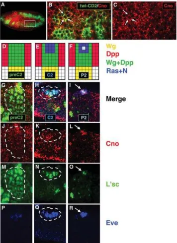
As the first step, we asked whether Cno is expressed in the mesoderm during progenitor specification. Cno expression was examined in embryos that carry atwi-CD2insertion, which drives
the expression of the transmembrane protein CD2 under the control of a mesoderm-specifictwist(twi) promoter. We observed that Cno was expressed in the mesoderm in a punctuate pattern close to the cell membrane (Fig. 1A–1C).
To investigate whether Cno was expressed when N and Ras signaling pathways interact to specify dorsal progenitors, we analyzed Cno expression with different progenitor markers. Cno was detected along with L’sc, the earliest and most general progenitor marker, in the dorsal region pre-patterned by Wg and Dpp (preC2). Cno was also detected along with the identity protein Eve in the dorsal Eve+ equivalence group (C2) and,
subsequently, in the progenitor (P2) (Fig. 1D–1R). Thus, Cno was expressed in the mesoderm during the time when Ras and N are regulating the specification of dorsal progenitors.
Figure 1.Cno is expressed in the mesoderm throughout progenitor specification. (A) Confocal immunofluorescence showing a lateral view of a late stage 10 embryo. Cno (red) is detected in the mesoderm (green). (B, C) Higher magnification of two hemisegments (636; inset in
A) reveals punctuate Cno expression at submembrane locations (arrows). (D–F) Diagrams show the most dorsal part of one hemiseg-ment and the signals involved throughout dorsal progenitor specifica-tion. (G–R) Confocal immunofluorescences showing high magnification (636) of the most dorsal part of one hemisegment. (D, G, J, M) Cno is
expressed in the dorsal mesodermal region pre-patterned by Wg and Dpp (preC2) along with L’sc. (E, H, K, N, Q) Cno is detected in the equivalence group in which Ras is locally activated (C2) restricting L’sc to this cluster and activating Eve. (F, I, L, O, R) Cno is expressed in the progenitor (P2) singled out from this cluster after N-promoted lateral inhibition.
cno zygotic mutant embryos show gain and loss of
Eve progenitors
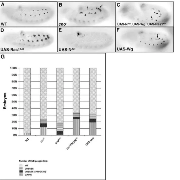
To investigate a potential function of Cno in Ras-N crosstalk during progenitor specification, we analyzed the dorsal Eve+
progenitors incno2zygotic null mutant embryos. Three different phenotypes were observed: (1) wildtype-like hemisegments, (2) hemisegments with extra Eve+progenitors and (3) hemisegments
displaying loss of Eve+ progenitors. Comparable results were
obtained with different cno mutant alleles (Fig. 2B and 2G). Likewise, the expression of other muscle progenitor markers and signaling proteins involved in muscle/heart progenitor
specifica-tion were, like Eve, both upregulated or downregulated in different hemisegments of cno2 zygotic null mutant embryos (Fig. S1A– S1R). These effects on muscle progenitor specification were reflected in the final muscle pattern where gain and loss of specific body muscles was observed (Fig. S1S–S1V). The progenitor markers (including Eve) and signaling proteins analyzed (Fig. S1) are under Ras and N regulation [11]. Indeed, the cno2 mutant phenotype resembled a mosaic of Ras and N mutant phenotypes (Fig. 2A, 2B, 2D and 2E) [11]. Given the reported relationship between Cno and Ras or N signaling pathways in different systems (see introduction), we hypothesized that Cno could be modulating both Ras and N signaling during muscle progenitor formation. Figure 2.cno2zygotic null mutants display gain and loss of progenitors. All panels show lateral views of stage 11 embryos stained with ana-Eve antibody. (A) Eve wildtype (wt) expression in a subset of dorsal muscle/heart progenitors and founder cells (2–3 cells) per hemisegment. (B)cno2 mutants show gain (arrow), loss (arrowhead) or a wildtype number (asterisk) of Eve+progenitors in different hemisegments. (C) Embryos in which NAct, Wg and RasAct are simultaneously expressed in the mesoderm display acno2
-like phenotype: gain (arrow), loss (arrowhead) and wildtype number of Eve+progenitors (asterisk). (D) Overexpression of Ras1Act in the mesoderm induces the specification of extra Eve+ progenitors. (E) Overexpression of NActin the mesoderm inhibits Eve+progenitor specification. (F) Embryos in which Wg is overexpressed in the mesoderm show gain of Eve+progenitors in intersegmental regions (arrow). (G) Table shows % of embryos of the indicated genotype that display a wildtype number of Eve+progenitors, Eve+progenitors gain, loss and both (gain and loss).cno2
andcnomis1are null and hypomorph mutant alleles, respectively;Df(3R)6-7 removescno.cnogof phenotype (UAS-cno) is also variable. Legends for bars are shown below the table. At least 70 embryos of each genotype were counted. P,0.0001 for all genotypes except forcnomis1(P = 0.0017).
Thus, we examined whether Cno interacted with these signaling pathways during progenitor specification.
cno genetically interacts with N and Ras signaling
pathways during progenitor specification
A classical genetic approach to uncover gene interactions during biological processes is the analysis of transheterozygous mutant phenotypes. Thus, to determine if Cno was interacting with N signaling during progenitor specification, we examined Eve expression in embryos with reduced doses of cno and different components of the N signaling pathway, including Dl, the ligand of N required for progenitor specification, and N itself. For example, double heterozygotes of cno and Dl (cno2/DlX) showed a great expansion of Eve+progenitors, which was not observed in
the single heterozygotes cno2/+ and DlX/+ (Fig. 3B and 3U). Further genetic interactions (Fig. 3G, 3U and not shown) indicated that cno interacts with N signaling during muscle progenitor formation.
In a similar way, transheterozygous mutant analysis revealed strong genetic interactions between cno and Ras pathway components, includingpointed(pnt), which encodes an ETS-domain effector of Ras signaling [44] and Ras itself (Fig. 3 and not shown). For example, double heterozygotes for cno and pnt (cno2/pntD88) showed a great loss of Eve+progenitors, which was not observed in
the single heterozygotes cno2/+ and pntD88/+ (Fig. 3C and 3U). Additional genetic interactions (Fig. 3H, 3E, 3J, 3U and not shown) indicated that Cno functions with Ras signaling during progenitor specification. In summary, our data revealed genetic interactions between Cno and both N and Ras-MAPK signaling pathways.
cno interacts genetically with Wg signaling pathway
The canonical Wg signaling pathway is another critical compo-nent for dorsal muscle/heart progenitor specification. wg lof mutants show a loss of Eve+ progenitors, whereas Wg
over-expression in the mesoderm induces extra Eve+progenitors; these
additional progenitors frequently appear in intersegmental regions [10,45] (Fig. 2F). Intriguingly, the extra Eve+progenitors specified
incno2null mutant embryos were often observed in intersegmental regions (Fig. 2B). Furthermore, Cno has been shown to interact genetically with some components of the Wg pathway during wing morphogenesis [24,46]. Hence, we investigated whether cno
genetically interacted with components of the Wg pathway during progenitor specification. Transheterozygotes for wg and cno
(wgCX4/+;cno2/+) revealed a strong interaction (Fig. 3D and 3U). Additional genetic interactions were found between cno and components of the canonical Wg pathway that further supported a functional relationship between Cno and Wg pathway during progenitor specification (Fig. 3I, 3U and Fig. 4, see below).
Cno inhibits N, Ras and Wg signaling pathways
Our genetic data indicated that Cno interacts with Wg, Ras and N signaling pathways throughout the process of dorsal muscle progenitor specification. These three signaling pathways act as positive (Wg and Ras) and negative (N) signals during progenitor formation [10]. We hypothesized that Cno regulates the relative thresholds of those positive and negative signals throughout progenitor specification. To further understand how Cno affects Wg, Ras and N signaling pathways during this process, we analyzed in more detail the functional relationships between Cno and each of these pathways.
Several experiments revealed an inhibitory effect of Cno on N signaling. For example, the cno overexpression phenotype was
enhanced when N activity was reduced (DlX heterozygous background) (Fig. 4). In addition, the N effector and transcrip-tional repressor, E(spl)-m8, was upregulated incno2null mutants (Fig. S2C and S2D). Thus, Cno showed an antagonist effect on N signaling.
Additional genetic interactions between Cno and the RTK-MAPK pathway also suggested an inhibitory effect of Cno on Ras-MAPK signaling during progenitor specification. For example, the RasAct overexpression phenotype was enhanced in acno2 homo-zygous background (Fig. 3T). In addition, examples of synergism between Cno and negative regulators of Ras signaling were found (Fig. 3E, 3J and Fig. S3). These genetic data supported a repressive effect of Cno on Ras-MAPK signaling.
Lastly, our experiments suggested that Cno, under wildtype conditions, also antagonized canonical Wg signaling. For example, we found that cno lof dominantly enhanced the overexpression phenotype of Wg, Dsh and Armadillo (Arm)/b-catenin [47] (Fig. 4). In addition, cuticles fromcno2 mutant embryos showed a naked phenotype, characteristic of Wg overexpression (not shown). These genetic interactions, in addition to the cno2 null mutant phenotype, suggested that Cno has an inhibitory effect on canonical Wg signaling.
Altogether, our genetic data supported an inhibitory effect of Cno on N, Ras-MAPK and Wg signaling pathways. We thus expected an increase in activity of all these signaling pathways in
cno2mutants. Indeed, the simultaneous overexpression of Wg, NAct and RasAct in the mesoderm phenocopied the cno2 null mutant phenotype (Fig. 3F, 3K, and 3P). Furthermore, the phenotypes observed by reducing N, Ras or Wg signaling in cno2 mutants resembled those found by simultaneously overexpressing Wg-Ras1Act, NAct-Wg or NAct-RasAct, respectively (compare Fig. 3G, 3L, 3Q; 3H, 3M, 3R and 3I, 3N, 3S). The inhibitory effect of Cno on N, Ras and Wg signaling pathways may seem in conflict with the phenotypes observed in some of the genetic combinations for
cno and components of the N, Ras or Wg pathways (Fig. 3). However, the upregulation of both positive and negative signals in
cno2mutant embryos can explain the net effect on Eve expression found in the different genetic combinations. For example, incno2/ cno2, pntD88embryos (Fig. 3H and 3M), there is an increase in all three signals: the two positive (Ras, Wg) and one negative N signal. However, the Ras pathway is impaired in these embryos (pntD88/+
genetic background). In addition, N signaling, which down-regulates the Ras pathway [11], increases in cno2/cno2, pntD88
embryos. Hence, the net effect is a great reduction of the positive Ras signal and thus, a great reduction of Eve expression.
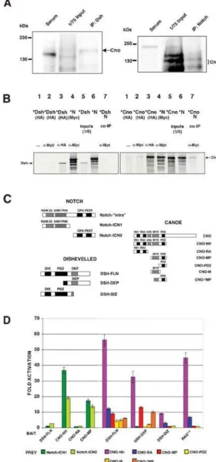
Cno physically interacts with N and Dsh
We carried out coimmunoprecipitations (co-IPs) using both embryonic lysates and in vitro translated proteins. These experi-ments showed that Cno coimmunoprecipitates with both N and Dsh (Fig. 5A, 5B and Fig. 7I). The co-IPs from embryonic lysates showed different sizes of the Cno proteins coimmunoprecipitated with either Dsh or N (Fig. 5A). We suggest that some of these different forms of Cno are due to different post-translational
modifications in each of those complexes, different isoforms of Cno or/and specific breakdown products of Cno (See also Fig. S5). To characterize the domains implicated in the binding, diverse deletion constructs of Cno, N and Dsh were tested in yeast two-hybrid assays (Fig. 5C and 5D). Only Cno constructs comprising the PDZ domain strongly interacted with both proximal (Notch-ICN1) and distal (Notch-ICN2) regions of the N intracellular Figure 3.cnointeracts genetically with N, Ras and Wg signaling pathways. (A–J and P–T) Lateral views of stage 11 embryos stained with ana-Eve antibody. (B, G) Extra Eve+progenitors are detected both incno2
/DlXtransheterozygotes and incno2/cno2,DlXembryos. (C, H) Bothcno2/pntD88 double heterozygotes andcno2, pntD88/cno2
embryos show loss of Eve+progenitors. (E, J) An increase in Eve+progenitors is observed both incno2
/ aosD7transheterozygotes and incno2,aosD7/cno2
embryos [74]. (D, I) Gain and loss of Eve+progenitors are detected both inwgCX4
/+;cno2/+double
heterozygotes and inwgCX4/
+;cnomis1/cno2
embryos. (F–I, K–N, P–S): (P) Simultaneous mesodermal overexpression of NAct-Wg-RasAct, (Q) Wg-RasAct, (R) NAct-Wg and (S) NAct-RasActhave similar phenotypes thancno2
/cno2(F, K),cno2/cno2,DlX(G, L),cno2/cno2,pntD88(H, M) andwgCX4/+;cnomis1/cno2(I, N) embryos, respectively. Arrows and arrowheads indicate gain and loss of progenitors, respectively. (K–O) Diagrammatic representation of the effect on Eve expression ofcnolof (K) andcnolof in combination with reduced doses of N (L), Ras (M) and Wg (N) signaling components, or with increased doses of Ras signal (O). (T) The phenotype of Ras1Actmesodermal overexpression is enhanced in acno2
mutant background. (U) Table shows % of embryos of the indicated genotype that display a wildtype number of Eve+progenitors, Eve+progenitors gain, loss and both (gain and loss). Legends for bars are shown right to the table. At least 70 embryos of each genotype were counted. P,0.0001 for all genotypes except forcno2/cno2,aosD7
(P = 0.0239) andwgCX4
/+;cno2/cnomis1(P = 0.006).
domain (approximately 40 and 20 fold activation, respectively). The Cno-Dsh interaction, however, mapped to two different domains of Cno: the PDZ domain and the Ras-binding domain (Fig. 5C and 5D). To distinguish which domains in Dsh bound the Ras-associating and PDZ motifs of Cno, different Dsh constructs were tested. A Dsh fragment containing the DIX (DIshevelled, aXin) and PDZ domains (DSH-DIZ) only bound Cno constructs containing the Ras-binding domain, as did the Ras positive control (Fig. 5C and 5D). In contrast, the Dsh fragment containing the DEP (Dsh, egl-10, pleckstrin) domain (DSH-DEP) only bound Cno constructs containing the PDZ domain plus some of the adjacent Myosin-V (Myo)-like domain (Fig. 5C and 5D). Thus, these data indicated that Cno physically interacted with both N and Dsh. These physical interactions could lead to the inhibitory effects of Cno on N and Wg-Dsh signaling pathways.
Cno links Wg-Dsh and Dl-N signaling pathways
Dsh has been shown to interact physically with N and to inhibit its signaling duringDrosophila wing bristle development [49]. We found that, genetically, Dsh also acted antagonistically to N signaling during muscle/heart progenitor specification (Fig. S2A, S2B and Fig. 4). For example, the phenotype of Dsh mesodermal overexpression was enhanced by reducing N signaling activity (DlX/+ genetic background) (Fig. 4). However, the mechanism underlying the Dsh-N antagonism is unclear: the reported Dsh-N (Notch-ICN2) physical interaction was very weak under our conditions (approximately 3 fold activation, Fig. 5D; see also Discussion) [49,50]. Moreover, Dsh failed to co-immunoprecipi-tate in vitro with N under our conditions (Fig. 5B). Hence, although Dsh had an antagonistic effect on N signaling during muscle progenitor specification, our data did not support a strong physical interaction between Dsh and N as the means by which Dsh inhibits N signaling. Dsh has been shown to co-immunopre-cipitate with N in vivo [51]. This interesting finding supports that Dsh and N are forming part of a complex during embryogenesis. However, it is not a proof of a direct physical interaction between both proteins. Since (1) Cno had a negative effect on N signaling, Figure 4.Cno inhibits Wg-Dsh pathway and links it with Dl-N pathway. Table shows % of embryos and hemisegments (Hs) for each indicated genotype that display extra Eve+ cells. At least 70 embryos/770 hemisegments were counted in each case. P,0.001 for all genotypes except for UAS-wg;cno2
/+, ‘‘% Embryos bar’’ (P = 0.0027); UAS-armS10, cno2
/+, ‘‘% Embryos bar’’ (P = 0.0337); UAS-cno,Dlx/+, ‘‘% Embryos bar’’
(P = 0.0177); UAS-dsh, UAS-cno, Dlx
/+, ‘‘% Embryos bar’’ (P = 0.0472); UAS-dsh, UAS-cno, ‘‘% Embryos bar’’ (P = 0.0115), ‘‘% Hs bar’’ (P = 0.0002).
doi:10.1371/journal.pone.0000066.g004
Figure 5.Cno physically interacts with Dsh and N. (A) Coimmunopre-cipitations from embryonic lysates. Left panel: Cno is coimmunopreci-pitated with endogenous Dsh. Right panel: Cno is coimmunoprecipi-tated with endogenous N. Different sizes of the Cno protein coimmunoprecipitated with Dsh and N (see text). A Rat and Rb sera without the Dsh or N antibodies were used as negative controls. (B) In vitro coimmunoprecipitations (see also Fig. 7I). Left panel: HA-tagged Dsh (DSH-FLN) does not coimmunoprecipitate with N. Right panel: HA-tagged Cno (CNO-NH) coimmunoprecipitates with Myc-HA-tagged N (Notch-‘‘intra’’). In each panel, lanes 1 and 2:35S-labelled (*) protein, HA-tagged, without adding any antibody (Ab) (lane 1) or addinga-Myc Ab (lane 2), as negative controls; lanes 3 and 4: *proteins HA-tagged (lane 3) or Myc-tagged (lane 4) tested with the corresponding Ab; lanes 5 and 6: translation products; lane 7: coimmunoprecipitation of the *HA-tagged protein with the Myc-tagged protein by ana-Myc-Ab. (C) N intracellular domain (Notch-‘‘intra’’), Dsh and Cno constructs used in the yeast two-hybrid and co-IP assays (see also ‘‘Materials and methods’’). (D) Yeast two-hybrid: averaged results of at least four independent protein extracts inb-galactosidase quantitative liquid assays. The fold activation determined with respect to the corresponding empty vector is represented. Bars indicate s.e.m. DSH-FLN, CNO-NH, CNO-RA and CNO-MP were tested against Notch constructs: ICN1 and Notch-ICN2. DSH-FLN, DSH-DEP, DSH-DIZ and RASV12 were tested against different Cno constructs (C).
(2) Cno coimmunoprecipated with N in vivo, (3) Cno and N proteins directly interacted in vitro and in yeast two-hybrid assays and (4) Cno interacted physically with Dsh, we reasoned that Cno could mediate the repressive effect of Dsh on N signaling.
To test this function of Cno as a mediator of the inhibitory effect of Dsh on N, we performed several genetic experiments. For example, the phenotype of Dsh overexpression under conditions of reduced N activity (DlX/+ genetic background, see above) was further enhanced by the simultaneous overexpression of Cno (Fig. 4). This result supported a function of Cno in mediating the N repression by Dsh. The relevance of Cno-Dsh interaction to inhibit N signaling was further supported by using specific deletion constructs of Dsh (DshDDEP) and Cno (CnoDN) that cannot interact with each other. Whereas the simultaneous mesodermal overexpression of wildtype forms of Dsh and Cno led to a great increase of Eve+ cells (a N lof phenotype), the simultaneous
overexpression of DshDDEP (lacks the DEP domain) [52] and CnoDN (lacks the RA1 and RA2 domains) [53] (see also Fig. 5) showed a much weaker effect, consistent with the existence of more N activity in the mesoderm of these embryos (Fig. 4; compare with overexpression of wildtype forms of Dsh and Cno). The DEP domain of Dsh is required to relocate Dsh at the membrane [54]. Given the physical interaction between Cno and the region of Dsh that contains the DEP domain, we next examined whether Cno was affecting Dsh cell localization. Cno overexpression in the mesoderm led to a striking accumulation of both Dsh and N close to the membrane of Eve+interacting cells
(arrows in Fig. 6B, 6D and 6F; compare to 6A, 6C, and 6E). This observation suggested that the negative effect of Cno on N and canonical Wg pathway could be related to the accumulation of Dsh and N at the cell membrane (see Discussion). Taken together with the genetic and physical interaction data, these results supported a cross-regulatory interaction between N and Dsh mediated by Cno during progenitor formation.
Cno links Wg-Dsh and Ras-MAPK signaling pathways
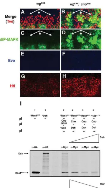
Wg signaling provides mesodermal cells competence to respond to Ras signaling, in part through the requirement of Wg to maintain the expression of different components of the Ras pathway [55]. For example, in wg mutant embryos, we detected reduced expression of the Drosophila FGF receptor Htl, as well as no expression of diP-MAPK, a measure of Ras activation (Fig. 7C and 7G; [56]). However, the molecular mechanism by which this competence is achieved, either by direct regulation or indirectly Figure 7. Wg-Dsh signaling releases Cno-mediated Ras repression. (A–H) Lateral views at high magnification (636) of stage 11 embryos
showing three hemisegments. (A, C, E) InwgCX4
mutants neither diP-MAPK (green) nor Eve (blue) expression is detected in the mesoderm, which is marked by Twi (red). (G) The FGFR Htl (red) is also downregulated inwgCX4
mutants. (B, D, F, H) Both diP-MAPK (B, D) and Htl (H) expression are rescued inwgCX4; cnomis1double mutants; Eve expression is not rescued (F). tp, tracheal pits. (I) Dsh competes with Ras for binding Cno. Proteins were translated in vitro in presence (*) or absence of35S-Methionine. Lane 1 and 2: *RasV12and *Dsh, both HA-tagged, were immunoprecipitated with ana-HA antibody (Ab). Lane 3: Both *RasV12and *Dsh were co-IP with Cno by ana-Myc Ab. Lane 4, 5 and 6: The addition of increasing amounts of Dsh leads to a decrease in the amount of *RasV12co-IP with Cno.
doi:10.1371/journal.pone.0000066.g007
Figure 6.Overexpression of Cno leads to the stabilization of Dsh and N close to the membranes of Eve+interacting cells. The most dorsal part of two hemisegments of stage 11 embryos are shown in all panels (636
magnification). Eve marks a dorsal equivalence group in the mesoderm (blue). (A, C, E, G) In wildtype (WT) embryos, Dsh (green) and N (red) colocalize at some regions in Eve+cell clusters (arrows). (B, D, F, H) Cno overexpression (UAS-cno) leads to an increase of Dsh and N close to the membranes of Eve+cells (arrows).
through unknown mediators, is not completely understood. Since Cno bound and inhibited Ras, and the Wg effector Dsh bound the Cno Ras-associating domain, we hypothesized that Wg signaling would release Cno-mediated Ras inhibition through competition between Dsh and Ras for the Cno Ras-binding domain. Therefore, we predicted that some Ras activity would be rescued inwgmutants in which Cno is no longer present to repress Ras. To test this hypothesis, we analyzed MAPK activity and the expression of proteins under Ras regulation in wgCX4; cnomis1
double mutants. DiP-MAPK expression, which is lost in wgCX4
mutants (Fig. 7A and 7C, compare to Figure S3A and S3B) [55], was partially rescued inwgCX4; cnomis1double mutants (Figure 7B and 7D). This was the same level of rescue obtained in wgCX4
mutant embryos by overexpression of RasAct [55]. As expected, Eve expression was not rescued inwgCX4; cnomis1mutant embryos (Fig. 7A, 7B, 7E and 7F) nor is it rescued inwgCX4mutant embryos by RasAct, as Eve expression, in addition to Ras, requires a direct
transcriptional input from Wg signaling [10,55,57]. Moreover, the expression of the Drosophila FGF receptor Htl [58], which is downregulated in wgCX4 mutant embryos, was also rescued in
wgCX4; cnomis1double mutants (Fig. 7G and 7H) [55]. Since Ras-MAPK upregulates Htl expression [11], the increase of Htl expression inwgCX4; cnomis1double mutants was consistent with the rescue of MAPK activity found in these embryos. These results suggested that Wg signaling releases the Ras repression mediated by Cno.
To further investigate how Wg signaling releases the Ras repression mediated by Cno, we examined the possible function of Dsh, which is activated upon Wg signaling, in this process. We found that diP-MAPK expression was lost in dshV26 lof mutants and expanded indsh gof mutants (Fig. S4). Since Dsh physically interacted with the Ras-binding domain of Cno (Fig. 5, see also Fig. 8A), we next tested whether Dsh competes with Ras for binding Cno. We found that addition of increasing amounts of Dsh led to reduced amounts of RasV12 (RasAct) co-immunopre-cipitated with Cno (Fig. 7I). Thus, Dsh was able to compete with Ras for binding Cno. Altogether, these experiments strongly suggested that Wg-Dsh signaling activateseveduring muscle/heart progenitor specification both by direct binding of dTCF/Arm to theevemuscle enhancer [55] and by releasing the Ras repression mediated by Cno.
DISCUSSION
Elucidating crosstalk mechanisms and regulation is fundamental to the understanding of how cells integrate the complex information that they receive from their environment to generate precise cell responses [1]. In this study, we argue that the PDZ-domain containing protein Cno/AF-6 is a key modulator of Ras-MAPK, N and Wg/Wnt signaling pathways cross-communication: (1) Cno is expressed during muscle progenitor formation, (2) Cno colocalizes in vivo and genetically interacts with components of these three pathways, which are critical to muscle progenitor specification and (3) Cno represses each of these pathways through physical interactions with RasAct, Dsh and N. We propose that
Cno, through those interactions, coordinates and integrates the information required by cells to adopt the progenitor fate.
Cno represses Ras-MAPK, N and canonical Wg/Wnt
signaling pathways
A body of evidence suggests that Cno/AF-6 represses the Ras-MAPK signaling pathway [34,35,59,60]. In this work, we present further data that strongly support the inhibition of Ras-MAPK signaling by Cno in vivo. The repression of Ras-MAPK signaling
by Cno does not account, however, for all the phenotypes detected incno2mutant embryos, such as cases of Eve+progenitor losses (i.e.
if Cno were merely inhibiting Ras, incno2mutant embryos there would be more positive Ras signaling and, thus, only more Eve+
progenitors should be found). In this work, we provide data that support an inhibitory effect of Cno on N signaling during muscle and heart progenitor specification. Since N inhibits progenitor fate, the cases of Eve+progenitors losses found incno2
mutants can be explained by the increase in N signaling in cno2 mutants. Indeed, an increase inE(spl)-m8expression, a main effector of N signaling, was detected incno2mutants (Fig. S2C, S2D). Given that Cno interacted physically with N and that the overexpression of Cno led to an accumulation of N at the membrane, one possibility is that Cno inhibits N signaling by impeding the cleavage of N from the membrane. Other possibilities are that Cno interferes with N endocytosis and vesicular trafficking, both key processes in Figure 8. Summary of physical interactions and working model. (A) Diagram summarizing the physical interactions between Cno and Nintra, RasAct and Dsh. (B) Working model. The modulation of the information conveyed by three signaling pathways, Wg, N and Ras-MAPK, is critical for muscle progenitor specification. Our data show that Cno interacts physically and genetically with these three pathways, negatively regulating the information flow to key progenitor transcrip-tional targets, L’sc and Eve. In this model, the Cno/Dsh interaction is pivotal to modulate signaling thresholds. Cno/Dsh interaction facilitates the release of Cno repression on RasActsignaling and the inhibition of N signaling by Cno. Dsh recruitment by Cno leads to canonical Wg signal inhibition. Arr, Arrow; Fz2, Frizzled2.
N signaling regulation [61,62]. Further experiments are required to test these hypotheses.
The repression of both Ras-MAPK and N signaling by Cno still cannot account for all the phenotypes observed in cno2mutants, such as the extra Eve+ cells that appeared in intersegmental
regions. This phenotype is characteristic of Wg overexpression suggesting that, in wildtype conditions, Cno is also repressing Wg signaling. Indeed, we present data in this work that support a Cno inhibitory effect on canonical Wg signaling. Since Dsh and Cno interacted physically, one possibility is that Cno interrupts Wg signaling at the level of Dsh, perhaps by sequestering Dsh away from other pathway components or by diverting it to other non-canonical roles.
Hence, a repressive effect of Cno on both negative (N) and positive (Wg and Ras-MAPK) signals required for progenitor fate could ultimately explain the variable phenotypes found incnolof and gof mutants (Fig. 2). For example, incno2mutants there will be more positive Ras and Wg signals, but also more negative N signal (Fig. 3K); and Cno overexpression will lead to less positive Ras and Wg signals, but also to less repressive N signal. Thus, the final net balance of positive and negative signals would determine the variable phenotype observed incno2zygotic null mutant embryos. A maternal contribution of cno has been reported [63]. This maternal contribution might be masking the complete lof phenotype of cno(i.e. a ‘‘not-variable’’ phenotype). However, we favour the hypothesis that the variable phenotype observed incno
zygotic mutants is due to the Cno regulation of both positive and negative signals required for progenitor fate. Thus, we would expect that different thresholds of Cno protein (from the complete lof condition to a gof condition) would cause a variable phenotype. Indeed both a hypomorph allele ofcno(cnomis1) andcnogof showed that variable phenotype (Fig. 2G). Given the reported relationships between Cno and Ras, N or Wg in different developmental contexts, Cno could be a general link and modulator of these signaling pathways throughout development.
How is Cno linking Ras, N and Wg signaling
pathways?
We propose a working model to explain how Cno is mediating Ras, N and Wg cross-talk during progenitor specification (Fig. 8B). First, just before Wg signaling begins, Cno directly interacts with RasActand inhibits its ability to signal. As a result of Wg signal activation, L’sc starts to be expressed in the mesoderm [10]. Then, L’sc upregulates Dl expression in the mesoderm (A.C., un-published data, not shown in the model), which in turn activates N in nearby cells, initiating the process of lateral inhibition in the equivalence groups. At the same time, Dsh activation by Wg would result in a competition between Dsh and Ras for the Cno Ras-binding domain. Consequently, Ras repression by Cno is released; this leads to the upregulation of L’sc and to the activation of the identity geneeve, which requires both Ras-MAPK and Wg signaling for its expression. The binding of Cno to the Dsh DEP domain facilitates the localization of both Dsh and Cno at the membrane. This would have two consequences: (1) the in-activation of the Wg signaling through the canonical pathway and (2) the repression of N signaling by Cno physical interaction. Whether Dsh is phosphorylated or unphosphorylated when forming part of the Cno-Dsh complex is something that will be pursued in future studies.
Thus, Cno links Ras and N pathways through its binding to Dsh: Dsh-Cno interaction would facilitate both Ras signaling activation and N signaling inhibition. By directly interacting with RasAct, Dsh and N and by locating these proteins in close
proximity, Cno would actively coordinate Wg with Ras and N signaling pathways, allowing their cross-communication and modulating their relative signal thresholds throughout progenitor specification. Our data imply that Wg/Dsh is not only a critical permissive signal for the initial mesoderm patterning, but also this signal would play an important role later, during the process of lateral inhibition, in combination with Ras and N. The complexes Cno-Ras, Cno-Dsh, and Cno-N would be in a dynamic equilib-rium. The thresholds and relative affinities of all these proteins, their spatio-temporal control and, ultimately, the specific cell context would dictate the binding state and activity of Cno.
Cno role as a mediator between Wg-Dsh and N
signaling pathways
Dsh has been shown to bind N by using yeast two-hybrid assays [49,50]. Dsh has also been shown to coimmunoprecipitate with N in vivo [51]. However, both our yeast two-hybrid and in vitro co-IP experiments did not support a direct Dsh-N interaction. Our experiments showed that Cno physically interacts with N by using both yeast two-hybrid and co-IP assays. Thus, the described inhibitory effect of Dsh on N signaling [49] is likely to be mediated through Cno, by directly binding N. Yet, other possibilities could explain our observations. For example, a weak physical interaction between Dsh and N may require the presence of Cno to strengthen it; this could be achieved by the simultaneous binding of Cno to Dsh and N. Another possibility is that the complex Cno-Dsh facilitates a post-translational modification of Cno-Dsh required for binding N, which is not occurring under our experimental conditions. Indeed, Dsh is a phosphoprotein that becomes hyperphosphorylated upon Wg signaling [64]. Our working model takes into account the following observations: Cno bound both Dsh and N, Dsh did not clearly bind N, both Dsh and Cno interacted antagonistically with N and the interaction between Dsh and Cno was important for N inhibition. Additional experiments are required to further clarify the functional relation-ships among these proteins.
Conclusions
Multiple points of cross-communication among signaling pathways are crucial to ensure a fine-tuned regulation of signaling networks. Integration of signals at the enhancer level has been shown to be an essential mechanism to achieve specific and coordinated responses [55]. In this study we show how regulation at the mem-brane level through Cno and Dsh, two PDZ domain-containing proteins, is also critical to elicit signaling pathways integration. Multiprotein complex formation around PDZ-based scaffolds at specific sub-membrane locations is known to be decisive for signal transduction rate and fidelity [29,30]. Thus, PDZ proteins are excellent candidates as points of crosstalk at the membrane level. Here, we show a dynamic role for a PDZ protein, modulating the thresholds of multiple signal transduction pathways in vivo. Future investigation will allow a further understanding of how these proteins organize and regulate the complex cell networks of signaling pathways.
MATERIALS AND METHODS
Drosophila strains and genetics
The following mutant stocks were used:cno2,cnomis1[24],Df(3L)6-7,
DlX,aosD7,pntD88,wgCX4, dshV26, Ras1e2F, NX81K1. Ectopic expres-sion was achieved with the GAL4-UAS system [65] and the following fly lines:twi-GAL4,Dmef2-GAL4,UAS-Ras1Act,UAS-NAct,
UAS-dshDDEP[52]. TwoUAS-cno transgenes were used in com-bination to analyze the effect on Dsh expression. All the crosses Gal4-UAS were carried out at 29uC.twi-GAL4 andDmef2-GAL4 drivers were used in combination for all overexpression experi-ments, except for overexpression of Ras1 in acno2 homozygous background; in this case, only thetwi-GAL4 driver was employed. Other lines used in this work were:twi-CD2[66] anddsh-GFP[52].
yw was used as the reference wildtype strain. Balancer chromo-somes containing different lacZ transgenes were used for identification of homozygous mutant embryos.
Immunohistochemistry, in situ hybridization and
microscopy
Embryo fixation, antibody staining and in situ hybridization were carried out by standard protocols [10]. The following primary antibodies were used: rabbita-Cno, 1/500 [25]; rata-Cno [53], mousea-CD2, 1/800 (Serotec); rata-L’sc, 1/800 [67]; rabbita -Eve, 1/3000 [68]; mousea-Eve, 1/25 (Hybridoma Bank); guinea piga-Eve, 1/200 [69]; guinea piga-Kr, 1/2000 [69]; mousea-Dl, 1/40 (M. Muskavitch); rabbit a-Htl 1/1000 [42]; rabbit a-b -galactosidase 1/100000 (Cappel); mousea-b-galactosidase 1/8000 (Promega); mouse a-diP-MAPK, 1/400 (Sigma); rabbit a-Twi, 1/1000 (S. Roth); mouse a-N 1/100 (Hybridoma Bank); mouse a-GFP, 1/400 (Clontech); rata-Dsh, 1/500 [70], mAb 3E2 [5]. For immunostaining with thea-Cno antibody, embryos were fixed by using the heat-methanol method [71]. L’sc and diP-MAPK signals were enhanced by use of Tyramide Signal Amplification reagents (New England Nuclear). Fluorescent images were recorded by using a Zeiss LSM 510 Axiovert 100M microscope and assembled by using Adobe Photoshop. Blinded scoring of Notch and Dsh staining in the wildtype and cno overexpression studies (Fig. 6) was performed.
Statistical analysis
The phenotypic effects of individual mutations in various genetic backgrounds were tested for significance (P,0.05) with pairwise comparisons of genotypes by a standard Fisher test.
Yeast two-hybrid assays and cloning
Yeast transformations and b-galactosidase quantitative liquid assays were carried out following Clontech User Manual protocols. Dsh constructs (DSH-FLN and DSH-DEP, in the bait vector pEG202/LexA) as well as N constructs (ICN1 and ICN2, in the prey vector PJG-4/pB42AD) were previously described [49]. DSH-DIZ comprising amino acids (aa) 1 to 393 was subcloned in the same bait vector, LexA. Cno-NH, in the prey vector pACT2, and RasV12, in the bait vector pAS2-1, were previously described [35]. Other Cno constructs used in this work were: Cno-RA (containing the two Ras-associating domains, aa 1-355), Cno-MP (comprising Myo-like and PDZ domains, aa 503-935), Cno-PDZ (aa 836-935), Cno-M (aa 503-754) and Cno-*MP (comprising a truncated Myo-like domain and the whole PDZ domain, aa 521-935). All these Cno fragments were PCR amplified, subcloned into the prey vector pACT2 and verified by sequence analysis. Western Blots confirmed expression of the fusion proteins (not shown). Cno NH, Cno-RA and Cno-MP were also subcloned in the bait vector LexA. Yeast strains L40 [72] or PJ69-4A [73] were transformed with the corresponding bait vector. Four to six colonies were tested for basal b-galactosidase activity and one picked for further transformation with the prey vector (with the insert and without any insert as control). Six colonies, harboring bait and prey vectors, were assayed forb-galactosidase activity. This assay was repeated at least four times with independent protein extracts. The
activity was represented as fold activation with respect to the empty vector.
Coimmunoprecipitation assays
Co-IP’s from embryonic lysates: Proteins extracts from 0–7 h-old fly embryos were homogenized and incubated for 45 min at 4uC in lysis buffer (50 mM Tris pH 8, 150 mM NaCl, 1 mM EDTA, 1% Triton X-100, 0.1% SDS) containing phosphatase and protease inhibitors (1 mM NaF, 100 mM NaVO4, 1 mM PMSF
and protease inhibitor complete cocktail from Roche). Protein extracts were incubated overnight at 4uC with the appropriate antibody (Rat a-Dsh or Rb a-Notch) 1/1000. Extracts were filtered and incubated with Protein A beads (Sigma) 2 h at room temperature (RT). Beads were washed 2 times (15 min each) and bound proteins were separated on SDS gels and immunoblotted. Rat (or Rb) a-Cno antibody and HRP-coupled secondary antibodies were used 1/5000 and 1/2000, respectively. Co-IP’s with in vitro translated proteins: Proteins were translated using the TNT Coupled Reticulocyte Lysate Systems (Promega) according to the manufacturer’s instructions, in presence or absence of L-[35S]-Methionine (Amersham). The in vitro coimmunoprecipi-tations were carried out following the directions of BD Biosciences Matchmaker Co-IP Kit User Manual. A N construct containing the whole intracellular domain was used for the co-IP’s (Notch-‘‘intra’’; Fig. 5C). Proteins were Myc (Cno and N) or HA tagged (Dsh, RasV12and Cno for the co-IP experiment with N-Myc).a -HA and a-Myc antibodies were from BD Biosciences. For the competition assay, in vitro translated proteins Cno and 35 S-Methionine-labeled RasV12 were incubated at RT for one hour. Then, different amounts of in vitro translated Dsh were added. After one hour at RT, 10 ml of c-Myc Monoclonal Antibody was added; the reactions were incubated overnight at 4uC for co-IP Cno-Dsh/Cno-RasV12complexes. These complexes were resolved on 10% SDS-PAGE minigels. The amount of35 S-Methionine-labeled RasV12coimmunoprecipitated with Cno was detected by autoradiography (Kodak BioMax MR).
SUPPORTING INFORMATION
Figure S1 The expression of different proteins required for progenitor specification is both upregulated and downregulated in cno2 zygotic null mutants. (A–R) Panels show 636magnifications of three hemisegments, in a lateral view, of stage 11 embryos. (A) L’sc (green) and Eve (red) colocalize in a dorsal progenitor singled out in three adjacent hemisegments. In wildtype embryos, only L’sc is expressed in the preC2 (inset; see also Fig. 1). (E) In cno2 mutants, L’sc is maintained in the extra progenitors specified and Eve is detected along with L’sc in the preC2 (inset). (B, F) L’sc expression in dorsal progenitors (arrows) also fails in some cno2 mutants (arrowheads in F) (NS, nervous system). (C) Kr (green) and Eve (red) colocalize in a dorsal muscle founder cell per hemisegment (yellow cells); (G) in cno2 mutants extra Kr+/Eve+
also affected (arrows). (U, V) 206(U) and 406(V) magnifications of a stage 17 cno2 mutant embryo, in a lateral view, stained with mAb 3E2 (stains all muscles). In wildtype hemisegments (asterisk), four lateral transverse (LT) muscles are detected. In some hemisegments LT muscles are lost (arrowhead) and in other hemisegments LT muscles are duplicated (arrows).
Found at: doi:10.1371/journal.pone.0000066.s001 (5.29 MB PDF)
Figure S2 The N effector E(spl)-m8 is upregulated in dshV26 null mutants and in cno2 null mutant embryos. (A, B) The most dorsal mesoderm of three hemisegments of stage 11 embryos is shown. (A) In situ hybridization analysis reveals expression of E(spl)-m8 in the embryonic mesoderm. (B) In dshV26 null mutants (germ line clones), E(spl)-m8 is widely expressed in the mesoderm. (C, D) The most dorsal mesoderm of four hemisegments of stage 11 embryos is shown. Eve protein appears in brown and E(spl)-m8 mRNA in blue. (C) In wildtype (WT) embryos E(spl)-m8 is detected in Eve+cells (arrows). (D) In cno2 null mutants, E(spl)-m8 is expressed in more cells (arrows). In one hemisegment (arrowhead), the increase in E(spl)-m8 correlates with the loss of Eve expression. as, amnioserosa; tp, tracheal pits.
Found at: doi:10.1371/journal.pone.0000066.s002 (7.11 MB PDF)
Figure S3 Cno represses Ras-MAPK signaling pathway. Three hemisegments (lateral views) of stage 11 embryos are shown at high magnification (636). (A–D) In wildtype embryos, diP-MAPK expression (green) is restricted along with Eve (blue) to the progenitor. (E–L) An expansion of diP-MAPK expression is detected both in cno2, aosD7 (E–H) and in cno2, DlX (I–L) transheterozygotes (arrows). The transcription factor Twist (Twi, red) marks mesoderm. tp, tracheal pits.
Found at: doi:10.1371/journal.pone.0000066.s003 (3.37 MB PDF)
Figure S4 Dsh is required for MAPK activity. A 636 magnification of the dorsal mesoderm of three hemisegments is
shown. Eve appears in red, diP-MAPK in green and colocalization of both in yellow. (A) In dshV26 null mutants, diP-MAPK expression is not detected in the dorsal mesoderm. Asterisks (*) mark the normal position of Eve+/diP-MAPK+ cells. (B) DshDDEP overexpression in the mesoderm leads to expansion of diP-MAPK (arrows). DshDDEP contains the region that bound the Ras-associating domain of Cno. tp, tracheal pits. (Wildtype diP-MAPK/Eve expression is shown in Fig. S3).
Found at: doi:10.1371/journal.pone.0000066.s004 (2.76 MB PDF)
Figure S5 Two independently generated Cno anti-sera coim-munoprecipitate Cno protein with Dsh (double arrow). Both Cno anti-sera recognize three specific bands (arrows in the ‘‘Input line’’). The Rabbita-Cno anti-serum recognizes an additional, non specific band, which is not detected by the Rata-Cno anti-serum. The presence of multiple bands for Cno in the input line has been already published (Takahashi et al., 1998, Mech. Dev. 78, 97–111; see Figure 11 A).
Found at: doi:10.1371/journal.pone.0000066.s005 (0.60 MB PDF)
ACKNOWLEDGMENTS
We thank K. Takahashi and D. Yamamoto, J. Axelrod, K. Matsuno, T. Uemura, U. Gaul, B. Boettner and L. Van Aelst, the Bloomington DrosophilaStock Center and the Hybridoma Bank for kindly providing fly strains, antibodies and cDNAs; K. Gonza´lez and E. Sa´nchez-Heras for advice in the binding assays and D. Soffar for technical assistance. We are grateful to K. Anderson, M. Bate, K. Beckett, L. Garcı´a-Alonso, S. Gisselbrecht, E. Lai, T. Lieber, A. Martı´nez-Arias, A. Michelson and N. Tolwinski for critically reading the manuscript.
Author Contributions
Conceived and designed the experiments: AC. Performed the experiments: AC. Analyzed the data: AC. Wrote the paper: AC. Other: Helped in the design of the competition assay, contributed to writing the paper: MB Carried out the co-IP’s from embryonic lysates: SS.
REFERENCES
1. Jordan JD, Landau EM, Iyengar R (2000) Signaling networks: the origins of cellular multitasking. Cell 103: 193–200.
2. Bate M (1993) The mesoderm and its derivatives. In: Arias MBAM, editor. The Development of Drosophila melanogaster: Cold Spring Harbor Laboratory Press. pp. 1013–1090.
3. Baylies MK, Michelson AM (2001) Invertebrate myogenesis: looking back to the future of muscle development. Curr Opin Genet Dev 11: 431–439. 4. Frasch M (1999) Controls in patterning and diversification of somatic muscles
during Drosophila embryogenesis. Curr Opin Genet Dev 9: 522–529. 5. Carmena A, Bate M, Jimenez F (1995) Lethal of scute, a proneural gene,
participates in the specification of muscle progenitors during Drosophila embryogenesis. Genes Dev 9: 2373–2383.
6. Carmena A, Murugasu-Oei B, Menon D, Jimenez F, Chia W (1998a) Inscuteable and numb mediate asymmetric muscle progenitor cell divisions during Drosophila myogenesis. Genes Dev 12: 304–315.
7. Ruiz Gomez M, Bate M (1997) Segregation of myogenic lineages in Drosophila requires numb. Development 124: 4857–4866.
8. Baylies MK, Bate M, Ruiz Gomez M (1998) Myogenesis: a view from Drosophila. Cell 93: 921–927.
9. Su MT, Fujioka M, Goto T, Bodmer R (1999) The Drosophila homeobox genes zfh-1 and even-skipped are required for cardiac-specific differentiation of a numb-dependent lineage decision. Development 126: 3241–3251. 10. Carmena A, Gisselbrecht S, Harrison J, Jimenez F, Michelson AM (1998b)
Combinatorial signaling codes for the progressive determination of cell fates in the Drosophila embryonic mesoderm. Genes Dev 12: 3910–3922.
11. Carmena A, Buff E, Halfon MS, Gisselbrecht S, Jimenez F, et al. (2002) Reciprocal regulatory interactions between the Notch and Ras signaling pathways in the Drosophila embryonic mesoderm. Dev Biol 244: 226–242. 12. Cooper MT, Bray SJ (2000) R7 photoreceptor specification requires Notch
activity. Curr Biol 10: 1507–1510.
13. Fitzgerald K, Harrington A, Leder P (2000) Ras pathway signals are required for notch-mediated oncogenesis. Oncogene 19: 4191–4198.
14. Weijzen S, Rizzo P, Braid M, Vaishnav R, Jonkheer SM, et al. (2002) Activation of Notch-1 signaling maintains the neoplastic phenotype in human Ras-transformed cells. Nat Med 8: 979–986.
15. zur Lage P, Jarman AP (1999) Antagonism of EGFR and notch signalling in the reiterative recruitment of Drosophila adult chordotonal sense organ precursors. Development 126: 3149–3157.
16. Culi J, Martin-Blanco E, Modolell J (2001) The EGF receptor and N signalling pathways act antagonistically in Drosophila mesothorax bristle patterning. Development 128: 299–308.
17. Ikeya T, Hayashi S (1999) Interplay of Notch and FGF signaling restricts cell fate and MAPK activation in the Drosophila trachea. Development 126: 4455–4463. 18. Sundaram MV (2005) The love-hate relationship between Ras and Notch.
Genes Dev 19: 1825–1839.
19. Berset T, Hoier EF, Battu G, Canevascini S, Hajnal A (2001) Notch inhibition of RAS signaling through MAP kinase phosphatase LIP-1 during C. elegans vulval development. Science 291: 1055–1058.
20. Shaye DD, Greenwald I (2002) Endocytosis-mediated downregulation of LIN-12/Notch upon Ras activation in Caenorhabditis elegans. Nature 420: 686–690. 21. Rohrbaugh M, Ramos E, Nguyen D, Price M, Wen Y, et al. (2002) Notch activation of yan expression is antagonized by RTK/pointed signaling in the Drosophila eye. Curr Biol 12: 576–581.
22. Yoo AS, Bais C, Greenwald I (2004) Crosstalk between the EGFR and LIN-12/ Notch pathways in C. elegans vulval development. Science 303: 663–666. 23. Hasson P, Egoz N, Winkler C, Volohonsky G, Jia S, et al. (2005) EGFR
signaling attenuates Groucho-dependent repression to antagonize Notch transcriptional output. Nat Genet 37: 101–105.
24. Miyamoto H, Nihonmatsu I, Kondo S, Ueda R, Togashi S, et al. (1995) canoe encodes a novel protein containing a GLGF/DHR motif and functions with Notch and scabrous in common developmental pathways in Drosophila. Genes Dev 9: 612–625.
regulation of the jun N-terminal kinase pathway in Drosophila morphogenesis. Mech Dev 78: 97–111.
26. Prasad R, Gu Y, Alder H, Nakamura T, Canaani O, et al. (1993) Cloning of the ALL-1 fusion partner, the AF-6 gene, involved in acute myeloid leukemias with the t(6;11) chromosome translocation. Cancer Res 53: 5624–5628.
27. Furuse M, Itoh M, Hirase T, Nagafuchi A, Yonemura S, et al. (1994) Direct association of occludin with ZO-1 and its possible involvement in the localization of occludin at tight junctions. J Cell Biol 127: 1617–1626.
28. Yamamoto T, Harada N, Kano K, Taya S, Canaani E, et al. (1997) The Ras target AF-6 interacts with ZO-1 and serves as a peripheral component of tight junctions in epithelial cells. J Cell Biol 139: 785–795.
29. Ponting CP, Phillips C, Davies KE, Blake DJ (1997) PDZ domains: targeting signalling molecules to sub-membranous sites. Bioessays 19: 469–479. 30. Sheng M, Sala C (2001) PDZ domains and the organization of supramolecular
complexes. Annu Rev Neurosci 24: 1–29.
31. Fanning AS, Anderson JM (1999) PDZ domains: fundamental building blocks in the organization of protein complexes at the plasma membrane. J Clin Invest 103: 767–772.
32. Ponting CP (1995) AF-6/cno: neither a kinesin nor a myosin, but a bit of both. Trends Biochem Sci 20: 265–266.
33. Ponting CP, Benjamin DR (1996) A novel family of Ras-binding domains. Trends Biochem Sci 21: 422–425.
34. Kuriyama M, Harada N, Kuroda S, Yamamoto T, Nakafuku M, et al. (1996) Identification of AF-6 and canoe as putative targets for Ras. J Biol Chem 271: 607–610.
35. Matsuo T, Takahashi K, Kondo S, Kaibuchi K, Yamamoto D (1997) Regulation of cone cell formation by Canoe and Ras in the developing Drosophila eye. Development 124: 2671–2680.
36. Gaengel K, Mlodzik M (2003) Egfr signaling regulates ommatidial rotation and cell motility in the Drosophila eye via MAPK/Pnt signaling and the Ras effector Canoe/AF6. Development 130: 5413–5423.
37. Boutros M, Mlodzik M (1999) Dishevelled: at the crossroads of divergent intracellular signaling pathways. Mech Dev 83: 27–37.
38. Cadigan KM, Nusse R (1997) Wnt signaling: a common theme in animal development. Genes Dev 11: 3286–3305.
39. Logan CY, Nusse R (2004) The Wnt signaling pathway in development and disease. Annu Rev Cell Dev Biol 20: 781–810.
40. Willert K, Jones KA (2006) Wnt signaling: is the party in the nucleus? Genes Dev 20: 1394–1404.
41. Buff E, Carmena A, Gisselbrecht S, Jimenez F, Michelson AM (1998) Signalling by the Drosophila epidermal growth factor receptor is required for the specification and diversification of embryonic muscle progenitors. Development 125: 2075–2086.
42. Michelson AM, Gisselbrecht S, Zhou Y, Baek KH, Buff EM (1998) Dual functions of the heartless fibroblast growth factor receptor in development of the Drosophila embryonic mesoderm. Dev Genet 22: 212–229.
43. Vassin H, Bremer K, Knust E, Campos-Ortega J (1987) The neurogenic gene Delta of Drosophila melanogaster is expressed in neurogenic territories and encodes a putative transmembrane protein with EGF-like repeats. Embo J 6: 3431–3440.
44. Brunner D, Ducker K, Oellers N, Hafen E, Scholz H, et al. (1994) The ETS domain protein pointed-P2 is a target of MAP kinase in the sevenless signal transduction pathway. Nature 370: 386–389.
45. Lawrence PA, Bodmer R, Vincent JP (1995) Segmental patterning of heart precursors in Drosophila. Development 121: 4303–4308.
46. Yamamoto D (1996) Molecular Dynamics in the Developing Drosophila Eye. New York: AustinTex: Chapman & Halln˜ RG Landes Co.
47. Peifer M, Wieschaus E (1990) The segment polarity gene armadillo encodes a functionally modular protein that is the Drosophila homolog of human plakoglobin. Cell 63: 1167–1176.
48. Songyang Z, Fanning AS, Fu C, Xu J, Marfatia SM, et al. (1997) Recognition of unique carboxyl-terminal motifs by distinct PDZ domains. Science 275: 73–77. 49. Axelrod JD, Matsuno K, Artavanis-Tsakonas S, Perrimon N (1996) Interaction between Wingless and Notch signaling pathways mediated by dishevelled. Science 271: 1826–1832.
50. Ramain P, Khechumian K, Seugnet L, Arbogast N, Ackermann C, et al. (2001) Novel Notch alleles reveal a Deltex-dependent pathway repressing neural fate. Curr Biol 11: 1729–1738.
51. Hayward P, Brennan K, Sanders P, Balayo T, DasGupta R, et al. (2005) Notch modulates Wnt signalling by associating with Armadillo/beta-catenin and regulating its transcriptional activity. Development 132: 1819–1830. 52. Axelrod JD (2001) Unipolar membrane association of Dishevelled mediates
Frizzled planar cell polarity signaling. Genes Dev 15: 1182–1187.
53. Boettner B, Harjes P, Ishimaru S, Heke M, Fan HQ, et al. (2003) The AF-6 homolog canoe acts as a Rap1 effector during dorsal closure of the Drosophila embryo. Genetics 165: 159–169.
54. Axelrod JD, Miller JR, Shulman JM, Moon RT, Perrimon N (1998) Differential recruitment of Dishevelled provides signaling specificity in the planar cell polarity and Wingless signaling pathways. Genes Dev 12: 2610–2622. 55. Halfon MS, Carmena A, Gisselbrecht S, Sackerson CM, Jimenez F, et al. (2000)
Ras pathway specificity is determined by the integration of multiple signal-activated and tissue-restricted transcription factors. Cell 103: 63–74. 56. Gabay L, Seger R, Shilo BZ (1997) MAP kinase in situ activation atlas during
Drosophila embryogenesis. Development 124: 3535–3541.
57. Knirr S, Frasch M (2001) Molecular integration of inductive and mesoderm-intrinsic inputs governs even-skipped enhancer activity in a subset of pericardial and dorsal muscle progenitors. Dev Biol 238: 13–26.
58. Gisselbrecht S, Skeath JB, Doe CQ, Michelson AM (1996) heartless encodes a fibroblast growth factor receptor (DFR1/DFGF-R2) involved in the directional migration of early mesodermal cells in the Drosophila embryo. Genes Dev 10: 3003–3017.
59. Radziwill G, Erdmann RA, Margelisch U, Moelling K (2003) The Bcr kinase downregulates Ras signaling by phosphorylating AF-6 and binding to its PDZ domain. Mol Cell Biol 23: 4663–4672.
60. Yamamoto T, Harada N, Kawano Y, Taya S, Kaibuchi K (1999) In vivo interaction of AF-6 with activated Ras and ZO-1. Biochem Biophys Res Commun 259: 103–107.
61. Le Borgne R, Bardin A, Schweisguth F (2005) The roles of receptor and ligand endocytosis in regulating Notch signaling. Development 132: 1751–1762. 62. Maitra S, Kulikauskas RM, Gavilan H, Fehon RG (2006) The tumor
suppressors Merlin and Expanded function cooperatively to modulate receptor endocytosis and signaling. Curr Biol 16: 702–709.
63. Bellotto M, Bopp D, Senti KA, Burke R, Deak P, et al. (2002) Maternal-effect loci involved in Drosophila oogenesis and embryogenesis: P element-induced mutations on the third chromosome. Int J Dev Biol 46: 149–157.
64. Yanagawa S, van Leeuwen F, Wodarz A, Klingensmith J, Nusse R (1995) The dishevelled protein is modified by wingless signaling in Drosophila. Genes Dev 9: 1087–1097.
65. Brand AH, Perrimon N (1993) Targeted gene expression as a means of altering cell fates and generating dominant phenotypes. Development 118: 401–415. 66. Dunin-Borkowski OM, Brown NH (1995) Mammalian CD2 is an effective
heterologous marker of the cell surface in Drosophila. Dev Biol 168: 689–693. 67. Martin-Bermudo MD, Martinez C, Rodriguez A, Jimenez F (1991) Distribution and function of the lethal of scute gene product during early neurogenesis in Drosophila. Development 113: 445–454.
68. Frasch M, Hoey T, Rushlow C, Doyle H, Levine M (1987) Characterization and localization of the even-skipped protein of Drosophila. Embo J 6: 749–759. 69. Kosman D, Small S, Reinitz J (1998) Rapid preparation of a panel of polyclonal
antibodies to Drosophila segmentation proteins. Dev Genes Evol 208: 290–294. 70. Shimada Y, Usui T, Yanagawa S, Takeichi M, Uemura T (2001) Asymmetric colocalization of Flamingo, a seven-pass transmembrane cadherin, and Dishevelled in planar cell polarization. Curr Biol 11: 859–863.
71. Tepass U (1996) Crumbs, a component of the apical membrane, is required for zonula adherens formation in primary epithelia of Drosophila. Dev Biol 177: 217–225.
72. Vojtek AB, Hollenberg SM, Cooper JA (1993) Mammalian Ras interacts directly with the serine/threonine kinase Raf. Cell 74: 205–214.
73. James P, Halladay J, Craig EA (1996) Genomic libraries and a host strain designed for highly efficient two-hybrid selection in yeast. Genetics 144: 1425–1436.