Abstract—Human mesenchymal stem cells (MSCs) are capable of being used in human therapeutic procedures such as tissue repair and gene therapy due to their multipotency. However, the expansion of MSCs is limited by the restricted use of fetal bovine serum (FBS) as the nutritional supplement. Hence, contamination by pathogens from the xenogenic component can be eliminated if a serum-free media were used.
In this paper, we identified the human growth receptors (HGRs) and corresponding signaling pathways expressed in undifferentiated proliferative MSCs by analyzing available microarray data. The microarray datasets were generated by Affymetrix GeneChips and Illumina BeadArrays. The MSC samples used were cultured in both fetal bovine serum (FBS) and autologous serum (AS) at passage one. Results from the different platforms indicated 94 similarly expressed human growth receptors. Affymetrix was also able to detect 129.19% more HGRs than Illumina. We also discovered that HGR expressions across different types of serum were similar, indicating that type of serum had little impact on proliferation mechanisms in early passages. Next, Ingenuity Pathway Analysis identified 11 signaling pathways that were expressed in all four MSC cultures. In particular, the Notch Signaling Pathway and Transforming Growth Factor – Beta Signaling Pathway were consistently expressed in all MSC cultures. These 11 signaling pathways and 94 HGRs are important in understanding the maintenance of MSC undifferentiated proliferative state, and the corresponding growth factors are possibly required minimally in developing a serum-free medium for MSC expansion.
Index Terms—Mesenchymal stem cells, microarray, signaling pathway, growth factors, growth receptors
I. INTRODUCTION
Human mesenchymal stem cells (MSC) are most commonly isolated from bone marrow and are expanded in vitro easily. Attributable to their multipotency, MSCs harbour much potential to be used in various human therapeutic procedures. They are capable of differentiating into bone [1,2], cartilage [3], fat [4], and a few other tissue types [5, 6, 7, 8], hence they might be key in treating genetic disorders [9].U. S. Food and
Drug Administration approved the first clinical application of MSC in children with osteogenesis imperfecta [10], and a few other therapeutic applications are currently undergoing trials, for example metachromatic leukodystrophy [19] and breast cancer [20]. While most research is centered on MSC cell fate determination – the differentiation of MSCs into various tissue lineages, there has been hardly any research into the mechanisms that regulate MSC self-renewal and proliferation.
Although MSCs are multipotent and the tissues that they can regenerate compares poorly with the pluripotency of embryonic stem cells (ESC) [15], MSCs do have some advantage over ESCs. MSCs are obtained from an initial culture of the patient’s own cells for expansion. Hence, most ethical and legal issues pertaining to sourcing are avoided. In addition, since MSCs are multipotent, directing them to a desired fate is less challenging compared to embryonic stem cells [9].
Because MSCs are present in spare amounts in the bone marrow, expansion is necessary before performing research and clinical tests [12]. In this aspect, the expansion and culture of MSCs is limited, technically and with respect to clinical potential, by the restricted use of fetal bovine serum (FBS) as nutritional supplement [15]. Some studies have reported success with autologous serum (AS) [13, 16] as an alternative for MSC culture. However, all current techniques of MSC expansion in vitro still contain in some part animal serum because the growth factors that are minimally required to fully support the growth of MSCs are still unknown [11].
The therapeutic potential of MSCs lies in the transplantation of differentiated cell types [9, 17], thus it is vital to eliminate pathogenic contamination from the xenogenic component and allow large-scale culture. Current serum media restricts human therapeutic transplantations due to xenogenic contaminants, large variability in MSCs from lot to lot which results in variable experimental results, increased work load and limited large-scale culture due to resource-intensive serum production. One in six patients with osteogenesis imperfecta who received expanded MSCs demonstrated an immunological anti-FBS response [10]. Therefore the development of a serum-free
Towards A Serum-Free Medium: Growth
Receptors And Signaling Pathways That
Regulate Multipotency In Human Mesenchymal
Stem Cells
clinical potential of MSCs in treatment [14].
In this study, we explore the development of a serum-free medium by identifying the signaling pathways and human growth receptors (HGRs) that regulate proliferation and maintain multilineage differentiation potential in MSCs. Microarray analysis was performed on available microarray datasets of undifferentiated MSCs cultured in FBS and AS at passage 1. The microarray datasets were generated using Affymetrix GeneChips and Illumina BeadArrays. Results from the different serum conditions and microarray platforms were analyzed and consolidated. HGR expression was compared across different microarray platforms and types of serum because the signaling pathways regulating MSC self-renewal and maintenance of multipotency are universally defined. All expressed HGRs and their corresponding signaling pathways were identified. A total of 11 signaling pathways and 94 common HGRs were expressed in undifferentiated proliferative MSCs. The Notch Signaling Pathway and Transforming Growth Factor-Beta (TGF-β) Signaling Pathway were reported to be consistently expressed in undifferentiated proliferative MSCs by analysis using Ingenuity Pathway Analysis (Ingenuity® Systems, www.ingenuity.com). These signaling pathways and their corresponding growth factors may be valuable for developing methods to maintain and propagate MSCs in a serum-free medium as well as to regulate their differentiation into different mesenchymal lineages.
II. MATERIALS AND METHODS
A. MSC culture and Microarray Data
In this study, microarray data from four MSC samples were used. The Affymetrix GeneChips datasets which consist of one MSC sample cultured in FBS and one MSC sample cultured in AS were obtained from the public repository ArrayExpress
(http://www.ebi.ac.uk/arrayexpress/). These microarray assays
were performed by Shahdadfar et al. [12]. The unpublished Illumina BeadArray microarray assays of the other two MSC samples cultured in FBS were performed by Invitrogen Corporation.
For the microarray assays performed by Affymetrix GeneChips, cRNA from the samples were hybridized to the HG-U133A array containing 22,284 probes, which corresponds to roughly 14,500 genes. Agilent Gene Array Scanner (Affymetrix) was used to scan the arrays. The resultant gene expression data was not processed using manufacturer software.
Similarly, the gene expression data from the Illumina BeadArray platform was not processed using manufacturer software.
B. Human growth receptors (HGRs) expression
Data processing was conducted by Microsoft Excel. The raw intensity of individual samples was normalized and
scaled. The intensity-based log ratio median method was employed for data normalization across the four gene expression datasets. Genes from the datasets met the signal intensity cut-off of fifty were considered to be expressed and used in analysis.
All known HGRs that were expressed in each individual microarray analysis were identified. HGR expression was compared between microarray platforms, as well as between types of serum. Although the two microarray platforms did not produce entirely similar results due to their different microarray technologies, the gene expression data from the two platforms were comparable especially after normalization.
Finally, HGRs that were expressed across all four microarray analyses were identified.
C. Ingenuity Pathway Analysis
Pathway analysis was performed by Ingenuity Pathway Analysis.
The HGRs that were considered to be expressed across all four microarray analyses were input into Ingenuity Pathway Analysis. The canonical pathways in the Ingenuity Pathways Knowledge Base that were associated with these expressed HGRs were identified. Out of the 11 common signaling pathways reported, we decided to focus on the overall expression of Notch Signaling Pathway and TGF-beta Signaling Pathway across MSC datasets because there were slightly more HGRs expressed in those pathways.
III. RESULTS
The HGR expression profiles of the four MSC cultures were reviewed using established procedures for Affymetrix GeneChips and Illumina BeadArrays.
A. Type of serum does not affect HGR expression in Passage 1
Table I: HGRs expression across different microarray platforms and serum types Number of expressed
HGRs
Number of similar HGRs expressed across each platform
Affymetrix FBS 310
Affymetrix AS 318
308
Illumina FBS 1 138
Illumina FBS 2 136
124 Common HGRs across all
analyses
94 -
B. Affymetrix GeneChips detected 129.19% more HGRs than
Illumina BeadArrays
Affymetrix platform reported 310 and 318 expressed HGRs in MSC samples cultured in FBS and AS respectively. Illumina platform reported 138 and 136 expressed HGRs in the MSC samples cultured in FBS. On average, Affymetrix detected 314 HGRs compared to Illumina that detected 137 HGRs. Hence, Affymetrix detected 129.19% more HGRs than Illumina.
C. Common HGR expression
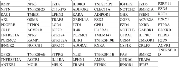
A total of 94 human growth receptors met the signal intensity cut-off of fifty consistently across the all four microarray analyses (Table II).
D. Signaling Pathways
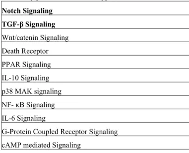
The 94 expressed HGRs were input into Ingenuity Pathways Analysis. Canonical pathways that were associated with at least 1 of the 94 HGRs in Ingenuity Pathways Knowledge Base were identified. Table III shows the 11 signaling pathways that consisted of at least one of the above 94 human growth receptors. The signaling pathways reported were limited to Ingenuity Pathways Knowledge Base. These signaling pathways and their corresponding growth factors are important in understanding how MSCs maintain and proliferate as well as in developing a serum-free medium for expansion.
E. Notch Signaling Pathway and TGF- β Signaling Pathway
Overall Expression
Ingenuity Pathway Analysis was used to determine the
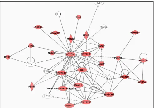
expression of the Notch Signaling Pathway and TGF-β Signaling Pathway across each of the four global gene expression datasets. Both pathways were consistently expressed across all four microarray analyses. Fig. 1 shows the overall expression of the Notch Signaling Pathway in the AS culture with Affymetrix as the microarray platform. Fig. 2 shows the overall expression of the TGF-β Signaling Pathway in the FBS culture with Illumina as the microarray platform. The figures show all objects associated in that particular pathway. The shade of each object represents the expression intensity. Higher levels of expression are represented by darker intensities.
The Notch Signaling pathway is commonly involved in intercellular signaling, proliferation, and cell fate determination during development. Ligands of the Notch Signaling Pathways belong to two subclasses of the Delta and Serrate-Lag2 family. In the human, there are two members from the Serrate family (JAG1 and JAG2) and three members from the Delta family (DLL1, DLL3 and DLL4). These ligands can bind with four Notch receptors (NOTCH1, NOTCH2, NOTCH3, NOTCH4). From Fig. 1, it can be seen that only 3 ligands were expressed (JAG1, JAG2, DLL3) and all four Notch receptors were expressed.
Table II: HGRs expressed across all microarray platforms and serum types
BZRP NPR3 FZD7 IL10RB TNFSF5IP1 IGFBP2 FZD6 P2RY11
NPTN TNFRSF25 C11orf75 ADIPOR2 CLEC11A NOTCH2 BMPR1A FZD5
RAC1 TMED1 LPHN2 RARA ADIPOR1 GHR PSEN1 ROR1
AXL OSMR TRAF5 GRINL1A FZD2 OGFR ACVR2A P2RY5
PDGFRB PTPRN LGR4 FZD1 GPR1 FZD4 RXRB PTPRA
CRLF1 ACVR1B IGF2R IL4R IL13RA1 NOTCH3 GABBR1 BDKRB1
TNFRSF1A NPR2 GPR124 PGRMC1 TMEM147 GFRA1 IL17RC PILRB
PGRMC2 RAMP1 GPR172A IL1R1 TNFRSF10B GRM4 CMKOR1 FZD9
IFNGR2 NOTCH1 GPR175 ADORA1 RXRA CSF1R CRLF3 ACVR1
OPRS1 TNFRSF6B PTPRG NLE1 TNFRSF11B FAS BMPR2
Table III: Signaling pathways expressed across all microarray platforms and serum types
Notch Signaling
TGF-β Signaling
Wnt/catenin Signaling Death Receptor PPAR Signaling IL-10 Signaling p38 MAK signaling NF- κB Signaling IL-6 Signaling
G-Protein Coupled Receptor Signaling cAMP mediated Signaling
IV. DISCUSSION
Although there have been successful reports that support proliferation of MSCs using human serum [16, 18], which effectively eliminates risk of the xenogenic contamination, there has been no significant breakthrough that supports the proliferation of MSCs in serum-free medium [14]. The increasing therapeutic applications of MSCs accentuate the need for a serum-free supplement in order to support large-scale expansion and reproducible laboratory-to-laboratory conditions. The discovery of growth factors involved in MSC proliferation is key in developing the serum-free medium.
This study reported 94 HGRs and their corresponding 11 signaling pathways expressed during undifferentiated MSC proliferation. The 11 signaling pathways have been generally reported to be associated with cell proliferation and apoptosis. However, the exact role of each HGR and pathway in regulating cell proliferation and maintaining multipotency in MSC is unclear, although their consistent expressions across all four MSC cell cultures indicate key functions. The next step would be to investigate each HGR and signaling pathway individually in order to determine
more accurately the function of the pathways and consequently the role of each growth factor.
Besides growth factors, conventional FBS serum also contains a range of macromolecules, carrier proteins for lipoid substances and trace elements, attachment and spreading factors, low molecular weight nutrients, and hormones [21]. These other components are critical for MSC proliferation and have to be defined in addition to the growth factors in order to develop a universally defined serum-free medium.
In this study, microarray data from three MSC cultures supplemented with FBS and one MSC culture supplemented with AS. It has been previously reported that serum supplement is a determinant of gene expression, with differential expression reflected from passage 4 [12]. When microarray analyses of MSC cell cultures were performed at passage 1, the difference in HGR expression was insignificant. Hence, it is possible that differential expression occurs only at later passages.
This study also reviewed the HGRs that were in agreement between the two platforms. This was done to ensure that the most expressed HGRs were represented. Another analysis involving all HGRs detected across each platform should be done. It would also be interesting to investigate the HGRs that were detected by the Affymetrix platform but not by the Illumina platform. While the expression levels of Notch ligands and receptors in undifferentiated MSCs are identified, it is helpful to compare these expression levels with differentiated cells further down the MSC lineage. We would be able to establish which ligands and receptors are differently regulated, and hence the function of the Notch Signaling pathway in MSCs.
Figure 1: Overall expression of the Notch Signaling Pathway in the MSC sample cultured in AS and analyzed by Affymetrix
REFERENCES
[1] Bruder SP, Kurth AA, Shea Metal. Bone regeneration by implantation of purified, culture-expanded human mesenchymal stem cells. J Orhtop RES 1998;16:155-162
[2] De Kok I, Peter SJ, Archambault Metal. Investigation of allogeneic mesenchymal stem cell-based alveolar bone formation: preliminary findings. ClinOral Implants Res 2003;14:481-489
[3] Wakitani S, Goto T, Pineda SJ et al. Mesenchymal cell-based repair of large, full thickness defects of articular cartilage. J Bone Joint Surg AM 1994;76:579-592
[4] Pittenger MF, Mackay AM, Beck SC et al. Multilineage potential of adult human mesenchymal stem cells. Science 1999;284:143-147 [5] Muraglia A, Cancedda R, Quarto R. Clonal mesenchymal progenitors
from human bone marrow differentiate in vitro according to a hierarchical model. J Cell Sci 2000;113:1161-1166
[6] Murphy JM, Fink DJ, Hunziker EB et al. Stem cell therapy in a caprine model of osteoarthritis. Arthritis Rheum 2003;95:9-20
[7] Woodbury D, Schwarz EJ, Prockop DJ et al. Adult rat and human bone marrow stromal cells differentiate into neurons. J Neurosci Res 2000;61:364-370
[8] Kopen GC, Prockop DJ, Phinney DG. Marrow stromal cells migrate throughout forebrain and cerebellum, and they differentiate into astrocytes after injection into neonatal mouse brains. Proc Natl Acad Sci U S A 1999;96:10711-10716
[9] Deans RJ, Moseley AB. Mesenchymal stem cells: biology and potential clinical uses. Exp Hematol 2000;28:875-884.
[10] Horwitz EM, Gordon PL, Koo WK et al. Isolated allogeneic bone marrow-derived mesenchymal cells engraft and stimulate growth in children with osteogenesis imperfecta: Implications for cell therapy of bone. Proc Natl Acad Sci.U.S.A 2002;99:8932-8937.
[11] Kuznetsov SA, Mankani MH, Robey PG. Effect of serum on human bone marrow stromal cells: ex vivo expansion and in vivo bone formation. Transplantation 2000;70:1780-1787.
[12] Shahdadfar A, Fronsdal K, Haug T, Reinholt F P and Brinchmann J E, In Vitro Expansion of Human Mesenchymal Stem Cells: Choice of Serum Is a Determinant of Cell Proliferation, Differentiation, Gene Expression, and Transcriptome Stability. Stem Cells 2005;23:1357-1366
[13] Stute N, Holtz K, Bubenheim M et al. Autologous serum for isolation and expansion of human mesenchymal stem cells for clinical use. Exp.Hematol. 2004;32:1212-1225.
[14] Matti K. Culture of human mesenchymal stem cells in serum-free conditions: no breakthrough yet. Eur J Haematol. 2006;78:167 [15] Caplan A I, Mesenchymal Stem Cells in Lanza R, et. al. (eds.),
Handbook of Stem Cells Volume 2 Adult and Fetal. Elsevier Acad Press 2004; 299-305
[16] Muller I, Kordowich S, Holzwarth C, Spano C, Isensee G, Staiber A, Viebahn S, Gieseke F, Langer H, Gawaz MP, Horwitz EM, Conte P, Handgretinger R, Dominici M. Animal serum-free culture conditions for isolation and expansion of multipotent mesenchymal stromal cells from human BM. Cytotherapy 2006;8(5):437-44.
[17] Nardi N B and da Silva Meirelles L, Mesenchymal stem cells: Isolation, in vitro expansion and characterization. Handb. Exp. Pharmacol 2006;174:249-282
[18] Romanov Y A, Darevskaya A N, Kabaeva N V and Antonova O A, Optimum conditions for culturing of human bone marrow and adipose tissue mesenchymal precursor cells. Bull of Exp Biol and Med 2006;142:515-520
[19] Koc ON, Day J, Nieder M, Gerson SL, Lazarus HM, Krivit W. Allogeneic mesenchymal stem cell infusion for treatment of metachromatic leukodystrophy (MLD) and Hurler syndrome (MPS-IH). Bone Marrow Transplant 2002;30:215-222
[20] Koc ON, Gerson SL, Cooper BW, Dyhouse SM, Haynesworth SE, Caplan AI, Lazarus HM. Rapid hematopoietic recovery after coinfusion of autologous-blood stem cells and culture-expanded marrow mesenchymal stem cells in advanced breast cancer patients receiving high-dose chemotherapy. J Clin Oncol 2000;18:307-316 [21] Gstraunthaler G. Alternatives to the use of fetal bovine serum: