Multiplex-PCR-Based Screening and
Computational Modeling of Virulence Factors
and T-Cell Mediated Immunity in
Helicobacter pylori
Infections for Accurate
Clinical Diagnosis
Sinem Oktem-Okullu1,2, Arzu Tiftikci3,4, Murat Saruc3,4, Bahattin Cicek3,4, Eser Vardareli3,4, Nurdan Tozun3,4, Tanil Kocagoz2, Ugur Sezerman5, Ahmet Sinan Yavuz6, Ayca Sayi-Yazgan1*
1Department of Molecular Biology and Genetics, Istanbul Technical University, Maslak, Istanbul, Turkey,
2Department of Medical Microbiology, School of Medicine, Acibadem University, Atasehir, Istanbul, Turkey,
3Department of Internal Medicine, School of Medicine, Acibadem University, Atasehir, Istanbul, Turkey,
4Department of Gastroenterology, Acibadem Hospital Group, Istanbul, Turkey,5Department of Biostatistics and Medical Informatics, School of Medicine, Acibadem University, Atasehir, Istanbul, Turkey,
6Molecular Biology, Genetics and Bioengineering Program, Faculty of Engineering and Natural Sciences, SabancıUniversity, Tuzla, Istanbul, Turkey
Abstract
The outcome ofH.pyloriinfection is closely related with bacteria's virulence factors and host immune response. The association between T cells andH.pyloriinfection has been identified, but the effects of the nine majorH.pylorispecific virulence factors;cagA,vacA,
oipA,babA,hpaA,napA,dupA,ureA,ureBon T cell response inH.pyloriinfected patients have not been fully elucidated. We developed a multiplex- PCR assay to detect nineH.
pylorivirulence genes with in a three PCR reactions. Also, the expression levels of Th1, Th17 and Treg cell specific cytokines and transcription factors were detected by using qRT-PCR assays. Furthermore, a novel expert derived model is developed to identify set of fac-tors and rules that can distinguish the ulcer patients from gastritis patients. Within all viru-lence factors that we tested, we identified a correlation between the presence ofnapA
virulence gene and ulcer disease as a first data. Additionally, a positive correlation between theH.pylori dupAvirulence factor and IFN-γ, andH.pylori babAvirulence factor and IL-17
was detected in gastritis and ulcer patients respectively. By using computer-based models, clinical outcomes of a patients infected withH.pylorican be predicted by screening the patient'sH.pylori vacA m1/m2,ureAandcagAstatus and IFN-γ(Th1), IL-17 (Th17), and
FOXP3 (Treg) expression levels. Herein, we report, for the first time, the relationship betweenH.pylorivirulence factors and host immune responses for diagnostic prediction of gastric diseases using computer—based models.
OPEN ACCESS
Citation:Oktem-Okullu S, Tiftikci A, Saruc M, Cicek B, Vardareli E, Tozun N, et al. (2015) Multiplex-PCR-Based Screening and Computational Modeling of Virulence Factors and T-Cell Mediated Immunity in
Helicobacter pyloriInfections for Accurate Clinical Diagnosis. PLoS ONE 10(8): e0136212. doi:10.1371/ journal.pone.0136212
Editor:Jian Zhang, The Ohio State University, UNITED STATES
Received:March 5, 2015
Accepted:July 30, 2015
Published:August 19, 2015
Copyright:© 2015 Oktem-Okullu et al. This is an open access article distributed under the terms of the
Creative Commons Attribution License, which permits unrestricted use, distribution, and reproduction in any medium, provided the original author and source are credited.
Data Availability Statement:All relevant data are within the paper.
Funding:The financial support of this work was provided by Istanbul Technical University through Scientific Research Project Funds with the grant number 36243.
Introduction
Helicobacter pylori (H.pylori)is a gram negative, microaerophilic, spiral-shaped bacteria that col-onize in human gastric mucosa and if not treated, it persists through lifespan.H.pyloriinfection has been implicated as the main cause of chronic gastritis, peptic ulcers and gastric adenocarci-noma [1,2].H.pyloriis carried by more than half of the world’s population with prevalence as high as 90% in developing countries [3]. Although most infected individuals remain asymptom-atic, 15–20% ofH.pyloripositive individuals develop at least one of the associated diseases at some point in their lives [4]. The clinical outcome ofH.pyloriinfection is determined by multiple factors includingH.pylorirelated virulence factors, host genetic predisposition and immune response [5,6]. Many putative virulence genes have been described to determine the clinical out-come ofH.pyloriinfection. In the recent studies, it is well established that virulence factors with potential value for specific pathologies are the cytotoxin associated gene A (cagA), vacuolating cytotoxin gene A (vacA), outer inflammatory protein A (oipA), the blood group antigen binding adhesin gene A (babA), the putative neuraminyllactose-binding hemagglutinin homolog (hpaA), neutrophil-activating protein A (napA), duodenal ulcer promoting gene A (dupA), urease A (ureA), and urease B (ureB) [7,8]. Although virulence factors are important in conditioning the clinical outcome of theH.pylori–driven infection, the local immune response mechanisms have been claimed to play a role in the pathogenesis of the disease [9]. Regarding host immune response, CD4+ T cells including T- helper cells (Th) and regulatory T-cells (Tregs) are shown to play critical roles in the pathogenesis ofH.pyloriinfection. T helper 1 (Th1) cells contribute to the host defense against pathogens by secreting various cytokines and through their effector functions. Recently described T helper 17 (Th17) cells are shown to play a role in host defense againstH.pyloriinfection by mediating the recruitment of neutrophils and macrophages into infected tissues.H.pyloriinfection may induce a mixed Th1/Th17 response. Tregs have been identified as the major regulatory component of the adaptive immune response and they have been involved inHelicobacter pylori-related inflammation and bacterial persistence [10–14].
In previous studies, single or multiplex-PCR assays have been developed to detect theH.
pylorivirulence factors. However, these PCR assays are not sufficient to detect the most impor-tantH.pylorivirulence factors simultaneously. Herein, we describe a sensitive and specific multiplex-PCR with a wide range of virulence factors which can detect nine virulence genes in three PCR reactions. This may help to better understand the correlation between the virulence factors and different clinical forms of gastric lesions and therefore the pathogenesis of each of these factors. Development of the gastric disease duringH.pyloriinfection depends on an acti-vated adaptive immune response controlled by T helper (Th) cells. However, the relative con-tributions of the Th1 and Th17 subsets and Tregs to gastric diseases and correlation of Th1, Th17 and Tregs withH.pylorivirulence factors are not well understood. In this study, we investigate the association of theH.pylorivirulence factors with Th1, Th17 and Treg cells by determining the cytokines and transcription factor specific for these T cells in mRNA expres-sion levels. Herein this report, it is the first time that an expert derived model was developed to show the relationship betweenH.pylorivirulence factors and host immune response for diag-nostic prediction of gastric diseases.
Materials and Methods
Ethical Approval
Patients Selection
Biopsy specimens were obtained from the patients who underwent endoscope because of gas-troduodenal diseases at the Gastroenterology Department of Acibadem Hospital Groups, in Istanbul, Turkey. In total, 80 patients were selected who fulfilled the following criteria for obtaining expulsion: under 18 years, 65 years or older patients with active infection, cancer patients, patients with inflammatory disease, patients who have had gastrointestinal bleeding within the last month and who have had previous gastrointestinal surgery, patients diagnosed with chronic liver failure, known renal failure, diabetic patients, pregnant women and patients who have previously receivedH.pylorieradication treatment, immunosuppressive therapy (including steroids), patients who have had NSAIDs and / or antibiotic treatment in the last three weeks, antisecretory therapy in the last two weeks and patients who refused to sign the informed consent form voluntarily. 18H.pylorinegative individuals were included to the study as a control group.
Gastric biopsy specimens
Two gastric biopsy specimens were taken from the antrum and corpus part of the patients’
stomach during endoscopy, one for DNA isolation and the other for RNA isolation. Fresh biopsy specimens were placed into the RNAlater solution (Ambion, RNAlater RNA Stabiliza-tion SoluStabiliza-tion) and kept at + 4°C for overnight, then put at–80°C deep freezer until DNA and RNA isolation.
RNA extraction
To study gene expression, RNA was isolated from RNALater-stabilized human tissue speci-mens. Frozen biopsy specimens were homogenized by using a high efficiency homogenization system (SpeedMill PLUS, Analytikjena) and total RNA was isolated by using RNA isolation kit, following the protocol recommended by the manufacturer (innuSPEED tissue RNA kit Analy-tikjena). All RNA samples were quantified using NanoDrop (ND-2000, ROCHE) and main-tained in–80°C deep freezer until cDNA synthesis. cDNA was synthesized using 1μg of RNA
through a reverse transcription reaction (High capacity cDNA Reverse Transcription Kit, Applied Biosystems).
DNA extraction
To study virulence factors ofH.pylori, DNA was extracted immediately after RNA isolation by using a DNA isolation kit (Quick g-DNATM, ZYMO RESEARCH) following the manufactur-er’s description. All DNA samples were quantified using NanoDrop (ND-2000, ROCHE) and maintained in–80°C deep freezer.
Primer design
(Table 2). A BLAST search was performed to confirm the specificity of the DNAsequences of all the primers (http://www.ncbi.nlm.nih.gov/BLAST/).
Multiplex-PCR assay
Genomic DNA isolated fromH.pyloriG27 strain used in this study were a kind gift from Prof. Dr. Anne Mueller from University of Zurich, Institute of Molecular Cancer Research, Switzer-land. Genomic DNA ofH.pyloriG27 strain and gastric specimens were used as target DNAs in multiplex-PCR assays. The amplification reactions were carried out in a total volume of 25μl and the multiplex-PCR reaction mixture consisted of 0.65 U of Dream Taq DNA
poly-merase (Thermo Scientific), 2.5μl from 10X DreamTaq Buffer (includes 20 mM MgCl2),
20μM of forward and reverse primers, 200μM of each dNTP and 3μl DNA. Amplification
programme included an initial denaturation step at 95°C for 3 min followed by 45 cycles of
Table 1. Multiplex-PCR primers designed for the amplification ofH.pylorivirulence genes.
DNA region(s) amplified Primer Name Sequences (50!3) PCR Product Size (bp) References
ure A ure A-F TGATGGGACCAACTCGTAACCGT 244 This study
ure A ure A-F CGCAATGTCTAAGCGTTTGCCGAA 244 This study
ure B ure B-F AGTAGCCCGGTGAACACAACATCCT 645 This study
ure B ure B-F ATGCCTTTGTCATAAGCCGCTTGG 645 This study
cag A cag A-F AGAGCAAGCGTTAGCCGATCTCAA 415 This study
cag A cag A-R TTTCCCTACACCACCCAAACCACT 415 This study
hpa A hpa A-F TAGTGGGATGCAGCCCGCATATTA 534 This study
hpa A hpa A-R CGCTATGGCTTGAATGGGTGGTTT 534 This study
oip A oip A-F GTTTTTGATGCATGGGATTT 401 [15]
oip A oip A-R GTGCATCTCTTATGGCTTT 401 [15]
bab A bab A-F AATCCAAAAAGGAGAAAAAGTATGAAA 832/601 [16,17]
bab A bab A-R TGTTAGTGATTTCGGTGTAGGACA 832/601 [16,17]
dup A dup A-F TGAGCGTGGTAGCTCTTGAC 584 This study
dup A dup A-R GAGCGCGTTAGCGATATAGG 584 This study
nap A nap A-F GAATGTGAAAGGCACCGATT 304 This study
nap A nap A-R ATCGTCCGCATAAGTTACGG 304 This study
vac A s1/s2 vac A s1/s2-F ATGGAAATACAACAAACACAC 259/286 [1]
vac A s1/s2 vac A s1/s2-R CTGCTTGAATGCGCCAAAC 259/286 [1]
vac A m1/m2 vac A m1/m2-F CAATCTGTCCAATCAAGCGAG 567/642 [18]
vac A m1/m2 vac A m1/m2-R GCG TCTAAATAATTCCAAGG 567/642 [18]
doi:10.1371/journal.pone.0136212.t001
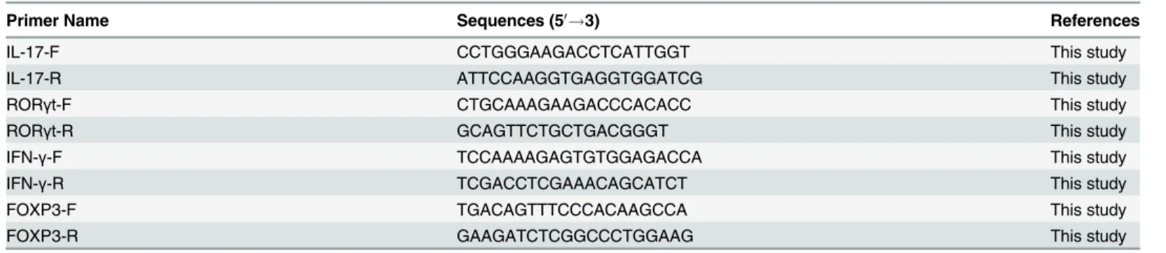
Table 2. Quantitative RT-PCR primers designed for the detection of Th1, Th17 and Treg cell response.
Primer Name Sequences (50!3) References
IL-17-F CCTGGGAAGACCTCATTGGT This study
IL-17-R ATTCCAAGGTGAGGTGGATCG This study
RORγt-F CTGCAAAGAAGACCCACACC This study
RORγt-R GCAGTTCTGCTGACGGGT This study
IFN-γ-F TCCAAAAGAGTGTGGAGACCA This study
IFN-γ-R TCGACCTCGAAACAGCATCT This study
FOXP3-F TGACAGTTTCCCACAAGCCA This study
FOXP3-R GAAGATCTCGGCCCTGGAAG This study
denaturation at 95°C for 45 s, primer annealing at 60°C for 45 s and primer extension at 72°C for 2 mins, with final extension step at 72°C for 5 mins. The PCR products were subjected to electrophoresis on agarose gels and stained with SYBR Gold (Invitrogene). The specificity of the primer pairs was confirmed by employing a positive control.
Quantitative RT-PCR assay
Optimal annealing temperatures of the qRT-PCR primer pairs and expected product size were determined with conventional RT-PCR. SYBR Green qRT-PCR amplifications were performed using a LightCycler 480 Real-Time Detection System (ROCHE). All qRT-PCR experiments were carried out in duplicate with a reaction volume of 10μl, using 96-well optical grade PCR
plates (ROCHE) covered with optical-quality sealing film (ROCHE). The efficiency of qRT-PCR amplification was optimized for each primer pair, using various dilution series of cDNA. The reaction mixtures for the optimized LightCycler 480 SYBR Green I Master Mix based assays consisted of 5μl SYBR Green I Master Mix, 0.5μM from each primer, 1.5μl PCR
grade water and 2.5μl target cDNA. The amplification reaction was performed with
prelimi-nary denaturation for 10 min at 95°C, followed by 40 amplification cycles of denaturation at 95°C for 15 s, primer extension at 58°C for 1 min, extension at 72°C for 1, 30 min and addi-tional final extension at 72°C 5 min. A final cooling step was performed at 4°C. The percentage of expression of the cytokines and the transcription factors are calculated from quantitative RT-PCR results based on the cycle threshold (Ct). Relative quantification method was used to analyze the PCR. Relative quantification describes the change in expression of the target gene relative to the Hp negative control group. 2-ΔΔCT Method is used to calculate relative changes in the gene expression determined by the quantitative RT-PCR and as an internal control 18S rRNA housekeeping gene primers were used to normalize the quantitative RT-PCR. Negative controls without cDNA and positive controls for gene were included
Statistical analysis
A two-tailed Fisher’s exact test was performed to examine the relationship betweenH.pylori
virulence factors and gastric disease type (i.e. Gastritis or Ulcer). Additionally, correspondence between virulence factor presence and immune response was assessed with Pearson product-moment correlation coefficients. Statistical significance of correlations between virulence fac-tors and immune response facfac-tors was assessed using a t-test. All calculations were performed using R (version 3.1.0, The R Foundation for Statistical Computing, Vienna, Austria;http:// www.r-project.org).
Expert-derived models for diagnostic prediction
Results
Detection of
H
.
pylori
virulence factors by multiplex-PCR
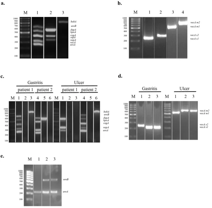
Initially, to optimize the concentrations of the conventional multiplex-PCR components, the specificity of each primer pair, and the thermocycling parameters for each virulence factor, genomic DNA isolated fromH.pyloriG27 strain were used as the target DNA. We optimized multiplex-PCR of genomic DNA derived fromH.pyloriG27 strain to detect nineH.pylori vir-ulence genes within a three PCR reaction;ureB,hpaA,cagA,napA,ureAin a single reaction,
dupA,oipA,vacAin a single reaction, andbabAin a single reaction (Fig 1(a) and 1(b)). Next, the multiplex-PCR was applied to genomic DNA derived from total of 80H.pyloripositive patients; 18 with ulcer and 62 with gastritis. Two representative multiplex-PCR data of nineH.
pylorivirulence genes obtained from these gastric specimens are shown inFig 1(c). In order to investigateH.pylori vacAsubtypes invacApositive gastric specimens, we performed four sepa-rate PCR reactions (Fig 1(d)). The genes encodingH.pyloriurease which areureAandureB, were also detected by a single multiplex-urease PCR assay to confirm the presence ofH.pylori
in gastric biopsy specimens which were detected by rapid urease test and pathological staining following endoscopy (Fig 1(e)). Hp-uninfected control group was used to check the multiplex-PCR primers for the virulence genes to monitor non-specific amplicons.
Correlation between the manifestations of gastric disease and bacterial
virulence factors
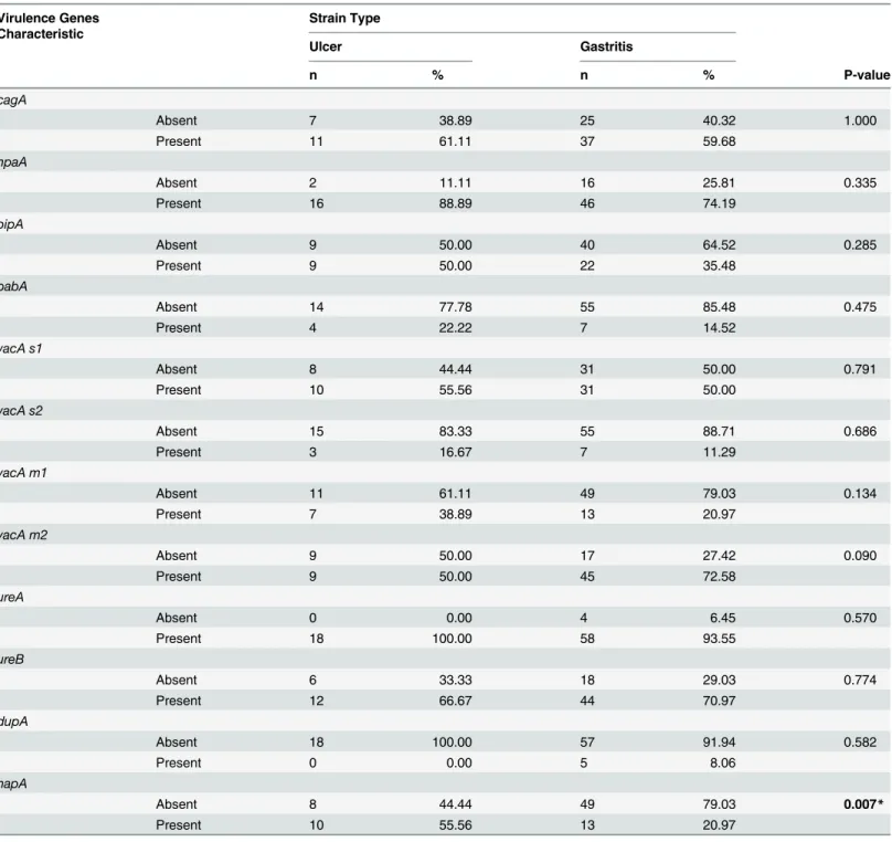
Based on multiplex-PCR results which were performed using 18 patients with ulcer and 62 patients with gastritis, no statistically significant difference was detected for the existence ofH.
pylori cagA,hpaA,oipA,babA,vacAs1,vacAs2,vacAm1,vacAm2,ureA,ureB anddupA viru-lence factors among patients with gastritis and ulcer (Table 3).H.pylori cagAgene amplifica-tion was found in 60% of all isolates; 61% of patients with ulcer and 60% of patients with gastritis. TheH.pylori vacA s1/m2genotypes were the most frequent allelic combination of the vacA gene detected both among gastritis and ulcer patients. TheH.pylori oipAgene prevalence was more frequent in ulcer patients than that of gastritis patients (50%, 35.48%). Also, there were no statistically significant difference between the patients with ulcer and gastritis for the presence ofH.pylori hpaAgene (88.89% in ulcer patients, 74.19% for in gastritis patients), for
babAgene (22.22% in ulcer patients, 14.52% for in gastritis patients), forureAgene (100% in ulcer patients, 93.55% for in gastritis patients) and forureBgene (66.67% in ulcer patients, 70.97% for in gastritis patients). Additionally, no ulcer patients were positive for dupA and only 8.06% of gastritis patients were positive for dupA gene.
However, the prevalence ofH.pylori napAvirulence factor was significantly higher in patients with ulcer than gastritis (Table 3).
Correlation between
H
.
pylori
virulence factors and involvement of Th1,
Th17 and Treg in H. pylori-induced gastric diseases
Fig 1. Multiplex-PCR Assay ForHelicobacter-specific Virulence Factors. (a)amplification of virulence genes ofH.pyloriG27 strain by multiplex PCR; Lane M, 100 bp- marker (ThermoSCIENTIFIC, Gene Ruler), Lane 1 (Reaction 1);ureA(244bp),ureB(645bp),hpaA(534bp),cagA(415bp),napA(384bp), Lane 2 (Reaction 2);dupA(584bp),oipA(401bp),vacA(333bp), and Lane 3 (Reaction 3);babA(832bp)(b)PCR inferred results ofs1/s2andm1/m2allelles ofvacAgene; Lane M, 100 bp- marker (ThermoSCIENTIFIC, Gene Ruler), Lane 1 (reaction 1);vacA s1(259bp), Lane 2 (Reaction 2);vacA s2(286 bp), Lane 3 (Reaction 3);vacA m1(567 bp), and Lane 4 (Reaction 4)vacA m2(642 bp).(c)Multiplex-PCR application of the biopsy samples taken from randomly selected patients with ulcer and gastritis; Lane M, 100 bp- marker (ThermoSCIENTIFIC, Gene Ruler), between Lane 1 to 6 are representing patients with gastritis or ulcer: Multiplex PCR reaction results of patient 1 and 2 (gastritis or ulcer) are shown between Lane 1 to 3 and Lane 4 to 6, respectively. Reaction 1 (Lane 1 and 4)ureA(244bp),ureB(645bp),hpaA(534bp),cagA(415bp),napA(384bp), Reaction 2 (Lane 2 and 5);dupA(584bp),oipA(401bp),vacA (333bp), and Reaction 3 (Lane 3 and 6);babA(832bp)(d)PCR inferred results ofs1/s2andm1/m2allelles ofvacAgene of randomly selected patients with gastritis and ulcer respectively; Lane M, 100 bp- marker (ThermoSCIENTIFIC, Gene Ruler), between Lane 1 to 3 are representing patients with gastritis (left side) or ulcer (right side): Patients with gastritis; Lane 1–3,vacA s1(259bp), andvacA s2(286 bp), patients with ulcer; Lane 1–3,vacA m1(567 bp), andvacA m2(642 bp).(e)Multiplex urease-PCR assay to detect theHelicobacterpositive and negative samples; Lane M, 100 bp- marker (ThermoSCIENTIFIC, Gene Ruler), Lane 1–2 are from randomly selected patients, and lane 3 is fromH.pyloripositive control strain G27. Sybr Gold (Invitrogene) was used for the gel in (a) and (c) and gel pictures were taken by using Observable Real Time Gel Electrophoresis System (Salubris Technica, Turkey).
individuals were used as a control group to verify the Th1 and Th17 immune response differ-ences between the Hp-infected and Hp-uninfected patients, and in order to normalize our method in quantitative RT-PCR. Our data demonstrated a positive correlation between theH.
pylori dupAvirulence factor and IFN-γin gastritis patients (r = 0.31, N = 62, p<0.05).
Table 3. Comparison of virulence genes ofH.pyloristrains isolated from patients suffering from gastritis and ulcer.
Virulence Genes Characteristic
Strain Type
Ulcer Gastritis
n % n % P-value
cagA
Absent 7 38.89 25 40.32 1.000
Present 11 61.11 37 59.68
hpaA
Absent 2 11.11 16 25.81 0.335
Present 16 88.89 46 74.19
oipA
Absent 9 50.00 40 64.52 0.285
Present 9 50.00 22 35.48
babA
Absent 14 77.78 55 85.48 0.475
Present 4 22.22 7 14.52
vacA s1
Absent 8 44.44 31 50.00 0.791
Present 10 55.56 31 50.00
vacA s2
Absent 15 83.33 55 88.71 0.686
Present 3 16.67 7 11.29
vacA m1
Absent 11 61.11 49 79.03 0.134
Present 7 38.89 13 20.97
vacA m2
Absent 9 50.00 17 27.42 0.090
Present 9 50.00 45 72.58
ureA
Absent 0 0.00 4 6.45 0.570
Present 18 100.00 58 93.55
ureB
Absent 6 33.33 18 29.03 0.774
Present 12 66.67 44 70.97
dupA
Absent 18 100.00 57 91.94 0.582
Present 0 0.00 5 8.06
napA
Absent 8 44.44 49 79.03 0.007*
Present 10 55.56 13 20.97
H.pyloristrains withnapAvirulence factor are found more frequently in patients with ulcer than gastritis. Statistical significance was assessed with
two-sided Fisher’s exact test.
Additionally, IL-17 gene expression was shown for the first time to significantly positively correlated with theH.pylori babAvirulence factor in ulcer patients (r = 0.74, N = 18, p<0.001).
Expert-derived models for diagnostic prediction of gastric diseases using
H
.
pylori
virulence factors and host immune responses
We created two expert-derived models (Fig 2(a) and 2(b)), which show the relationship between theH.pylorispecific virulence factors and Th1, Th17 and Treg response on the basis of their specific cytokines and transcription factor levels inH.pyloriinfected gastritis or ulcer patients. By the aid of expert-derived models, knowledge ofH.pylorivirulence factors:
vacAm1/m2,cagAandureAand IL- 17, FOXP3 and IFN-γexpression level, it is possible to pre-dict a patient’s clinical outcomes.
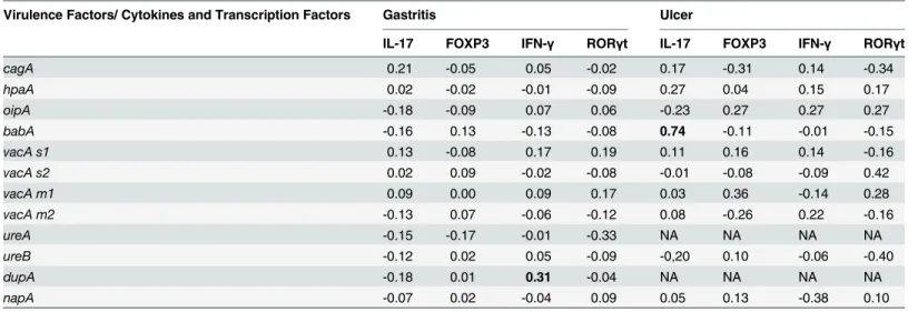
Table 4. Correlation between virulence factors and cytokines, transcription factors.
Virulence Factors/ Cytokines and Transcription Factors Gastritis Ulcer
IL-17 FOXP3 IFN-γ RORγt IL-17 FOXP3 IFN-γ RORγt
cagA 0.21 -0.05 0.05 -0.02 0.17 -0.31 0.14 -0.34
hpaA 0.02 -0.02 -0.01 -0.09 0.27 0.04 0.15 0.17
oipA -0.18 -0.09 0.07 0.06 -0.23 0.27 0.27 0.27
babA -0.16 0.13 -0.13 -0.08 0.74 -0.11 -0.01 -0.15
vacA s1 0.13 -0.08 0.17 0.19 0.11 0.16 0.14 -0.16
vacA s2 0.02 0.09 -0.02 -0.08 -0.01 -0.08 -0.09 0.42
vacA m1 0.09 0.00 0.09 0.17 0.03 0.36 -0.14 0.28
vacA m2 -0.13 0.07 -0.06 -0.12 0.08 -0.26 0.22 -0.16
ureA -0.15 -0.17 -0.01 -0.33 NA NA NA NA
ureB -0.12 0.02 0.05 -0.09 -0,20 0.10 -0.06 -0.40
dupA -0.18 0.01 0.31 -0.04 NA NA NA NA
napA -0.07 0.02 -0.04 0.09 0.05 0.13 -0.38 0.10
A Pearson’s correlation between virulence factors and cytokines were calculated using R (version 3.1.0, The R Foundation for Statistical Computing, Vienna, Austria;http://www.r-project.org).
doi:10.1371/journal.pone.0136212.t004
Fig 2. Expert-derived models for diagnostic prediction of gastric diseases usingH.pylorivirulence factors and host immune responses. (a)shows that a possible relationship prediction between thevacAm1/m2,cagAandureAand IL-17, FOXP3 and patient’s clinical outcomes and(b)shows that the possible relationship prediction betweenvacAm1/m2,cagAandureAand IL-17, FOXP3 and IFN-γand patient’s clinical outcomes.
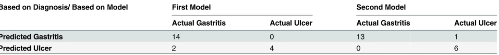
Performance evaluation using repeated test set sampling showed that first model (Fig 2(a)) has mean accuracy of 79% (standard deviation: 9%) in gastritis classification, 44% (standard deviation: 16%) in ulcer classification. Overall mean classification accuracy of this model was found as 69% (standard deviation: 8%). On the other hand, performance evaluations of the sec-ond model (Fig 2(b)) with repeated test set sampling resulted in a performance of 71% (stan-dard deviation: 1%) mean accuracy for gastritis predictions, 61% mean accuracy (stan(stan-dard deviation: 17%) for ulcer predictions, which corresponds to a 68% overall mean accuracy (stan-dard deviation: 9%). Confusion matrix of models for best performing test set samples can be found inTable 5.
Discussion
The virulence factors ofH.pylori, which are known to be directly correlated with the extreme degree of genetic heterogeneity inH.pylorigenomes, play a pivotal role in determining the out-come ofH.pyloriinfection. The immune response of the host including T cell activation has also been the subject of recent studies, together with specific virulence factors ofH.pylori[19]. Multiplex-PCR based genotyping systems have initially been developed to detect the Helicobac-ter pylori-specific virulence factors. However, these assays were not sufficient to detect many
Helicobacter pylori-specific virulence factors simultaneously that are thought to be associated with bacterial pathogenesis and increase the risk for developing severe clinical manifestations. In our present study, a genotyping system-based multiplex-PCR assay was developed to detect nine potential virulence genes (vacA,cagA,oipA,babA,hpaA,dupA,napA,ureA,ureB) within three PCR reactions directly from human gastric biopsies. This assay is able to detect both the presence and absence ofH.pyloribyureAandureBgenes, and identify the leading disease-associated virulence genes ofH.pyloristrains isolated from patients. Moreover, this PCR assay helps to better understand the correlation between the virulence factors and different clinical forms of gastric lesions and the pathogenesis of each of these factors.
Our multiplex-PCR assay results indicated no correlation between theH.pylorigenesvacA,
cagA,oipA,babA,hpaA,dupA,ureA,ureBandH.pylori-related gastritis and ulcer diseases. However, we identified a statistically significant correlation (p = 0.007) betweennapAgene andH.pylori-associated ulcer disease. In the literature it was indicated that activation of neu-trophil may contribute to the damage of stomach [20]. However, we could not find any research like in our study in which the relationship between thenapAgene and ulcer disease was showed clearly. It might be the first study to indicate the correlation betweenH.pylori
related-ulcer disease and presence ofnapAvirulence factor gene.
The type of host immune response, particularly driven by T cells, is crucial for the outcome ofH.pyloriinfection in humans. Given the increasing number of reports, correlations between the characteristics of the T helper cell responses, including Th1, Th17, and Treg cell response
Table 5. Confusion matrices for test set predictions.
Based on Diagnosis/ Based on Model First Model Second Model
Actual Gastritis Actual Ulcer Actual Gastritis Actual Ulcer
Predicted Gastritis 14 0 13 1
Predicted Ulcer 2 4 0 6
Test set samples with best overall classification accuracy was used in calculation of confusion matrices. The actual numbers were the numbers that represent the patients with gastritis and ulcer diagnosed based on clinical diagnosis. The predicted numbers were the number of patients with ulcer or gastritis based by the models.
and virulence factors should be of great interest in determining the outcomes ofH.pylori infec-tions. In previous studies, it was shown that Th1 cells contributed to inflammation and partici-pated in the pathogenesis ofH.pyloriinfection. However, the role of Th17 has not been clearly explicated. The efficacy of Th17 for the host protection against Gram-negative bacteria has also been shown [13]. In this study, we characterized the cell responses of T helper cells, particularly Th1 and Th17, in clinical isolates ofH.pyloriobtained from patients with gastritis and ulcer. One of the most prominent outcomes of the study is the identification of a higher Th17 cell response compared to Th1 response toH.pyloriinfection in patients with gastritis and ulcer. This finding has been shown previously in mice models [21], while this is the first time that it is identified in clinical samples. A possible reason for this outcome is that Th17 cells are clearly implicated in the pathogenesis ofH.pyloriinfection by promoting the mucosal infection and contributing to bacterial colonization. Also Th1 cells cause mucosal inflammation inH.pylori
infection, but our results suggested that Th17 cell responses might be induced earlier than Th1 cell responses inH.pyloriinfected ulcer and gastritis patients.
TheH.pylorispecific virulence factors are important for the pathogenesis of bacterium. These virulence factors facilitate the survival of the bacterium and immune response. In sup-port of this, we found that there was a correlation betweendupApositive strains and IFN-γ
(Th1-specific cytokine) expression level in patients with gastritis. Our results confirmed a pre-vious study that was indicated thatdupAcauses gastric inflammation and pathology either directly or indirectly over action upon infiltrating leukocytes rather than upon epithelial cells. It was also referred in the study thatdupApositiveH.pyloristrains increase the risk of disease by stimulating a more definite Th1 response [22].babAvirulence factor helps bacteria to adhere to the fucosylated Lewis b antigen on the surface of the gastric epithelial cells.H.pylori
adheres to the host and secretes effector molecules that can change function and viability of gastric epithelial cells. The production of various cytokines, gastric inflammation, and epithe-lial cell damage is enhanced by these changes. Also recent studies have indicated that there is severe mucosal injury in patients infected withbabApositiveH.pyloristrains [23]. Our data showed for the first time that there is a positive relationship betweenbabApositivity and inter-leukin-17 (IL-17) expression levels in patients with ulcer.
In this present study, we identified significant correlations between the virulence factors of
H.pylori, responses of T cells (Th1, Th17, Treg) and clinical outcomes ofH.pyloriinfections, and these eventually helped evolution of expert-derived models. Initially, first model (Fig 2(a)) was developed using the strategy outlined in the method section; however, low ulcer classifica-tion accuracy of this model indicated that there is a need for addiclassifica-tional factors to distinguish ulcer and gastritis patients more accurately. With this motivation, in transition to model 1 (Fig 2(a)) to model 2 (Fig 2(b)), we have used an additional factor (IFN-γ) that eventually improved the ulcer classification accuracy from 44% to 61% with a cost of only one misclassified gastritis patient. Further more in the first model predictions were done by using the results of the rela-tionship between the virulence factors, Th1 and Treg response while in the second model Th1 response also was added to these combinations. To our knowledge, this is the first report of expert-derived models that are based on the relationship betweenH.pylorivirulence factors and host immune responses. Using the proposed models, the likelihood for a patient to have gastritis or ulcer can be estimated by screening the presence ofH.pylorivirulence genes (i.e.
vacA m1/m2,ureAandcagA) and expression levels of IL-17, FOXP3 and IFN-γin the host. In the future, the model can be developed further to be utilized as a supporting tool in the diag-nostic prediction of life- threatening gastric diseases, such as ulcer.
In conclusion, our current data support the hypothesis that the relationships between theH.
infection. A better understanding of the relationship between virulence factors and T cell responses may help to guess the clinical outcomes ofH.pyloriinfection and prevents risk for gastric cancer development.
Author Contributions
Conceived and designed the experiments: AS. Yazgan TK SÖO. Performed the experiments: AS. Yazgan SÖO. Analyzed the data: AS. Yazgan SÖO US AS. Yavuz. Contributed reagents/ materials/analysis tools: AS. Yazgan SÖO. Wrote the paper: AS. Yazgan SÖO. Collected patient material: AT MS BC EV NT.
References
1. Atherton JC, Cao P, Peek RM Jr., Tummuru MK, Blaser MJ, Cover TL. Mosaicism in vacuolating cyto-toxin alleles of Helicobacter pylori. Association of specific vacA types with cytocyto-toxin production and peptic ulceration. The Journal of biological chemistry. 1995; 270(30):17771–7. Epub 1995/07/28. PMID:7629077.
2. Blaser MJ, Atherton JC. Helicobacter pylori persistence: biology and disease. The Journal of clinical investigation. 2004; 113(3):321–33. Epub 2004/02/03. doi:10.1172/jci20925PMID:14755326; PubMed Central PMCID: PMCPmc324548.
3. Frenck RW Jr., Clemens J. Helicobacter in the developing world. Microbes and infection / Institut Pas-teur. 2003; 5(8):705–13. Epub 2003/06/20. PMID:12814771.
4. Lima VP, de Lima MA, Ferreira MV, Barros MA, Rabenhorst SH. The relationship between Helicobacter pylori genes cagE and virB11 and gastric cancer. International journal of infectious diseases: IJID: offi-cial publication of the International Society for Infectious Diseases. 2010; 14(7):e613–7. Epub 2010/01/ 29. doi:10.1016/j.ijid.2009.09.006PMID:20106696.
5. Correa P, Piazuelo MB, Camargo MC. The future of gastric cancer prevention. Gastric Cancer. 2004; 7 (1):9–16. Epub 2004/03/31. doi:10.1007/s10120-003-0265-0PMID:15052434.
6. Peek RM Jr., Blaser MJ. Helicobacter pylori and gastrointestinal tract adenocarcinomas. Nature reviews Cancer. 2002; 2(1):28–37. Epub 2002/03/21. doi:10.1038/nrc703PMID:11902583.
7. Alm RA, Ling LS, Moir DT, King BL, Brown ED, Doig PC, et al. Genomic-sequence comparison of two unrelated isolates of the human gastric pathogen Helicobacter pylori. Nature. 1999; 397(6715):176–80. Epub 1999/01/29. doi:10.1038/16495PMID:9923682.
8. Tomb JF, White O, Kerlavage AR, Clayton RA, Sutton GG, Fleischmann RD, et al. The complete genome sequence of the gastric pathogen Helicobacter pylori. Nature. 1997; 388(6642):539–47. Epub 1997/08/07. doi:10.1038/41483PMID:9252185.
9. Sommer F, Faller G, Konturek P, Kirchner T, Hahn EG, Zeus J, et al. Antrum- and corpus mucosa-infil-trating CD4(+) lymphocytes in Helicobacter pylori gastritis display a Th1 phenotype. Infection and immunity. 1998; 66(11):5543–6. Epub 1998/10/24. PMID:9784570; PubMed Central PMCID: PMCPmc108696.
10. Eaton KA, Mefford M, Thevenot T. The role of T cell subsets and cytokines in the pathogenesis of Heli-cobacter pylori gastritis in mice. Journal of immunology (Baltimore, Md: 1950). 2001; 166(12):7456–61. Epub 2001/06/08. PMID:11390498.
11. Karttunen R, Karttunen T, Ekre HP, MacDonald TT. Interferon gamma and interleukin 4 secreting cells in the gastric antrum in Helicobacter pylori positive and negative gastritis. Gut. 1995; 36(3):341–5. Epub 1995/03/01. PMID:7698689; PubMed Central PMCID: PMCPmc1382441.
12. Bamford KB, Fan X, Crowe SE, Leary JF, Gourley WK, Luthra GK, et al. Lymphocytes in the human gastric mucosa during Helicobacter pylori have a T helper cell 1 phenotype. Gastroenterology. 1998; 114(3):482–92. Epub 1998/03/13. PMID:9496938.
13. Smythies LE, Waites KB, Lindsey JR, Harris PR, Ghiara P, Smith PD. Helicobacter pylori-induced mucosal inflammation is Th1 mediated and exacerbated in IL-4, but not IFN-gamma, gene-deficient mice. Journal of immunology (Baltimore, Md: 1950). 2000; 165(2):1022–9. Epub 2000/07/06. PMID: 10878379.
15. Versalovic J, Koeuth T, Lupski JR. Distribution of repetitive DNA sequences in eubacteria and applica-tion to fingerprinting of bacterial genomes. Nucleic acids research. 1991; 19(24):6823–31. Epub 1991/ 12/25. PMID:1762913; PubMed Central PMCID: PMCPmc329316.
16. Pavithran K, Doval DC, Pandey KK. Gastric cancer in India. Gastric Cancer. 2002; 5(4):240–3. Epub 2002/12/20. doi:10.1007/s101200200042PMID:12491084.
17. Abdollahi H, Shokoohi M, Savari M. The Prevalence of Helicobacter pylori babA2, iceA1 and iceA2 Genes and Their Association with Clinical Outcomes in Patients with Chronic Gastritis, Ulcerative Dis-eases and Non-Ulcer Dyspepsia in South East of Iran. Jundishapur J Microbiol. 2013; 6(4):e4739. Epub 2013-06-10. doi:10.5812/jjm.4739
18. Atherton JC, Cover TL, Twells RJ, Morales MR, Hawkey CJ, Blaser MJ. Simple and accurate PCR-based system for typing vacuolating cytotoxin alleles of Helicobacter pylori. Journal of clinical microbiol-ogy. 1999; 37(9):2979–82. Epub 1999/08/17. PMID:10449485; PubMed Central PMCID:
PMCPmc85427.
19. Kaebisch R, Mejias-Luque R, Prinz C, Gerhard M. Helicobacter pylori cytotoxin-associated gene A impairs human dendritic cell maturation and function through IL-10-mediated activation of STAT3. Jour-nal of immunology (Baltimore, Md: 1950). 2014; 192(1):316–23. Epub 2013/12/03. doi:10.4049/ jimmunol.1302476PMID:24293633.
20. Dundon WG, de Bernard M, Montecucco C. Virulence factors of Helicobacter pylori. International jour-nal of medical microbiology: IJMM. 2001; 290(8):647–58. Epub 2001/04/20. PMID:11310443.
21. Aujla SJ, Dubin PJ, Kolls JK. Th17 cells and mucosal host defense. Seminars in immunology. 2007; 19 (6):377–82. Epub 2007/12/07. doi:10.1016/j.smim.2007.10.009PMID:18054248; PubMed Central PMCID: PMCPmc2293278.
22. Hussein NR, Argent RH, Marx CK, Patel SR, Robinson K, Atherton JC. Helicobacter pylori dupA is poly-morphic, and its active form induces proinflammatory cytokine secretion by mononuclear cells. The Journal of infectious diseases. 2010; 202(2):261–9. Epub 2010/06/11. doi:10.1086/653587PMID: 20533870.