In silico
Structural Prediction of E6 and E7 Proteins
of Human Papillomavirus Strains
by Comparative Modeling
Satish Kumar1*, Lingaraja Jena1, Vidya W. Bhomale1, Sangeeta Daf2,3
1
Bioinformatics Centre
Mahatma Gandhi Institute of Medical Sciences Sevagram (Wardha) – 442 102, Maharashtra, India
E-mails: [email protected], [email protected], [email protected]
2
Datta Meghe Institute of Medical Sciences (Deemed University) Atrey Layout, Pratap Nagar, Nagpur – 440 022, Maharashtra, India
3
Mother Care Hospital
805, Sudampuri, Umrer Road, Nagpur – 440 009, Maharashtra, India E-mail: [email protected]
*
Corresponding author
Received: June 6, 2011 Accepted: July 12, 2012
Published: July 16, 2012
Abstract: More than 200 different types of Human papillomavirus (HPV) are identified, 40 transmit extensively through sexual contacts affecting the genital tract. HPV strains have been etiologically linked to vaginal, vulvar, penile, anal, oral and cervical cancer (99.7%) as a result of mutations leading to cell transformations due to interference of E6 and E7 oncoproteins with p53 and pRB tumor suppressor genes respectively, besides other cellular proteins. As structures of E6 and E7 proteins are not available, the simultaneous structural analysis of E6 and E7 proteins of 50 different HPV strains was carried out in detail for prediction and validation, using bioinformatics tools. E6 and E7 proteins sequences were retrieved in FASTA format from NCBI and their structures predicted by comparative modeling using modeller9v6 software. Further, most of the HPV strains showed good stereochemistry results in most favored regions when subjected to PROCHECK analysis and subsequently each protein was validated using ProSA-web tool. The work carried out on comparing and exploring the structural variations in these oncogenic proteins might help in genome based drugs and vaccines designing, beyond their limitations.
Keywords: Human papillomavirus, E6 and E7 oncoproteins, Comparative modeling, Modeller.
Introduction
Genome of Human papillomavirus consists of three regions such as Six Early ORFs (Open Reading Frames), Two Late ORFs and Upstream Regulatory Region (URR) [21]. E6 and E7 proteins encoded by E6 and E7 early ORFs, are transforming in nature [10, 11] and have strong binding affinity to p53 and pRB tumor suppressor genes respectively [6, 9, 11]. These viral oncoproteins are major contributors to neoplastic progression by interfering with cell cycle G1-S checkpoint. Among a variety of cellular targets, E6 binds and degrades TP53 protein by forming a complex with the ligase E6AP [14, 20], leading to genetic instability while E7 abrogates pRB protein function through its ubiquitination-mediated degradation, which leads to activation of E2F regulated genes and deregulates the progression through G1 phase of the cell cycle. Integration of viral sequences into the host genome interrupts E2 ORF, leading to the constitutive expression of E6/E7 in the transformed cells [14].
Interestingly, none of the structures of these two oncoproteins of different HPV strains are available; hence, we carried out this study of structure prediction and validation of possible available HPV strains whose genomes are completely sequenced.
Materials and methods
There are 50 strains of HPV whose genomes are completely sequenced and available at genome site of National Centre for Biotechnology Information (NCBI). Hence, all E6 and E7 protein sequences of HPV strains were retrieved in FASTA Format with their respective accession numbers. The Molecular Weight (MW) and Isoelectric Points (pI) of these proteins were calculated using tools available at ExPASy (Expert Protein Analysis System) proteomic server of Swiss Institute of Bioinformatics [5]. Then all the results were put in the tabular format separately for E6 and E7 protein sequences.
Each of these protein sequences is aligned based on multiple sequence protein alignment program, BLASTp [1] against Protein Data Bank for characterization at molecular level. Three dimensional structures of E6 and E7 proteins were predicted by comparative modeling using MODELLER9v6 software [15] and visualized in the RasMolv2.5 software [16]. The stereochemistry of each protein was evaluated through PROCHECK analysis [8] and validated using ProSA-web [19].
E6 & E7 Structure
HPV Stereochemical
Validation
Proteins Prediction
Genome Analysis
Results and discussion
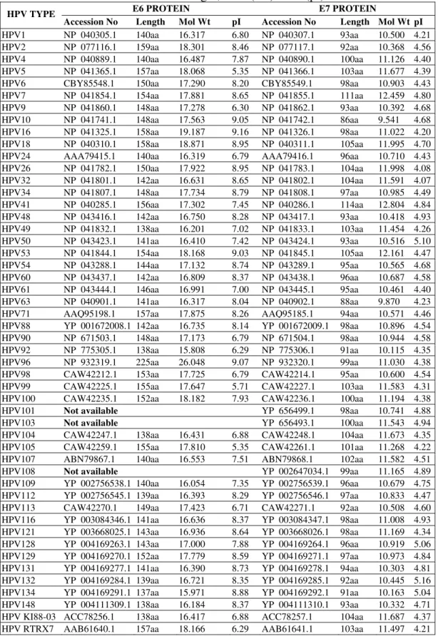
Although genome sequence of 50 different strains of HPV is available at NCBI, E6 proteins of HPV 101, 103 & 108 are not available. Accordingly, the comparative results of E6 proteins of 47 different strains of HPV as well as E7 proteins of 50 different strains of HPV were subjected to a tabular format, based on Sequence Length, MW and pI. pI and MW of all the proteins vary with protein sequences (Table 1). The lengths of E6 proteins of all HPV strains under study, range in between 137 to 159 amino acids, having MW in the range of 15.80 kD to 19.18 kD and pI from 5.35 to 9.16 except HPV 96 which has longest E6 protein having length of 225 amino acids, MW 26.04 kD and pI 9.07. However, the number of amino acids in E7 proteins of different strains of HPV varies from 86 to 114 having MW in the range of 9.54 kD to 12.8 kD and pI range of 4.07 to 5.16.
(NCBI) from NCBI Site) (Downloaded (Using
MODELLER9v6)
Table 1. Comparative analysis of E6 & E7 proteins of 50 different strains of HPV on the basis of length, MW (kD) & IP (pI).
E6 PROTEIN E7 PROTEIN
HPV TYPE
Accession No Length Mol Wt pI Accession No Length Mol Wt pI
HPV1 NP 040305.1 140aa 16.317 6.80 NP 040307.1 93aa 10.500 4.21
HPV2 NP 077116.1 159aa 18.301 8.46 NP 077117.1 92aa 10.368 4.56
HPV4 NP 040889.1 140aa 16.487 7.87 NP 040890.1 100aa 11.126 4.40
HPV5 NP 041365.1 157aa 18.068 5.35 NP 041366.1 103aa 11.677 4.39
HPV6 CBY85548.1 150aa 17.290 8.20 CBY85549.1 98aa 10.903 4.43
HPV7 NP 041854.1 154aa 17.881 8.65 NP 041855.1 111aa 12.459 4.80
HPV9 NP 041860.1 148aa 17.278 6.30 NP 041862.1 93aa 10.392 4.68
HPV10 NP 041741.1 148aa 17.563 9.05 NP 041742.1 86aa 9.541 4.68
HPV16 NP 041325.1 158aa 19.187 9.16 NP 041326.1 98aa 11.022 4.20
HPV18 NP 040310.1 158aa 18.871 8.95 NP 040311.1 105aa 11.995 4.70
HPV24 AAA79415.1 140aa 16.319 6.79 AAA79416.1 96aa 10.710 4.43
HPV26 NP 041782.1 150aa 17.922 8.95 NP 041783.1 104aa 11.998 4.08
HPV32 NP 041801.1 142aa 16.631 8.65 NP 041802.1 104aa 11.591 4.07
HPV34 NP 041807.1 148aa 17.734 8.79 NP 041808.1 97aa 10.985 4.49
HPV41 NP 040285.1 156aa 17.302 7.45 NP 040286.1 114aa 12.804 4.84
HPV48 NP 043416.1 142aa 16.750 8.28 NP 043417.1 93aa 10.418 4.93
HPV49 NP 041832.1 138aa 16.201 7.02 NP 041833.1 103aa 11.454 4.26
HPV50 NP 043423.1 141aa 16.410 7.42 NP 043424.1 93aa 10.516 5.10
HPV53 NP 041844.1 154aa 18.168 9.03 NP 041845.1 105aa 12.161 4.47
HPV54 NP 043288.1 144aa 17.132 8.74 NP 043289.1 95aa 10.565 4.68
HPV60 NP 043437.1 142aa 16.809 8.37 NP 043438.1 96aa 10.687 4.58
HPV61 NP 043444.1 146aa 16.991 7.00 NP 043445.1 95aa 10.461 4.40
HPV63 NP 040901.1 141aa 16.317 8.04 NP 040902.1 88aa 9.870 4.23
HPV71 AAQ95198.1 157aa 17.875 8.26 AAQ95185.1 94aa 10.571 4.46
HPV88 YP 001672008.1 142aa 16.735 8.14 YP 001672009.1 98aa 10.896 4.54
HPV90 NP 671503.1 148aa 17.173 6.79 NP 671504.1 98aa 10.944 4.58
HPV92 NP 775305.1 138aa 15.808 6.29 NP 775306.1 91aa 10.115 4.35
HPV96 NP 932319.1 225aa 26.048 9.07 NP 932320.1 99aa 11.030 4.38
HPV98 CAW42212.1 153aa 17.725 6.79 CAW42214.1 95aa 10.600 4.54
HPV99 CAW42225.1 155aa 17.647 5.71 CAW42227.1 103aa 11.583 4.31
HPV100 CAW42235.1 152aa 18.182 7.93 CAW42236.1 100aa 11.194 4.38
HPV101 Not available YP 656499.1 98aa 10.741 4.88
HPV103 Not available YP 656493.1 100aa 11.543 4.94
HPV104 CAW42247.1 138aa 16.431 6.88 CAW42248.1 104aa 11.673 4.35
HPV105 CAW42259.1 155aa 17.810 5.35 CAW42261.1 101aa 11.268 4.22
HPV107 ABN79867.1 140aa 16.553 7.51 ABN79868.1 102aa 11.582 4.51
HPV108 Not available YP 002647034.1 99aa 11.165 4.89
HPV109 YP 002756538.1 140aa 16.054 7.35 YP 002756539.1 96aa 10.679 4.75 HPV112 YP 002756545.1 139aa 16.393 8.29 YP 002756546.1 97aa 10.833 4.47
HPV113 CAW42270.1 149aa 17.423 6.71 CAW42271.1 92aa 10.508 4.60
Homology modeling
Three dimensional structure of each protein was predicted by homology modeling. In general, 30% sequence identity is required for generating useful 3D structure models [2, 4, 13]. Kaladhar et al. also predicted structures of E6 & E7 proteins along with other proteins of only HPV Type 92 using Swissmodel server but it seems that the predicted structures are not found validated [7]. In our study, we predicted structures of E6 proteins of 47 and E7 proteins of 50 different strains of HPV by comparative modeling using MODELLER9v6 software and visualized in the RasMolv2.5 software. However, the structure of E7 protein of HPV 41 was not predicted since it had only 27% percentage identity with known structure (PDB ID 1P5Q). Accordingly, total 96 structures of E6 and E7 proteins of 50 different HPV strains were generated in Modeller9v6 software. The E6 proteins of all HPV strains show close similarities with structures (2FK4) available at PDB except HPV 92 (3GE3), HPV 99 (1DQ3) and HPV 116 (1VZ3), while E7 proteins of all HPV strains show close similarities with the structures (2B9D) except HPV 2, 6, 10, 16, 18, 24, 26, 32, 53, 96, 99 and 109 (2EWL).
Evaluation of protein structure quality
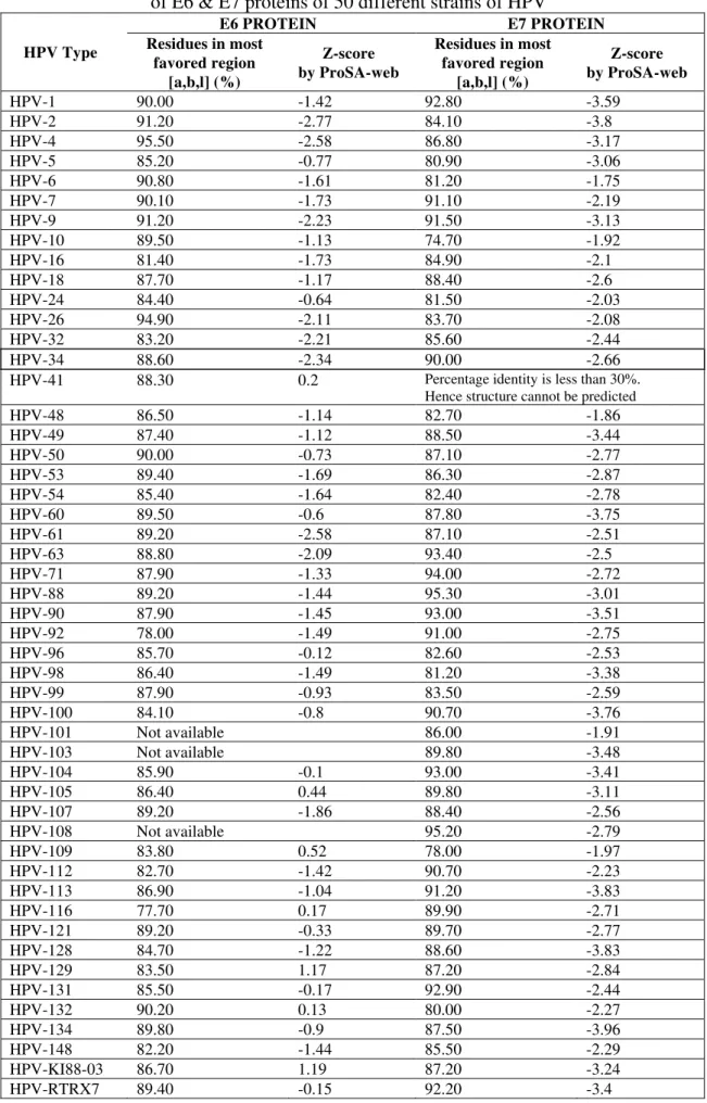
The stereo-chemical quality of these predicted structures were then evaluated through PROCHECK analysis. Remarkably the stereochemistry of E6 proteins of HPV 4 & 26 and E7 proteins of HPV 88 & 108 revealed that 94.90% to 95.50% residues were positioned in most favorable region of the Ramachandran plot. Some of E6 (HPV 1, 2, 6, 7, 9, 50, 132) & E7 proteins (HPV 1, 7, 9, 34, 63, 71, 90, 92, 100, 104, 112, 113, 131, RTRX7) show a good quality model range of 90 to 92% and 90 to 94% respectively. Most of E6 (HPV 5, 10, 16, 18, 24, 32, 34, 41, 48, 49, 53, 54, 60, 61, 63, 71, 88, 90, 96, 98, 99, 100, 104, 105, 107, 109, 112, 113, 121, 128, 129, 131, 134, 148, KI88-03 & RTRX7) & E7 proteins (HPV 2, 5, 4, 6, 16, 18, 24, 26, 32, 48, 49, 50, 53, 54, 60, 61, 96, 98, 99, 101, 103, 105, 107, 116, 121, 128, 129, 132, 134, 148, KI88-03) show the range of 85 to 89% and remaining E6 (92, 116) and E7 proteins (10, 109) show the range of 74 to 79% (Table 2).
Validation of 3D structures
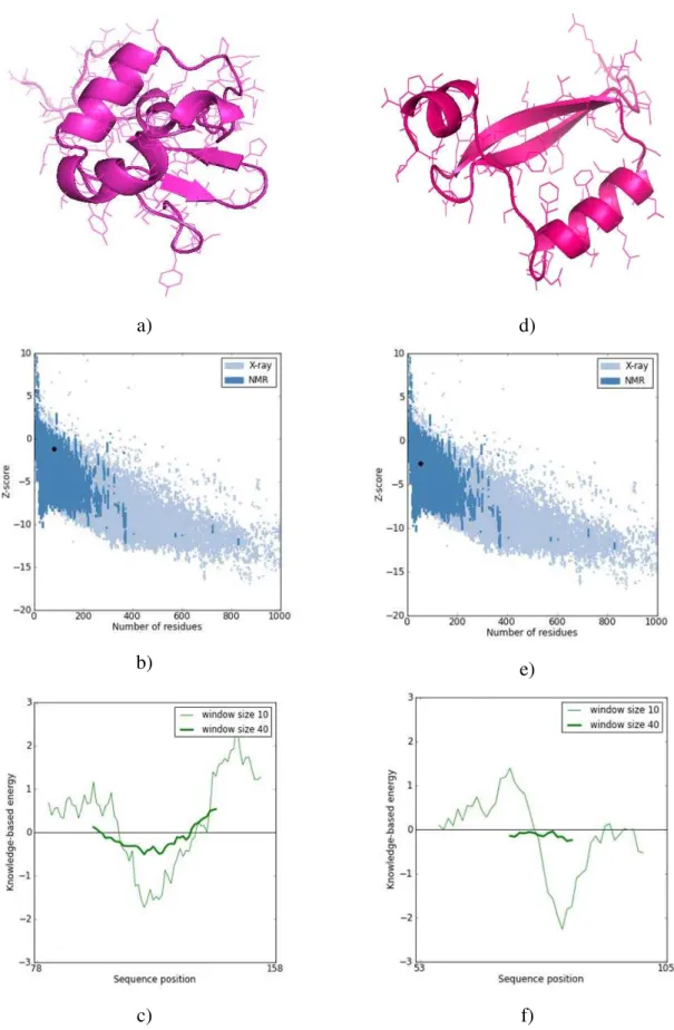
3D structures of each protein were subjected to validation using ProSA-web and when analyzed, it revealed a compatible Z-score value of residue energies (Table 2) within the range of native conformations of crystal structures. The ProSA-web based validation analysis showed largely negative Z-score in most of E6 proteins expect HPV 41, 105, 109, 116, 129, 132 and KI88-03, while negative Z-score in all E7 proteins. The residue energies including pair energy, combined energy and surface energy were all negative and had similar surface energy tendency with template.
Table 2. PROCHECK analysis and ProSA-web based validation of E6 & E7 proteins of 50 different strains of HPV
E6 PROTEIN E7 PROTEIN
HPV Type Residues in most
favored region [a,b,l] (%)
Z-score by ProSA-web
Residues in most favored region
[a,b,l] (%)
Z-score by ProSA-web
HPV-1 90.00 -1.42 92.80 -3.59
HPV-2 91.20 -2.77 84.10 -3.8
HPV-4 95.50 -2.58 86.80 -3.17
HPV-5 85.20 -0.77 80.90 -3.06
HPV-6 90.80 -1.61 81.20 -1.75
HPV-7 90.10 -1.73 91.10 -2.19
HPV-9 91.20 -2.23 91.50 -3.13
HPV-10 89.50 -1.13 74.70 -1.92
HPV-16 81.40 -1.73 84.90 -2.1
HPV-18 87.70 -1.17 88.40 -2.6
HPV-24 84.40 -0.64 81.50 -2.03
HPV-26 94.90 -2.11 83.70 -2.08
HPV-32 83.20 -2.21 85.60 -2.44
HPV-34 88.60 -2.34 90.00 -2.66
HPV-41 88.30 0.2 Percentage identity is less than 30%. Hence structure cannot be predicted
HPV-48 86.50 -1.14 82.70 -1.86
HPV-49 87.40 -1.12 88.50 -3.44
HPV-50 90.00 -0.73 87.10 -2.77
HPV-53 89.40 -1.69 86.30 -2.87
HPV-54 85.40 -1.64 82.40 -2.78
HPV-60 89.50 -0.6 87.80 -3.75
HPV-61 89.20 -2.58 87.10 -2.51
HPV-63 88.80 -2.09 93.40 -2.5
HPV-71 87.90 -1.33 94.00 -2.72
HPV-88 89.20 -1.44 95.30 -3.01
HPV-90 87.90 -1.45 93.00 -3.51
HPV-92 78.00 -1.49 91.00 -2.75
HPV-96 85.70 -0.12 82.60 -2.53
HPV-98 86.40 -1.49 81.20 -3.38
HPV-99 87.90 -0.93 83.50 -2.59
HPV-100 84.10 -0.8 90.70 -3.76
HPV-101 Not available 86.00 -1.91
HPV-103 Not available 89.80 -3.48
HPV-104 85.90 -0.1 93.00 -3.41
HPV-105 86.40 0.44 89.80 -3.11
HPV-107 89.20 -1.86 88.40 -2.56
HPV-108 Not available 95.20 -2.79
HPV-109 83.80 0.52 78.00 -1.97
HPV-112 82.70 -1.42 90.70 -2.23
HPV-113 86.90 -1.04 91.20 -3.83
HPV-116 77.70 0.17 89.90 -2.71
HPV-121 89.20 -0.33 89.70 -2.77
HPV-128 84.70 -1.22 88.60 -3.83
HPV-129 83.50 1.17 87.20 -2.84
HPV-131 85.50 -0.17 92.90 -2.44
HPV-132 90.20 0.13 80.00 -2.27
HPV-134 89.80 -0.9 87.50 -3.96
HPV-148 82.20 -1.44 85.50 -2.29
HPV-KI88-03 86.70 1.19 87.20 -3.24
a) d)
b) e)
c) f)
Fig. 1 ProSA-web Z-scores of modeled protein in PDB with respect to their protein length. Z-score is represented in black dot. The energy plots presented with window size 10 & 40.
Conclusion
The variation of causing nongenital cutaneous, nongenital mucosal and anogenital diseases by different HPV types could always remain a challenge to find out the cause behind it. These variations may be in their genomic content leading deviation in their proteomic structures, causing different types of infections as an outcome. As proteins of HPV are directly involved in causing the infection in human, so, it may be of significant interest to explore and analyze their protein structures. In this study, we made an effort to predict the 3D structures of E6 and E7 oncoproteins of 50 different strains of HPV. This study provides simultaneous predicted and validated structures of these HPV proteins. The outcome of this study might provide a platform for simultaneous structural comparative analysis of these proteins and help in finding out variations in their structures so as to explore why different strains of HPV cause different type of cancers. Further, this might also help in exploring for designing specific drugs or vaccines against specific strains of HPV.
Acknowledgement
Authors thank Department of Biotechnology, Ministry of Science & Technology, Government of India for financial support to Bioinformatics Centre wherein this study has been carried out. Authors also thank Shri D. S. Mehta, President, Kasturba Health Society, Dr. B. S. Garg, Dean, Mahatma Gandhi Institute of Medical Sciences, Sevagram and Dr. B. C. Harinath, Director, JBTDRC & Coordinator, BIC for their encouragement.
References
1. Altschul S. F., W. Gish, W. Miller, E. W. Myers, D. J. Lipman (1990). Basic Local Alignment Search Tool, J Mol Biol, 215, 403-410.
2. Brooks W. H., K. G. Daniel, S. S. Sung, W. C. Guida (2008). Computational Validation of the Importance of Absolute Stereochemistry in Virtual Screening, J Chem Inf Model, 48, 639-645.
3. Contreras A., I. Martínez, E. Cruz, M. Lizano (2007). Role of HPV18 E6 in PKB Signal Transduction Pathways, BMC Cancer, 7(1), A7.
4. Friesner R. A., J. L. Banks, R. B. Murphy, T. A. Halgren, J. J. Klicic, D. T. Mainz, M. P. Repasky, E. H. Knoll, M. Shelley, J. K. Perry, D. E. Shaw, P. Francis, P. S. Shenkin (2004). Glide: A New Approach for Rapid, Accurate Docking and Scoring. 1. Method and Assessment of Docking Accuracy, J Med Chem, 47(7), 1739-1749.
5. Gasteiger E., C. Hoogland, A. Gattiker, S. Duvaud, M. R. Wilkins, R. D. Appel, A. Bairoch (2005). Protein Identification and Analysis Tools on the ExPASy Server. In: John M. Walker (Ed.), The Proteomics Protocols Handbook, Humana Press, 571-607. 6. Hiller T., S. Poppelreuther, F. Stubenrauch, T. Iftner (2006). Comparative Analysis of
19 Genital Human Papillomavirus Types with Regard to p53 Degradation, Immortalization, Phylogeny, and Epidemiologic Risk Classification, Cancer Epidem Biomark Prevent, 15, 1262-1267.
7. Kaladhar D. S. V. G. K., T. Uma Devi, P. V. N. Rao (2010). An in silico Genome Wide Identification, Characterization and Modeling of Human Papilloma Virus strain 92, Int J Eng Sci Techn, 2(9), 4288-4291.
8. Laskowski R. A., M. W. MacArthur, D. S. Moss, J. M. Thornton (1993). PROCHECK: A Program to Check the Stereochemical Quality of Protein Structures, J Appl Cryst, 26, 283-291.
10. Mayers G., E. Androphy (1995). The E6 Protein, Human Papillomaviruses, 1995 Compendium, Part III, 47-57.
11. Munger K., A. L. Halpern (1997). HPV16 E7: Primary structure and biological properties. In Human Papillomaviruses 1997 Compendium, Part III, 17-36.
12. Nelson P. H., K. H. Vousden, N. L. Hubbert, D. R. Lowy, J. T. Schiller (1989). HPV16 E6 and E7 Proteins Cooperate to Immortalize Human Foreskin Keratinocytes, The EMBO Journal, 8, 3905-3910.
13. Plewczynski D., M. Hoffmann, M. von Grotthuss, K. Ginalski, L. Rychewski (2007).
In silico Prediction of SARS Protease Inhibitors by Virtual High Throughput Screening, Chem Biol Drug Des, 69, 269-279.
14. Rosty C., M. Sheffer, D. Tsafrir, N. Stransky, I. Tsafrir, M. Peter, P. Rochefordière, R. Salmon, T. Dorval, J. Thiery, J. Couturier, F. Radvanyi, E. Domany, X. Sastre-Garau (2005). Identification of a Proliferation Gene Cluster Associated with HPV E6/E7 Expression Level and Viral DNA Load in Invasive Cervical Carcinoma, Oncogene, 24(47), 7094-7104.
15. Sali A., T. L. Blundell (1993). Comparative Modelling by Statisfaction of Spatial Restraints, J Mol Biol, 234, 779-815.
16. Sayle R. A., E. J. Milner-White (1995). RASMOL: Biomolecular Graphics for All, Trends Biochem Sci, 20, 374-376.
17. Tungteakkhun S. S., M. Filippova, J. W. Neidigh, N. Fodor, P. J. Duerksen-Hughes (2008). The Interaction between Human Papillomavirus Type 16 and FADD is Mediated by a Novel E6 Binding Domain, J Virol, 82, 9600-9614.
18. Watts K. J., C. H. Thompson, T. E. Cossart, B. R. Rose (2001). Variable Oncogene Promoter Activity of Human Papillomavirus Type 16 Cervical Cancer Isolates from Australia, J Clin Microbiol, 35(5), 2009-2014.
19. Wiederstein M., M. J. Sippl (2007). ProSA-web: Inter-active Web Service for the Recognition of Errors in Three-dimensional Structures of Proteins, Nucleic Acids Res, 35, 407-410.
20. Xuemei J. I., E. M. Sturgis, C. Zhao, C. J. Etzel, Q. Wei, G. Li (2009). Association of p73G4C14-to-A4T14 Polymorphism with Human Papillomavirus Type 16 Status in Squamous Cell Carcinoma of the Head and Neck in Non-Hispanic whites, Cancer, 115(8), 1660-1668.
Prof. (Dr) Satish Kumar, M.D.
E-mail: [email protected]
MBBS from GMC, Jammu in 1990 and M.D. (Biochemistry) from MGIMS, Sevagram in 1994. Presently continuing as Professor, Biochemistry & Dy Coordinator, Bioinformatics Centre, MGIMS, Sevagram, India. Member, Board of Studies in Biotechnology & Bioinformatics, MUHS, Nashik; Member, Scientific Advisory Committee (SAC), ICMR-NIC National Database on Indian Medical Journals; Member, Faculty of Medicine, IMS, Banaras Hindu University, Varanasi; Editor-in-Chief of Journal of Pathology Research and Journal of BIOINFO Medicine; Member & Referee, National Advisory Board, JK Science, Journal of Medical Education & Research; Principal Investigator on CSIR funded two Projects on Immunodiagnostics in tuberculosis; Patent on M tb ES-31 isolation process (184510/410/BOM/99/2001); Coordinator, STP in Bioinformatics & Biotechnology; Introduced SEVA TB ELISA in 1993 for TB detection in patients attending our hospital; Continuing research work in immunodiagnostics in tuberculosis and in silico
cancer work; Co-Investigator on DBT funded project on BTISnet; Associate Editor & Publisher, SEVAMED Quarterly Journal; Involved in developing clinical databases; Establishing Moving Academy on Biomedical Communication; Convener, Online Health Informatics Course (OHIC); Executive Member, Bioinformatics Centre, Raipur; Organized 14 workshops/CMEs on Medical Informatics & Biomedical Communication; Recognized UG, PG and PhD teacher of MUHS, Nashik; On panel of UG, PG & PhD examination under many universities; Published 46 papers (7 International & 39 National); Served on many Institutional committees (HIS, NAAC, College Council etc) with an indomitable commitment.
Lingaraja Jena, M.Sc., Ph.D.
E-mail: [email protected]
Vidya W. Bhomale, M.Sc.
E-mail: [email protected]
University Gold Medalist in B.Sc. and College Topper in MSc Biotechnology from RSTM University, Nagpur in 2011. Carried out Project Work on Structural Comparison of E6 and E7 Proteins of different strains of HPV using Bioinformatics tools at Biochemistry, MGIMS, Sevagram. Undergone training in “Recent Agricultural Biotechnology” at Anand Niketan College of Agriculture, Anandwan, Warora. Areas of interest are Molecular biology, Genetic engineering, Pathogens & viruses microbiological studies, Plant biotechnology & breeding. Presently working in the area of protein structure analysis of different strains of HPV.
Prof. (Dr) Sangeeta Daf, M.D.
E-mail: [email protected]