http://doi.org/10.1590/0004-282X20160194
ARTICLE
Experimental neurocysticercosis: absence of
IL-4 induces lower encephalitis
Neurocisticercose experimental: ausência de IL-4 induz menos encefalite
Hidelberto Matos Silva1,2, Marina Clare Vinaud1, Ruy de Souza Lino Júnior1
Neurocysticercosis (NCC) is the most severe clinical manifestation of cysticercosis and is responsible for up to 29% of the epilepsy cases in Latin America, India and Africa1,2.
he NCC symptoms are caused by the host inlammatory response, immune response modulation, location of the par-asite, evolutionary stage of the parpar-asite, parasitic burden, as well as the mass efect induced by its growth, which results in a wide clinical and laboratory polymorphism challenging the diagnosis of this disease3,4.
Experimental models have been used as important tools in the study and comprehension of the host-parasite relationship in human cysticercosis in several locations including the neurological one5,6,7. The most-used parasite
in the experimental models is Taenia crassiceps cysticer-cus due to its rapid development cycle and its antigenic similarity with T. solium7,8. The intraperitoneal experimen-tal model is the most common and is very useful in the evaluation of the genetic factors involved in the host’s sus-ceptibility and resistance, with emphasis on the immu-nological mechanisms9. The T. crassiceps intraperitoneal infection induces an initial Th1 response accompanied by large amounts of interferon-gamma, classically activated macrophages (pro-inflammatory or M1), IgG2a antibod-ies and interleukin-12 (IL-12) leading to a low parasitic burden. Soon afterwards there is a strong Th2 polariza-tion due to the immunoregulatory capacity of the parasite
1Universidade Federal de Goiás, Instituto de Patologia Tropical e Saúde Pública, Goiânia GO, Brasil; 2Centro Universitário UNIRG, Faculdade de Medicina, Gurupi TO, Brasil.
Correspondence: Hidelberto Matos Silva; Centro Universitário UNIRG, Faculdade de Medicina; Av. Rio De Janeiro, 1585; 77403-090 Gurupi TO, Brasil; E-mail: [email protected]
Conflict of interest: There is no conlict of interest to declare. Received 02 May 2016; Accepted 31 October 2016.
ABSTRACT
Neurocysticercosis (NCC) is the most severe clinical manifestation of cysticercosis. One of the factors responsible for its symptomatology is the host inlammatory response. Therefore the inluence of interleukin 4 (IL-4) on the induction of encephalitis in experimental NCC was evaluated. Methods: BALB/c (WT) and BALB/c (IL-4-KO) mice were inoculated intracranially with Taenia crassiceps cysticerci and euthanized at 7, 30, 60 and 90 days later, the encephala removed and histopathologically analyzed. Results: The absence of IL-4 induced greater parasitism. In the initial phase of the infection, IL-4-KO showed a lower intensity in the inlammatory iniltration of polimorphonuclear cells in the host-parasite interface and intra-parenquimatous edema. The IL-4-KO animals, in the late phase of the infection, showed lower intensity of ventriculomegaly, encephalitis, and meningitis, and greater survival of the parasites in comparison with the WT animals. Conclusion: The absence of IL-4 induced lower inlammatory iniltration, ventriculomegaly and perivasculitis in experimental NCC.
Keywords: neurocysticercosis; Taenia crassiceps; encephalitis; interleukin-4.
RESUMO
A Neurocisticercose (NCC) é a manifestação clínica mais severa da cisticercose, e um dos fatores responsáveis pela sintomatologia é a resposta inlamatória do hospedeiro. Desta forma avaliou-se a inluência da interleucina 4 (IL-4) na indução de encefalite na NCC experimental. Métodos: Camundongos das linhagens BALB/c (WT) e BALB/c (IL-4-KO) foram inoculados intracranialmente com cisticercos de Taenia crassiceps e eutanasiados aos 7, 30, 60 e 90 dias após a infecção, os encéfalos foram removidos e analisados histopatologicamente. Resultados: A ausência da IL-4 induziu um maior parasitismo nos animais. Na fase inicial da infecção os animais IL-4-KO apresentaram menor intensidade tanto de iniltrado inlamatório de polimorfonucleares na interface parasito-hospedeiro quanto de edema intraparenquimatoso. Os animais IL-4-KO, na fase tardia, apresentaram menor intensidade de ventriculomegalia, encefalite, meningite e maior sobrevivência dos cisticercos em relação aos animais WT. Conclusão: A ausência da IL-4 induz menos iniltrado inlamatório, ventriculomegalia e perivasculite na NCC experimental.
increasing serum levels of IgG and IgE as well as IL-4, IL-13 and IL-5, which inhibits the initial Th1 response and favors the growth of parasitic burden7,8,10,11.
he experimental models exploring the neurological location of cysticerci are described using Mesocestoides corti5,12,13 and T. crassiceps6. he model carried out with
M. corti was used to describe the role of defense cells and cytokines in the host-parasite relationship13, the expres-sion of Toll-like receptors5, the migration of leucocytes to the central nervous system, the junction complex proteins and the immune system evasion strategies14. According to these studies, the immune response in the murine exper-imental model of NCC is associated with a predominant
h1 immune response directed by gamma-delta T lympho
-cytes, which arrive at the infection site at the beginning of the infection and remain in it during the whole course of the infection. hese cells induce an increase in the pro-inlammatory cytokine production and a greater local cellular response7, which does not eliminate the parasite, possibly due to its evasion strategies14. herefore there is an
increase in the parasitic burden and in the inlammatory cells within extraparenchymatous areas of the encephala, such as ventricles, meninges and subarachnoid space13. It is interesting to highlight that a low parasitic burden is able to promote an initial h1 response that is capable of eliminating the parasite5. As the infection progresses, para-sites and immune cells reach the brain parenchyma, which increases the inlammation intensity and results in more severe clinical symptoms13.
Taenia crassiceps is also used as an experimental model for NCC studies due to is antigenic similarities to
T. solium cysticerci8,15. It has also already been described that
T. crassiceps cysticerci are able to induce NCC in immuno-compromised16 and immunocompetent individuals17. In this model, the infected animals presented with tissue alterations and lesions such as encephalitis, perivasculitis, ventriculo-megaly, ependymitis, meningitis, microgliosis accompanied by the parasite’s death, and extraparenchymal and parenchy-mal inlammatory iniltration6,18.
Other experimental models have been proposed but instead of using T. crassiceps cysticerci they performed an intracranial inoculation of T. solium oncospheres in rats19 and in swine20. he rat NCC model reported that the infected
animals presented with epilepsy, inlammatory iniltration, perivascular iniltrate, angiogeneses, spongy changes and mass efect in the brain tissue19. In the swine NCC model, after four months of infection, it was possible to observe
cal-ciied cysticerci surrounded by an exacerbated inlamma
-tory response with lymphocyte iniltration and increased
inlammatory markers20.
he BALB/c mice are genetically susceptible to intraper
-itoneal infection with T. crassiceps cysticerci because they
present a h2 immune proile7,9. However the
immunologi-cal mechanisms associated with susceptibility or resistance
of this mice lineage to T. crassiceps cysticerci are not com-pletely understood.
he aim of this study was to describe and compare the inluence of IL-4 in the encephalic inlammatory response to the experimental T. crassiceps NCC infection in two lineages of BALB/c mice - wild type (WT) and IL-4 knockout (IL-4-KO).
METHODS
Maintenance of T. crassiceps
he T. crassiceps cysticerci (ORF strain) have been main-tained through intraperitoneal passages in female BALB/c mice8 in the animal facility of the Tropical Pathology and Public Health Institute (IPTSP) of the Federal University of Goias (UFG) since 2002.
Animals
Two lineages of female BALB/c mice were used: conven
-tional BALB/c also known as WT, and IL-4-KO BALB/c mice that have no gene for IL-4 production. he animals provided by the IPTSP/UFG animal facilities were eight to 12 weeks old and weighed 20 to 30 grams .
In this study the animals were divided into four groups with ive mice each as follows: group 1 - WT BALB/c mice inoculated with T. crassiceps cysticerci; group 2 - WT BALB/c
mice inoculated with sterile saline solution; groups 3 - IL-4-KO BALB/c mice inoculated with T. crassiceps cysticerci; and group
4 - IL-4-KO BALB/c mice inoculated with a sterile saline solu
-tion. his study was approved by the ethics committee for ani
-mal use (CEUA/UFG) protocol number 034/09.
Experimental infection
he intracranial infection of the animals was performed as described by Matos-Silva et al6. Briely, the animals were
weighed and anesthetized with 0.01ml/g of ketamine 10% and xilazine 2% intraperitoneal injection. After trichotomy of the superior portion of the head and antisepsis with topi-cal iodine, a longitudinal and median incision was made on the skin of the skull with a scalpel. he trepanation oriice was performed with a drill (44.5mm x 2mm) moved by a
micromotor (LB100-Beltec)in the topography of the right
parietal bone at 3mm from the median line (sagittal suture) and at 3mm posterior to the coronal suture and with 4mm of depth. he infected animals were intracranially inocu
-lated with three to ive cysticerci, following which the trep
-anation oriice was closed with sterile dental alginate and the incision sutured.
Removal of the encephala
Histopathological analysis
After the removal, the encephala were ixed in 10% buf
-ered formaldehyde for 24 hours. he encephala were ana
-lyzed and their macroscopic characteristics photographi-cally documented. he macroscopic analysis considered the presence of parenchymal or extra-parenchymal cysticerci, edema, hyperemia, tissue consistency and ventriculomegaly.
Afterwards the encephala were histologically processed, in which the encephala fragments were included in parain. After a 5 µm width microtomy the fragments were stained with hematoxilin-eosin6.
Analysis of the general pathologic processes and classification of the cysticerci into evolutionary stages
he general pathologic processes were evaluated within the parasite, in the host-parasite interface and in the host tis-sue as to: presence of cysticercus, anatomic location, evolution -ary stage of the cysticercus (initial - no buddings, translucent membrane and vesicular luid; larval - presence of buddings, translucent membrane and vesicular luid; inal - no buddings,
opaque membrane and vesicular luid), ventriculomegaly, par
-enchymatous and perivascular edema, gliosis, meningitis, epen-dymitis, choroiditis, perivasculitis, microgliosis, and the proile
of inlammatory iniltration cells (polymorphonuclear or mono
-nuclear cells). he pathologic processes detected were classiied
in a semi-quantitative form according to the following criteria: absent (score 0), discrete - with up to 25% of area commitment (score 1), moderate - 26% to 50% of area commitment (score 2), and accentuated - more than 50% of area commitment (score 3)6.
he score attributed to each analysis was counted and used to calculate mean ± standard deviation and the statistical analysis.
Statistical analysis
he statistical analysis was performed using the Sigma Stat 2.3 software. All variables were tested for normal distribution and homogenous variation. If they presented non-normal dis-tribution, the variables were analyzed by the nonparametric
Mann-Whitney test. he diferences were considered signii
-cant when p < 0.05.
RESULTS
Macroscopic alterations
he macroscopic alterations were more evident after 30 DPI in BALB/c WT animals, which presented with hyperemia, and decreased encephalic consistency accompanied by intraventric-ular cysticerci. Most of these animals presented with ventriculo-megaly, adjacent parenchymal hypotrophy and/or deviation of the medial line structures (Table 1, Figure 1).
Table 1. Macroscopic analysis of encephala from BALB/c WT and BALB/c IL-4-KO mice experimentally infected with Taenia crassiceps cysticerci.
Variables 07 DPI 30 DPI 60 DPI 90 DPI
WT IL-4-KO WT IL-4-KO WT IL-4-KO WT IL-4-KO
Location of cysticerci LV LV/3rd V LV LV LV LV LV LV
Edema + ++ + + + + + +
Ventriculomegaly + + + ++ ++ ++ +++ +++
WT: Wild type; IL-4-KO: IL-4 knockout gene; DPI: days post infection; -: absent; +: discrete; ++: moderate; +++: accentuated; LV: lateral ventricle; 3rd V: third
ventricle; n = 5 per each experimental day.
Figure 1. Mesoscopy of BALB/c WT (A) and IL-4-KO BALB/c (B) encephala showing ventriculomegaly and deviation of the medial line. It is possible to observe a cysticercus (arrow) inside the ventricle (scale bar = 1mm).
A
B
Cysticerci location and classification
When analyzing all the infected BALB/c animals, it was possible to visualize the cysticerci in 60% (n = 24). In BALB/c WT animals these occurred in 20% (n = 4) of the infected animals, while in BALB/c IL-4-KO infected animals the rate of cysticerci observation was 100% (n = 20). Also, 25% (n = 5) of BALB/c IL-4-KO infected animals showed more than one cysticercus in the encephala.
Regarding the cysticerci evolutionary stage classiica -tion, it was possible to observe initial stage parasites at
seven DPI, in both mice lineages. At 30 DPI, BALB/c WT ani
-mals showed initial stage cysticerci while BALB/c IL-4-KO animals showed larval stage ones. At 60 DPI, both lin-eages showed larval stage cysticerci. Finally, at 90 DPI, BALB/c WT animals showed inal stage cysticerci, while in
BALB/c IL-4-KO animals both larval and inal stage cysti
-cerci were observed.
As to location, most of the parasites were observed in the lateral ventricles (Table 1).
Host tissue response
At seven DPI, the BALB/c IL-4-KO animals showed
increased parenchymal inlammatory iniltration of mononu
-clear cells surrounding the parasite (Table 2, Figures 2A, 2B), gliosis (Figure 2C) and meningitis when compared to BALB/c WT animals (p < 0.05). While the BALB/c WT animals showed an intraventricular cysticercus with inlammatory iniltration of mononuclear cells in the host tissue (Figure 2D).
At 60 DPI, BALB/c WT animals showed higher inlammatory iniltration of polymorphonuclear cells and hyperemia when compared to BALB/c IL-4-KO animals (p < 0.05). In BALB/c WT animals, it was possible to observe an inlammatory iniltration of mononuclear cells inside the ventricles (Figure 2E).
At 90 DPI, BALB/c IL-4-KO animals showed higher hyper
-emia and meningitis when compared to BALB/c WT animals (p < 0.05) (Figure 2F).
he control group animals from both lineages showed hyperemia and edema in discrete intensity in the irst days after the inoculation of saline sterile solution.
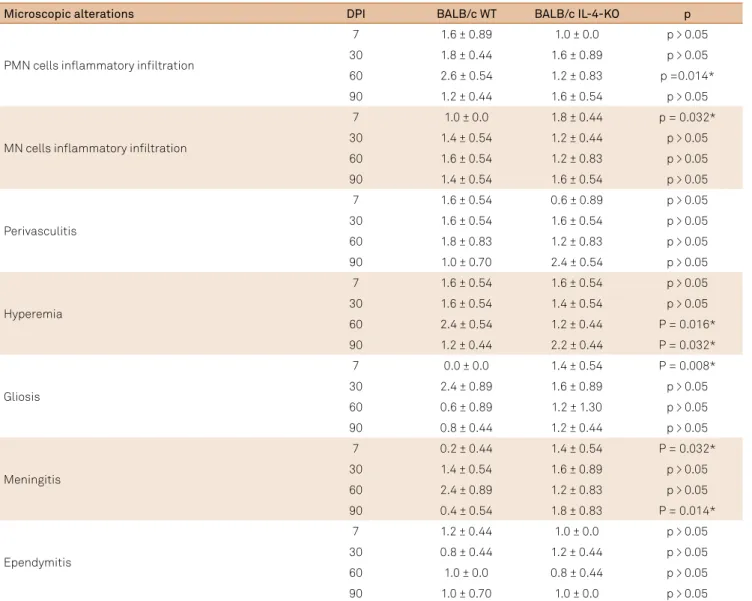
Table 2. Histopathologic analysis of host tissue from encephala of BALB/c WT and BALB/c IL-4-KO mice infected with Taenia crassiceps cysticerci. Results in mean ± standard deviation.
Microscopic alterations DPI BALB/c WT BALB/c IL-4-KO p
PMN cells inlammatory iniltration
7 1.6 ± 0.89 1.0 ± 0.0 p > 0.05
30 1.8 ± 0.44 1.6 ± 0.89 p > 0.05
60 2.6 ± 0.54 1.2 ± 0.83 p =0.014*
90 1.2 ± 0.44 1.6 ± 0.54 p > 0.05
MN cells inlammatory iniltration
7 1.0 ± 0.0 1.8 ± 0.44 p = 0.032*
30 1.4 ± 0.54 1.2 ± 0.44 p > 0.05
60 1.6 ± 0.54 1.2 ± 0.83 p > 0.05
90 1.4 ± 0.54 1.6 ± 0.54 p > 0.05
Perivasculitis
7 1.6 ± 0.54 0.6 ± 0.89 p > 0.05
30 1.6 ± 0.54 1.6 ± 0.54 p > 0.05
60 1.8 ± 0.83 1.2 ± 0.83 p > 0.05
90 1.0 ± 0.70 2.4 ± 0.54 p > 0.05
Hyperemia
7 1.6 ± 0.54 1.6 ± 0.54 p > 0.05
30 1.6 ± 0.54 1.4 ± 0.54 p > 0.05
60 2.4 ± 0.54 1.2 ± 0.44 P = 0.016*
90 1.2 ± 0.44 2.2 ± 0.44 P = 0.032*
Gliosis
7 0.0 ± 0.0 1.4 ± 0.54 P = 0.008*
30 2.4 ± 0.89 1.6 ± 0.89 p > 0.05
60 0.6 ± 0.89 1.2 ± 1.30 p > 0.05
90 0.8 ± 0.44 1.2 ± 0.44 p > 0.05
Meningitis
7 0.2 ± 0.44 1.4 ± 0.54 P = 0.032*
30 1.4 ± 0.54 1.6 ± 0.89 p > 0.05
60 2.4 ± 0.89 1.2 ± 0.83 p > 0.05
90 0.4 ± 0.54 1.8 ± 0.83 P = 0.014*
Ependymitis
7 1.2 ± 0.44 1.0 ± 0.0 p > 0.05
30 0.8 ± 0.44 1.2 ± 0.44 p > 0.05
60 1.0 ± 0.0 0.8 ± 0.44 p > 0.05
90 1.0 ± 0.70 1.0 ± 0.0 p > 0.05
DISCUSSION
Helminths such as T. crassiceps cysticerci have devel-oped complex mechanisms to evade or modulate the host response during infection. his study describes the inluence of an important cytokine such as IL-4 in response to helmin-thic infection. he use of genetically modiied mice lineages is an important tool in the understanding of the host-parasite relationship to the development of the parasitic infection and, consequently, the clinical manifestations of the infection7.
All the infected animals showed both micro- and mac-roscopic alterations, even when the cysticerci were not vis-ible on macroscopic analysis. All the control animals showed
discrete levels of edema and hyperemia due to the mechani-cal trauma of the inoculation of sterile saline solution. hese discrete alterations in the control group have also been observed in other experimental NCC studies6,13,18,21.
In the infected animals, the majority of the cysti-cerci were located inside the lateral ventricles accom-panied by a discrete-to-moderate intensity of inflamma-tory response. This fact was previously reported in studies that showed that the mere presence of the parasite’s anti-gens are responsible for the development of inflamma-tory alterations and ventriculomegaly, which may lead to cerebrospinal fluid obstruction22,23. Zepeda et al.21 showed that T. crassiceps antigens are able to induce inflammation
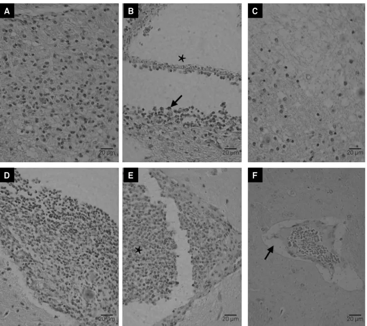
Figure 2. Microscopic alterations in encephala of intracranial infected BALB/c WT and BALB/c IL-4-KO mice. (A-F) hematoxilin-eosin stain, scale bar = 20µm. (A) parenchymal mononuclear cells inlammatory iniltration at seven days post-inoculation (DPI) in BALB/c IL-4-KO; (B) Intraventricular viable cysticercus (asterisk) and mononuclear cell inlammatory iniltration (arrow) in the host tissue at seven DPI in BALB/c WT; (C) Gliosis, at seven DPI in BALB/c IL-4-KO; (D) meningitis at 90 DPI in BALB/c IL-4-KO; (E) intraventricular polymorphonuclear cell inlammatory iniltration (asterisk) at 60 DPI in BALB/c WT; (F) hyperemia and perivascular edema (arrow) at 90 DPI in BALB/c IL-4-KO.
A
D
B
E
C
F
20 µm 20 µm
20 µm 20 µm
in the brain tissue and apoptosis of hippocampal cells. Histological studies have shown that viable cysticerci, such as the ones in our study, have little or no surrounding inflammation in humans and pigs; while degenerating or dead cysticerci, which were not found in our study, had an inflammatory response in their surroundings24.
In our study, all infected animals showed ependymitis, meningitis and perivasculitis. hese alterations have also been described in experimental NCC in rats19. In our study,
it was not possible to observe calciication of the cysticerci, or in the surrounding host tissue. However, the calciication of the cysticerci and host tissue was observed in autopsies of human intraventricular NCC25 and in a swine model of NCC2, probably because the material analyzed had a longer time of infection.
he initial stage cysticerci showed a discrete inten -sity of inlammatory iniltration surrounding the parasite. As the cysticerci die, the immune modulation is lost,
result-ing in an increase in the inlammatory intensity accompa
-nied by ibrosis. Alvarez et al.,5 studying experimental NCC,
reported that the parasite initiates the immunomodula-tion through complex glycoproteins present in the tegu-ment surface resulting in the viability of the parasite. he histopathologic analysis of human asymptomatic NCC shows viable cysticerci surrounded by discrete or absent
inlammatory iniltration that contributes to the chro
-nicity of the infection25. On the other hand, Fleury et al.25, studying cysticerci removed from human NCC, reported that the parasite is surrounded by human immunoglobulin such as IgG, IgM, IgA and IgE, which act as a kind of cam-oulage due to the presence of Fc receptors in the parasite tegument surface.
In BALB/c WT animals it was possible to observe that the peak evolution of the encephalitis was accompanied by a partial destruction of the cysticerci in the experimental days. In the BALB/c IL-4-KO animals, the evolution of the inflammatory process was not resolved due to the pres-ence of viable cysticerci until the end of the experimen-tal days. The absence of IL-4 resulted in difficulty in con-trolling the parasitic infection. A Th1 immune response is observed while the parasite is still viable, while a mixed
Th1/Th2 immune response is observed when the para
-site is being destroyed26. Experimental NCC studies have shown an inflammatory infiltration of macrophages and plasmocytes in the high levels of tissue IL-425 in rats and pigs. These findings show that the parasite maintains an equilibrium with the host immune response at the begin-ning of the infection and therefore a rapid Th1 response is observed, leading to the production of IgG. However, as the infection progresses the balance is modified to a mixed Th1/Th2 response, which may result in destruction of the cysticerci27.
When mononuclear cells from human asymptom
-atic NCC patients are stimulated in vitro, it is possible
to observe the increased production of IL-4, IL-5 and IL-13, which are Th2 cytokines27. However, when the mononuclear cells are collected from symptomatic NCC patients, the profile of the cytokines produced are quite different, with a predominance of IFN-gamma, IL-17, IL-23 and TNF-alpha, which are from the Th2 profile24. Asymptomatic NCC patients from endemic areas have shown a Th2 response with production of IgG4, IL-4, IL-5 and IL-1327.
In the M. corti experimental NCC model, it was observed that the h1 immune proile was related to a strong immune response capable of destruction of the parasite and tissue injury13. Matos-Silva et al.6 also found similar results using the T. crassiceps NCC model. Using the same model, Moura et al.18 described a predominance of the h2 immune proile cytokines at the end of the infection, while in the acute phase
of the infection there was a mixed h1/h17 proile accompa
-nied by high levels of IL-10.
BALB/c animals are naturally susceptible to T. crassiceps
infection, both in the intraperitoneal and intracranial locations, due to its natural development of a h2 proile response7,28. he main cytokine of the h2 proile is IL-4 as it
induces activation and efectiveness of T lymphocytes and also stimulates the change of lymphocyte B isotype into IgE production. As well, IL-4 suppresses the IFN-gamma activity of macrophages resulting in the inhibition of these cells7,26.
Therefore an immune Th2 response, which is consid-ered protective against intestinal helminthic infections, is harmful to the host when it favors the establishment and growth of tissue helminthes26. Greater parasitic bur-den was observed in Th2 immune profile animals stud-ied by Rodrigues-Sosa et al.26, as well as in our findings
in which we observed cysticerci in all BALB/c IL-4-KO infected animals.
In central nervous system infections, IL-4 plays an important role in the immune regulation due to stimu-lation and chemotaxis of eosinophils, mastocytes, baso-phils and other Th2 profile cells. Also, the survival of these cells are maintained by the influence of IL-429. Experimental studies have shown the influence of this cytokine as a neuroprotective factor on astrocytes and microglia29,30. Falcone et al.30, studying an
experimen-tal model of autoimmune encephalitis in IL-4-KO mice, reported that the animals without IL-4 showed a more severe form of clinical disease, with extensive involve-ment of spinal cord accompanied by increased produc-tion of pro-inflammatory cytokines.
According to the anatomopathological indings and the analysis of the inlammatory process in the infected animals, it is possible to conclude that the absence of IL-4 induced
less intense inlammatory alterations. his response prob
References
1. Cantey PT, Coyle CM, Sorvillo FJ, Wilkins PP, Starr MC, Nash TE. Neglected parasitic infections in the United States: cysticercosis. Am J Trop Med Hyg. 2014;90(5):805-9. http://doi.org/10.4269/ajtmh.13-0724
2. Nash TE. Parasitic diseases that cause seizures. Epilepsy Curr. 2014;14(1 suppl):29-34. http://doi.org/10.5698/1535-7511-14.s2.29
3. Del Brutto OH. Neurocysticercosis among international travelers to disease-endemic areas. J Travel Med. 2012;19(2):112-7. http://doi.org/10.1111/j.1708-8305.2011.00592.x.
4. Garcia HH, Rodriguez S, Gilman RH, Gonzalez AE, Tsang VC. Neurocysticercosis: is serology useful in the absence of brain imaging? Trop Med Int Health. 2012;17(8):1014-8. http://doi.org/10.1111/j.1365-3156.2012.03037.x
5. Alvarez JI, Mishra BB, Gundra UM, Mishra PK, Teale JM.
Mesocestoides corti intracranial infection as a murine model for neurocysticercosis. Parasitology. 2010;137(3):359-72. http://doi.org/10.1017/S0031182009991971
6. Matos-Silva H, Reciputti BP, Paula EC, Oliveira AL, Moura VB, Vinaud MC et al. Experimental encephalitis caused by Taenia crassiceps cysticerci in mice. Arq Neuropsiq. 2012;70(4):287-92. http://doi.org/10.1590/S0004-282X2012005000010
7. Terrazas LI The complex role of pro- and anti-inlammatory cytokines in cysticercosis: immunological lessons from experimental and natural hosts. Curr Top Med Chem. 2008;8(5):383-92.
http://doi.org/10.2174/156802608783790848
8. Vaz AJ, Nunes CM, Piazza RM, Livramento JA, Da Silva MV, Nakamura PM et al. Immunoblot with cerebrospinal luid from patients with
neurocysticercosis using antigen from cysticerci of Taenia solium and
Taenia crassiceps. Am J Trop Med Hyg. 1997;57(3):354-7.
9. Fragoso G, Lamoyi E, Mellor A, Lomelí C, Hernández M, Sciutto E. Increased resistance to Taenia crassiceps murine cysticercosis in Qa-2 transgenic mice. Infect Immun. 1998;66(2):760-4.
10. Villa OF, Kuhn RE. Mice infected with the larvae of Taenia crassiceps
exhibit a Th2-like immune response with concomitant anergy and downregulation of Th1-associated phenomena. Parasitology. 1996;112(6):561-70. http://doi.org/10.1017/S0031182000066142
11. Terrazas LI, Bojalil R, Govezensky T, Larralde C. Shift from an early protective Th1-type immune response to a late permissive Th2-type response in murine cysticercosis (Taenia crassiceps). J Parasitol. 1998;84(1):74-81. http://doi.org/10.2307/3284533
12. Toenjes SA, Spolski RJ, Mooney KA, Kuhn RE. The systemic immune response of BALB/c mice infected with larval Taenia crassiceps is a mixed Th1/Th2-type response. Parasitology. 1999;118(6):623-33. http://doi.org/10.1017/S0031182099004370
13. Cardona AE, Restrepo BI, Jaramillo JM, Teale JM. Development of an animal model for neurocysticercosis: immune response in the central nervous system is characterized by a predominance of gamma delta T cells. J Immunol. 1999;162(2):995-1002.
14. Alvarez JI, Teale JM. Differential changes in junctional complex proteins suggest the ependymal lining as the main source of leukocyte iniltration into ventricles in murine neurocysticercosis. J Neuroimmunol. 2007;187(1-2):102-13. http://doi.org/10.1016/j.jneuroim.2007.05.005
15. Abraham R, Pardini AX, Vaz AJ, Livramento JA, Machado LR.
Taenia antigens detection in the cerebrospinal luid of patients with neurocysticercosis and its relationship with clinical activity of the disease. Arq Neuropsiquiatr. 2004;62(3B):756-60. http://doi.org/10.1590/S0004-282X2004000500002
16. Heldwein K, Biedermann HG, Hamperl WD, Bretzel G, Löscher T, Laregina D et al. Subcutaneous Taenia crassiceps infection in a patient with non-Hodgkin’s lymphoma. Am J Trop Med Hyg. 2006;75(1):108-11.
17. Ntoukas V, Tappe D, Pfütze D, Simon M, Holzmann T. Cerebellar cysticercosis caused by larval Taeniacrassiceps tapeworm in immunocompetent woman, Germany. Emerg Infect Dis. 2013;19(12):2008-11. http://doi.org/10.3201/eid1912.130284
18. Moura VB, Lima SB, Matos-Silva H, Vinaud MC, Loyola PR, Lino RS. Cellular immune response in intraventricular experimental neurocysticercosis. Parasitology. 2016;143(3):334-42. http://doi.org/10.1017/S0031182015001572
19. Verastegui MR, Mejia A, Clark T, Gavidia CM, Mamani J, Ccopa F et al. Novel rat model for neurocysticercosis using Taenia solium. Am J Pathol. 2015;185(8):2259-68. http://doi.org/10.1016/j.ajpath.2015.04.015
20. Fleury A, Trejo A, Cisneros H, García-Navarrete R, Villalobos N, Hernández M et al. Taenia solium: development of an experimental model of porcine neurocysticercosis. PLoS Negl Trop Dis. 2015;9(8):e0003980. http://doi.org/10.1371/jornal.pntd.0003980
21. Zepeda N, Solano S, Copitin N, Chávez JL, Fernández AM, García F et al. Apoptosis of mouse hippocampal cells induced by Taenia crassiceps metacestode factor. J Helminthol. 2016 Mar 28. http://doi.org/10.1017/S0022149X16000146
22. Amelot A, Faillot T. Hydrocephalus and neurocysticercosis: cases illustrative of three distinct mechanisms. J Clin Neurol. 2014;10(4):363-6. http://doi.org/10.3988/jcn.2014.10.4.363
23. Garcia HH, Nash TE, Del Brutto OH. Clinical symptoms, diagnosis, and treatment of neurocysticercosis. Lancet Neurol. 2014;13(12):1202-15. http://doi.org/10.1016/S1474-4422(14)70094-8
24. Singh SK, Prasad KN. Immunopathogenesis of Neurocysticercosis: Role of cytokines. Immunome Res. 2015;11(2):96.
http://doi.org/10.4172/1745-7580.1000096
25. Fleury A, Cardenas G, Adalid-Peralta L, Fragoso G, Sciutto E. Immunopathology in Taenia solium neurocysticercosis. Parasite Immunol. 2016;38(3):147-57. http://doi.org/10.1111/pim.12299
26. Rodriguez-Sosa M, David JR, Bojalil R, Satoskar AR, Terrazas LI. Cutting edge: susceptibility to the larval stage of the helminth parasite Taenia crassiceps is mediated by Th2 response induced via STAT6 signaling. J Immunol. 2002;168(7):3135-9. http://doi.org/10.4049/jimmunol.168.7.3135
27. Chavarría A, Roger B, Fragoso G, Tapia G, Fleury A, Dumas M et al. TH2 proile in asymptomatic Taenia solium
human neurocysticercosis. Microbes Infect. 2003;5(12):1109-15. http://doi.org/10.1016/S1286-4579(03)00206-5
28. Freitas AA, Moura VB, Gonçalves SF, Rodrigues AA, Félix RM, Soares TP et al. Kinetics of the inlammatory response in subcutaneous cysticercosis induced in mice by Taenia crassiceps. J Comp Pathol. 2012;147(2-3):267-74. http://doi.org/10.1016/j.jcpa.2011.12.009
29. Gadani SP, Cronk JC, Norris GT, Kipnis J. IL-4 in the brain: a cytokine to remember. J Immunol. 2012;189(9):4213-9. http://doi.org/10.4049/jimmunol.1202246