Anatomopathological Session
A 50-year-old Caucasian male patient, a coppersmith by profession, born in Recife (State of Pernambuco) and living in São Paulo (SP), with a history of previous myocardial infarction, hypertension, diabetes and chronic renal failure was hospitalized with hypotension and decompensated heart failure.
At 42 years of age (year 2000), he presented with prolonged chest pain and was hospitalized with the diagnosis of acute myocardial infarction. Cineangiography at that time revealed a 50% lesion in the anterior interventricular branch, a 90% lesion in the circumflex branch which appeared to be recanalized, and occlusion of the right coronary artery. Ventriculography revealed inferolaterobasal akinesia.
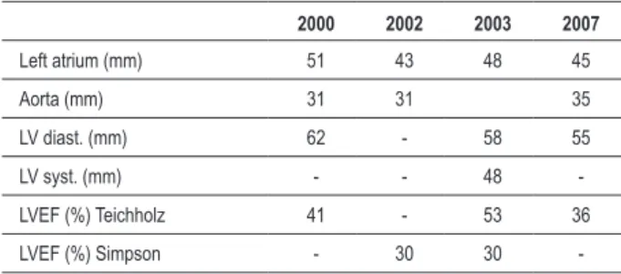
Echocardiography (February 2000) showed a left atrium with 51 mm, left ventricle with 62 mm, left ventricular ejection fraction of 41% (Teichholz), inferior and lateral wall akinesia, and moderate mitral regurgitation (Table 1).
After this episode, he developed dyspnea on exertion, which progressed to dyspnea at rest and orthopnea, accompanied by leg edema. Approximately two years after the infarction, he was hospitalized for right lobar pneumonia and empyema, which was drained (December 2001).
The patient was then referred to InCor for treatment of heart failure.
He also had hypertension and type II diabetes mellitus, and had been a smoker up until the time of infarction.
He was on glibenclamide 10 mg, isosorbide mononitrate 80 mg, spironolactone 100 mg, digoxin 0.25 mg, furosemide 120 mg, captopril 100 mg, and ASA 100 mg daily.
Physical examination (01/18/2002) revealed an emaciated patient, with a heart rate of 104 beats per minute, blood pressure of 120/86 mmHg , grade 3/4 jugular venous distension; pulses were normal and symmetrical to palpation;
there was a drain in his right hemithorax and crackles in the lower third of this hemithorax; cardiac auscultation showed accentuated intensity of the pulmonary component of the second heart sound, with no murmurs. The liver was enlarged and tender, and was palpated at 8 cm below the right costal margin. Grade 2+ pitting leg edema was present.
ECG (01/06/2002) revealed sinus rhythm, with a heart rate of 97 bpm; low-voltage QRS complexes in the frontal plane; an electrically inactive area in inferior wall; and ventricular repolarization abnormalities (Figure 1).
Laboratory tests showed anemia, hyperuricemia, and elevated glycated hemoglobin (Table 2).
Echocardiogram (February 2002) showed inferior, lateral and apical wall akinesia, pronounced left ventricular dilatation and dysfunction with ejection fraction of 30% (Simpson). There was mild left atrial dilatation and mild mitral regurgitation (Table 1). Carvedilol and insulin were started, and captopril was replaced by enalapril, with improvement of dyspnea, which was now triggered by moderate exertion.
Dobutamine-stress echocardiography revealed septal and anterior wall viability; anterior ischemia; and a healed area in lateral, posterior and mid-basal inferior walls.
Repeat catheterization (08/28/2005) revealed left ventricular pressures (Syst/Diast1/Diast2) of 140/19/40 mmHg. Coronary cineangiography showed right coronary occlusion, 40% lesion of the proximal portion of the anterior interventricular branch, and proximal occlusion of the circumflex branch; and presence of diffuse and severe left ventricular hypokinesia.
The patient had heart failure decompensations with dyspnea on minimal exertion in 2005 and 2007, the latter attributed to bronchopneumonia and worsening of renal failure, and was hospitalized again in July 2009 for decompensation and pneumonia.
Table 1 - Echocardiograms
2000 2002 2003 2007
Left atrium (mm) 51 43 48 45
Aorta (mm) 31 31 35
LV diast. (mm) 62 - 58 55
LV syst. (mm) - - 48
-LVEF (%) Teichholz 41 - 53 36
LVEF (%) Simpson - 30 30
-Case 6 − Late Cardiogenic Shock after Myocardial Infarction in a 50
Year-Old Hypertensive and Diabetic Man
Wilma Noia Ribeiro, Alice Tatsuko Yamada, Paulo Sampaio Gutierrez
Instituto do Coração (InCor) - HC-FMUSP, São Paulo, SP, Brazil
Keywords
Shock, cardiogenic; myocardial infarction; hypertension; diabetes mellitus.
Section Editor: Alfredo José Mansur ([email protected])
Associated Editors: Desidério Favarato ([email protected]) Vera Demarchi Aiello ([email protected])
Mailing Address: Vera Demarchi Aiello •
Av. Dr Enéas C. Aguiar, 44, Sub-solo, bloco I, Cerqueira César. Postal Code 05403-000, São Paulo, SP – Brazil
Renal replacement therapy for hemodyalisis became necessary as of mid-2008.
After one week of hypotension during the hemodialysis sessions and worsening of dyspnea, he was referred to the InCor emergency service.
On physical examination (on 01/19/2009) the patient was pale, eupneic, with mild jugular venous distension, heart rate of 88 bpm, blood pressure of 80/55 mmHg; pulmonary auscultation revealed crackles in both bases; cardiac auscultation revealed irregular rhythm, with normal heart sounds and no murmurs; the liver was enlarged and palpated at 4 cm below the costal margin, and there was moderate ascites; the extremities were cold and there was grade 2+ pitting leg edema, with no signs of deep venous thrombosis.
Laboratory tests revealed anemia, severe renal failure, increased count of band neutrophils, lymphopenia and thrombocytopenia (Table 2).
ECG (December, 19) revealed atrial fibrillation with a mean heart rate of 90 bpm; low voltage in the frontal plane; right bundle branch block; and an inactive area in the inferior wall (Figure 2).
Fluid, vasoactive amines and broad-spectrum antibiotics were administered.
The patient progressed with worsening of dyspnea and of peripheral blood flow, cyanosis and decreased level of consciousness, and required orotracheal intubation for respiratory support. During this procedure, the patient presented with bradycardia followed by cardiopulmonary arrest in pulseless electrical activity, with recovery within 2 min.
After intubation and recovery of the baseline rhythm (atrial fibrillation), there were diffuse ronchi and crackles in pulmonary bases; oxygen saturation was 96% with FIO2 of 100%. A few hours later he had another cardiac arrest in pulseless electrical activity preceded by bradycardia and recovered in 10 min. Post-arrest blood pressure was 117 x 69 mmHg, and heart rate of 120 bpm. He had three additional cardiac arrests and eventually died in asystole on the early morning of 12/21/2009.
Clinical aspects
We report the case of a 50-year-old patient with signs and symptoms of heart failure syndrome that appeared after an episode of acute myocardial infarction. The main working diagnosis for the case is ischemic cardiomyopathy.
Ischemic cardiomyopathy is defined by the occurrence of congestive heart failure in patients with coronary artery disease without concomitant hypertension, primary heart valve disease, ventricular aneurysm, or other known cause of cardiomyopathy. It is associated with severe diffuse coronary artery disease and multiple infarctions.
Obstructive coronary artery disease is currently the major cause of death worldwide, with seven million deaths per year, accounting for 12.8% of all deaths1. Its occurrence is
expected to increase in the coming years, as the incidence of cardiovascular risk factors such as population aging and the prevalence of metabolic syndrome also increase.
It is the major cause of systolic heart failure in Brazil, accounting for 29.7% of cases2. This patient had several risk
Anatomopathological Session
Ribeiro et al.Anatomopathological Session
factors that contributed to the development of coronary artery disease: male gender; systemic arterial hypertension; diabetes mellitus; smoking; and dyslipidemia.
According to the Framingham studies, the risk for individuals between 40-44 years of age to develop coronary artery disease within 10 years is of 7% for men and 2% for women. Although, based on the patient’s age, this risk was low, he presented with the other factors mentioned, which probably contributed to the early development of the atherosclerotic event3.
The clinical manifestations of ischemic cardiomyopathy are similar to those of other dilated cardiomyopathies of different etiologies. Among the clinical symptoms are dyspnea on exertion, orthopnea and paroxysmal nocturnal dyspnea. Chest pain may also be another common patient complaint. The clinical signs may be those of pulmonary congestion (crackles) or those of systemic venous congestion (jugular venous distension, liver enlargement and leg edema).
The first echocardiogram showed segmental wall motion abnormalities consistent with the findings of cineangiography, which showed right coronary artery occlusion and severe lesion in the circumflex branch, with signs of recanalization. These findings are common in ischemic cardiomyopathy and corroborate the diagnosis of previous acute myocardial infarction.
Acute myocardial infarction caused by occlusion of the circumflex artery is the one that is the most difficult to identify. Studies show that the anterior descending artery, right coronary artery, and circumflex artery are identified as the culprit vessels for the acute event in 40.3%, 43.3% and 14.8% of cases, respectively. This difficulty in the identification is due to lower sensitivity of electrocardiography to detect the abnormalities caused by the occlusion of this artery, which frequently prevents an early and effective reperfusion strategy4.
Among the differential diagnoses for this case is dilated chagasic cardiomyopathy. This patient had negative serologic tests for Chagas disease, which does not fully rule out Table 2 - Laboratory tests
2002 2004 2005 2007 2009
Hemoglobin (g/dL) 10,3 13,8 15,3 10,4
Hematocrit (%) 32 41 46 45
Leucocytes/mm³ 9.900 6.800 7.600 8.100
Neutrophils (%) 69 70 86 (26 bands)
Lymphocytes (%) 19 14 2
Platelets/mm³ 515.000 293.000 80.000
PT (INR) 1,1 1,0 2,1
APTT (rel) 1,07 1,01 1,28
Glucose (mg/dL) 103 200 263 157
Glycated hemoglobin (%) 8,6 9,9
BUN (mg/dL) 72 56 112 170 96
Creatinine (mg/dL) 1,1 1,4 2,5 2,77 5,27
Uric acid (mg/dL) 10,6 9,8 9,9
Total cholesterol (mg/dL) 156 262 198
HDL-c (mg/dL) 31 53 36
LDL-c (mg/dL) 96 − −
Triglycerides (mg/dL) 147 1.607 881
Total proteins (g/dL) 9,1
Albumin (g/dL) 3,1
TSH (µUI/mL) 6,3
Sodium (mEq/L) 139 135 138 141 138
Potassium (mEq/L) 4,8 5,9 6,6 5,3 4,5
Iron (µg/dL) 48
Total iron-binding cap. (µg/dL) 219
Serologic test for Chagas disease negative
BNP (pg/mL) 1.923
the diagnosis of the disease, because the Latin-American guideline on Chagas disease published in 2011 recommends two negative serologic test to rule out this diagnosis5. Other
aspects that make this diagnosis less feasible are the fact that the echocardiogram did not show conduction disturbances typical of this disease and also that the echocardiogram did not reveal diffuse hypokinesia, but rather left ventricular segmental motion abnormalities.
Although the patient had a history of hypertension, the hypothesis of maladaptive hypertensive cardiomyopathy in a more advanced stage with dilatation is less likely, because the echocardiogram showed ventricular dysfunction due to segmental abnormalities.
Idiopathic dilated cardiomyopathy should also be considered for this case. It typically affects individuals between 18-50 years of age, and may occur in children and the elderly. It is more common among males and Blacks, and at least 25% of cases are genetically transmitted. Genetic factors associated with alterations of the immunologic response and infectious factors are believed to act synergistically in the development of the structural abnormalities and subsequent onset of the clinical manifestations. Approximately 10-20% of cases of idiopathic dilated cardiomyopathy are estimated to be caused by a complication of a previous viral infection6.
In December 2002, the patient presented with a recurrent episode suggestive of decompensated heart failure with clinical signs of systemic and pulmonary venous congestion, which seemed to have been triggered by a pulmonary infectious disease.
In December 2009, the patient had another episode suggestive of decompensated heart failure with clinical signs of low cardiac output associated with systemic and pulmonary venous congestion. Clinical measures for compensation of this condition were started, including the use of vasoactive amines and antibiotics. However, the patient had clinical worsening and a cardiopulmonary arrest in asystole on 12/21/2009.
The main differential diagnoses for the final clinical picture are pulmonary thromboembolism, cardiac tamponade; cardiogenic shock; and mixed shock (septic/cardiogenic), which will be further discussed.
Renal failure and hypothyroidism are risk factors for the development of pericardial effusion, which makes the working diagnosis of cardiac tamponade plausible in this context. Its clinical presentation results from restricted ventricular filling during diastole. Hypotension is usually present, although the compensatory mechanisms are able to keep blood pressure at normal levels in the early stages. Pulsus paradoxus; tachycardia; and signs of right ventricular failure such as increased jugular venous pulse, Kussmaul sign and enlarged liver on physical examination are also common findings.
The electrocardiographic findings commonly observed in cardiac tamponade are low voltage – which was present in this case, and QRS electrical alternans. Echocardiography is the noninvasive method used to confirm the diagnosis7.
Although pericardial abnormalities had not been mentioned in the previous patient’s echocardiographic studies, this working diagnosis cannot be ruled out.
Figure 2 - ECG. Atrial ibrillation rhythm, low-voltage QRS-complexes in the frontal plane, right branch conduction disturbance, indirect signs of right atrial overload
Anatomopathological Session
Ribeiro et al.Anatomopathological Session
Pulmonary thromboembolism is a common event in heart failure, and its incidence is two-fold higher in patients with left ventricular systolic dysfunction. In the present context, this diagnosis is less easy to be made due to the similar clinical presentation of the two conditions, thus requiring the use of imaging methods for diagnostic confirmation8. Atrial fibrillation,
which was present in this case, is another condition that also increases the incidence of pulmonary thromboembolism.
The typical clinical manifestations of pulmonary thromboembolism consist of dyspnea and hypoxemia out of proportion with the finding of pulmonary congestion, in addition to worsening or onset of signs of right ventricular failure. In the present case, the patient presented with signs of left ventricular failure, which could explain the dyspnea as well as right ventricular dysfunction, probably secondary to the elevated left filling pressures. Although physical examination had revealed accentuated S2 in the pulmonic area, which is suggestive of pulmonary hypertension, there is no record of pressure levels in the pulmonary artery in the echocardiographic studies performed.
Finally, the most probable working diagnoses are cardiogenic shock or mixed shock (cardiogenic/septic). Countless factors can trigger an acute relapse of chronically compensated heart failure, especially the incorrect use of medications and the natural progression of cardiomyopathy. Another factor that may have triggered or contributed to the clinical worsening of the patient was the onset of atrial fibrillation. Atrial contraction accounts for 20% of the systolic volume in normal individuals, reaching 33% in those with coronary artery disease. Failure in this contribution may result in worsening of the cardiac function, especially in patients with already compromised myocardial function9.
The hypothesis of myocardial ischemia should also be considered, because the patient had risk factors for coronary artery disease, had previous acute ischemic syndrome, and his electrocardiogram showed new abnormalities (atrial fibrillation and right bundle branch block). The fact that this acute relapse had not occurred during angina is what makes this diagnosis less probable. In addition to an atherosclerotic ischemic event, an embolic ischemic event may not be ruled out, although it is an uncommon complication also reported in patients with advanced dilated cardiomyopathy10.
Based on the history presented, the main triggering factor for the final clinical picture of this patient seems to have been the infectious disease. This is corroborated by the elevated band neutrophil count and by the fact that the patient had advanced heart and renal diseases, which made him more susceptible to infectious diseases.
Septic shock is a severe syndrome characterized by hemodynamic abnormalities and organ dysfunction as a result of the interaction of bacteria-released products with the cells. Myocardial dysfunction occurs during sepsis in approximately 40% of cases, and its pathogenesis is not fully understood. Studies have demonstrated that this myocardial depression is triggered mainly by the action of endotoxins and endogenous inflammatory mediators such as the tumoral necrosis factor and interleukins and, sometimes, also by the direct action of bacteria11.
The patient in the present case report had previous ventricular dysfunction, which was probably aggravated during
the septic event, thus leading to mixed shock refractory to medical treatment and subsequent death in December 2009.
(Dr. Wilma Noia Ribeiro; Alice Tatsuko Yamada)
Working diagnoses: syndromic diagnosis: congestive heart failure; etiology: ischemic heart disease; final event: mixed shock (cardiogenic – septic). (Dr. Wilma Noia Ribeiro; Alice Tatsuko Yamada)
Necropsy
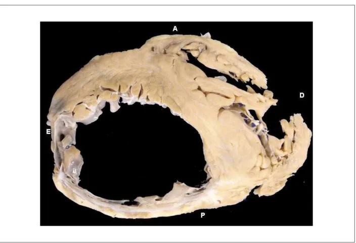
The main disease of our patient, which accounted for his death, was systemic atherosclerosis. There was a higher than 70% obstruction in the three main coronary artery branches, reaching 95% in the area of an old recanalized thrombus in the left circumflex branch; in the right coronary artery there was 95% obstruction by an atherosclerotic lesion and 98% in the site of an old recanalized thrombus (Figure 3). Noteworthy is the fact that this patient had an infarction while still young, at little more than 40 years of age (according to the records of a clinical summary of a previous hospital discharge, approximately 9 years prior to his death, therefore at approximately 41 years of age). More importantly, the area of ischemic necrosis was very extensive, reaching more than 40% of the left ventricular mass (Figure 4); in the past, such an extensive infarction was considered incompatible with life but, today, patients like the one reported manage to survive for a long time.
The patient also had benign nephrosclerosis associated with systemic arterial hypertension.
Death seems to have been a consequence of heart failure, with cardiogenic shock in the final stage.
Additionally, the microscopic study revealed the existence of giant cell inflammation affecting arteries of the intraparenchimal pulmonary area and intermediate coronary artery branches, as well as the myocardium itself (Figure 5). This is an uncommon pattern, because giant cell arteritis usually affects the temporal artery, the aorta or its major branches, or the pulmonary artery, but not the intraparenchimal branches of the latter, and even less frequently without the trunk being also affected. On the other hand, the association of arteritis with myocarditis is equally uncommon. Tests for infectious agents were negative, and the cause of the disease could not be established. It does not fit adequately any of the types of arteritis12, but it closer resembles Wegener’s granulomatosis,
which affects small-diameter vessels. It probably contributed to the clinical worsening of the patient.
Brain infarction was also observed in the right frontoparietal region. Although this probably resulted from atherosclerosis, the possibility of its being secondary to arteritis in the cerebral artery distribution cannot be ruled out, because the arteries of this region were not examined. This ischemic lesion did not play a significant role in the patient’s death. (Paulo Sampaio Gutierrez)
Anatomopathological diagnosis: healed myocardial
infarction resulting from coronary atherosclerosis. Cause of death: cardiogenic shock.
Figure 3 - Histological sections of right coronary artery. A - 2º. cm: 95% obstruction by a ixed atherosclerotic lesion; B - 5º. cm: 98% obstruction by atherosclerosis
complicated by old recanalized thrombus. Hematoxylin and eosin staining; objective magniication: 1x.
Figure 4 - Cross-section of heart showing extensive areas of thinning and ibrosis replacement in left ventricular posterior (inferior, diaphragmatic) and lateral walls. A:
Anatomopathological Session
Ribeiro et al.Anatomopathological Session
Figure 5 - Pictures showing histological sections of the arteries (A to E) and heart (F to H). A - intramyocardial coronary branch, with presence of giant cells. B
-intrapulmonary branch, with the same characteristics. C - intrapulmonary branch showing ibrointimal thickening which corresponds to the inner area of the internal
elastic lamina (IEL), stained in black. D - aorta, with no signiicant abnormalities. E - pulmonary artery trunk, with no signiicant abnormalities. F - myocarditis, with
presence of giant cells. G - myocardium with negative tests for fungi. H - myocardium with negative tests for alcohol-acid resistant bacilli. Hematoxylin and eosin (A, B,
1. World Health Organization (WHO). Top 10 causes of death. WHO Fact sheet Nº 310, updated June 2011. [Internet] [Cited in 2012 Jun 12]. . Available from http://www.who.int/mediacentre/factsheets/fs310/en/index.html.
2. Bochi EA, Marcondes-Braga FG, Bacal F, Ferraz AS, Albuquerque D, Rodrigues D, et al. Sociedade Brasileira de Cardiologia. Atualização da Diretriz brasileira de insuficiência cardíaca crônica – 2012. Arq Bras Cardiol. 2012;98(1 supl. 1):1-33.
3. Wilson PW, D’Agostino RB, Levy D, Belanger AM, Silbershatz H, Kannel WB. Prediction of coronary heart disease using risk factor categories. Circulation. 1998;97(18):1837-47.
4. Krishnaswamy A, Lincoff AM, Menon V. Magnitude and consequences of missing the acute infarct-related circumflex artery. Am Heart J. 2009;158(5):706-12.
5. Andrade JP, Marin Neto JA, Paola AA, Villas-Boas F, Oliveira GM, Bacal F, et al.; Sociedade Brasileira de Cardiologia. I Diretriz Latino Americana para o diagnóstico e tratamento da cardiopatia chagásica. Arq Bras Cardiol. 2011;97(2 supl.3):1-47.
6. Baboonian C, Treasure T. Meta-analysis of the association of enteroviruses with human heart disease. Heart. 1997;78(6):539-43.
7. Spodick DH. Acute cardiac tamponade. N Engl J Med. 2003;349(7):684-90.
8. Piazza G, Goldhaber SZ. Pulmonary embolism in heart failure. Circulation. 2008;118(15):1598-601.
9. Hamby RI, Noble WJ, Murphy DH, Hoffman I. Atrial transport function in coronary disease: relation to left ventricular function. J Am Coll Cardiol. 1983;1(4):1011-7.
10. Canali G, Girardi P, Barbieri E. [Coronary embolus and acute myocardial infarction in a patient with dilated cardiomyopathy and chronic atrial fibrillation]. G Ital Cardiol (Rome). 2006;7(5):365-8.
11. Fernandes Junior CJ, Iervolino M, Neves RA, Sampaio EL, Knobel E, Sustovich DR. [The myocardium in sepsis: anathomo-pathologic aspects]. Arq Bras Cardiol. 1988;50(3):175-8.
12. Jennette JC, Falk RJ, Andrassy K, Bacon PA, Churg J, Gross WL, et al. Nomenclature of systemic vasculitides. Proposal of an international consensus conference. Arthritis Rheum. 1994;37(2):187-92.